

81fa7bca-64fc-4282-8af9-2bd9c1d6cbcb.jpeg
Answer all the inentificaion questions under forensic medicine 5 mark for exam purpose add flow chart and pictures if needed add neet point wise
Reading File
The image shows Identification topics in Forensic Medicine for a 5-mark exam. The topics are: Gustafson Method, DNA Fingerprinting, Dactylography, Superimposition, Tattoo Marks, Medicolegal Importance of Age, and Ossification Centres.
Searching sources...
Finding Sources
Finding Sources
Finding Sources
Searching Images
Gustafson method dental age estimation forensic
Finding Sources
Searching Images
fingerprint ridge patterns dactylography forensic identification
Searching Images
ossification centres age estimation bones radiology
Finding Sources
Finding Sources
Finding Sources
Finding Sources
Finding Sources
Finding Sources
Reading File
Reading File
Reading File
Reading File
Reading File
Reading File
I now have comprehensive textbook content. Let me also fetch the key images from the textbook to include in the answer.
Loading Image
Loading Image
Loading Image
Loading Image
Loading Image
I now have all the key content and images. Let me compile the comprehensive exam answer.
FORENSIC MEDICINE - IDENTIFICATION: 5-Mark Questions (NEET/Exam Prep)
Source: The Essentials of Forensic Medicine and Toxicology, 36th Edition (2026) | Forensic Anthropology - A Comprehensive Introduction
1. GUSTAFSON'S METHOD (Age Estimation from Teeth)
Definition
Gustafson's method is an objective approach for estimating age from teeth in deceased individuals, based on six physiological changes occurring in teeth with advancing age.
Mnemonic: APSRTC (easy to remember!)
| Change | What Happens |
|---|---|
| A - Attrition | Wear of occlusal surface - enamel > dentin > pulp exposed |
| P - Periodontosis | Recession of gums and periodontal tissue; teeth loosen and fall |
| S - Secondary dentin | Deposition of secondary/irregular dentin; pulp cavity narrows |
| R - Root resorption | Root tip gets absorbed; more in old age |
| T - Transparency | Root tip becomes translucent with age (best single indicator) |
| C - Cementum apposition | Cementum increases in thickness, especially near root end |
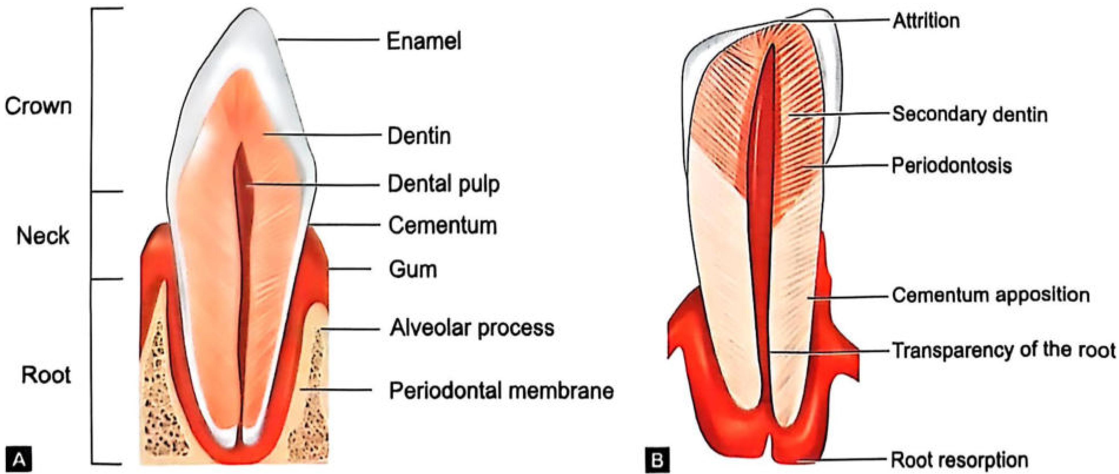
Scoring System
Stage 0 = No change
Stage 1 = Beginning of change
Stage 2 = Obvious changes
Stage 3 = Maximum change
Each change scored 0-3 → Total score correlates with age
Procedure
- Estimate periodontosis before extraction
- Grind tooth to ~1 mm (for transparency)
- Further grind to 0.25 mm (for microscopy)
- Score each of the 6 changes
- Total score plotted on regression graph → age estimated with ±4-7 years error
Key NEET Points
- Best teeth: Anterior teeth (incisors most suitable; molars least)
- Third molar: Quite unsuitable
- Changes absent: Below 15 years of age
- Error increases: Above 50 years of age
- Johanson's modification: More accurate than original Gustafson
- Boyde's method: Uses daily incremental lines (cross striations) in enamel - useful for dead infants
- Stack's method: Uses weight and height of erupting teeth in infants
2. DNA FINGERPRINTING
Definition
DNA fingerprinting (DNA profiling/genetic fingerprinting) is a technique to identify an individual using their unique DNA pattern. Discovered by Sir Alec Jeffreys in 1984 (UK).
Flowchart: DNA Fingerprinting Process
Biological sample (blood/semen/saliva/hair/bone)
           ↓
    Extract DNA
           ↓
   Cut with Restriction Endonucleases (RE)
           ↓
  DNA fragments of varying sizes
           ↓
  Gel Electrophoresis (separation by size)
           ↓
  Southern Blotting (transfer to nitrocellulose membrane)
           ↓
  Add radioactive DNA probe (binds to VNTR/STR sequences)
           ↓
  Autoradiography (X-ray film exposed)
           ↓
  Banding pattern = DNA "fingerprint"
           ↓
  Compare with suspect/reference sample
VNTR - Variable Number Tandem Repeats
- Highly polymorphic regions of DNA
- Unique to each individual (except identical twins)
- STR (Short Tandem Repeats) - used in modern analysis (PCR-based, faster)
Uses in Forensic Medicine
- Identification of criminals from crime scene samples
- Paternity disputes - establish biological parentage
- Identification of decomposed/disaster victims
- Rape cases - match semen to suspect
- Immigration disputes - family relationship confirmation
Key NEET Points
- Gold standard of identification
- Unique for every individual EXCEPT identical (monozygotic) twins
- Smallest sample can yield results (PCR amplification)
- Mitochondrial DNA (mtDNA) - used when nuclear DNA unavailable; inherited maternally
- Y-chromosome analysis - for paternal lineage
- Degraded samples - STR more sensitive than RFLP
- PCR (Polymerase Chain Reaction) amplifies tiny DNA samples
3. DACTYLOGRAPHY (Fingerprinting)
Definition
Dactylography is the study of fingerprints for identification purposes. Also called Dactyloscopy.
Historical Points (NEET Favourite!)
- Sir Francis Galton - first to classify fingerprints scientifically (1892)
- Sir Edward Henry - introduced fingerprint classification system used by Scotland Yard (1897)
- Juan Vucetich - developed first fingerprint classification in Argentina
- India: First fingerprint bureau established at Calcutta (Kolkata) in 1897
Galton's Properties (Why fingerprints are used)
- Uniqueness - No two fingers have identical prints (even twins differ)
- Permanence - Patterns formed by 4th month of fetal life and remain until decomposition
- Reproducibility - Can always be re-taken
Fingerprint Patterns
         FINGERPRINTS
        /     |      \
     ARCH   LOOP    WHORL
    (5%)   (65%)   (30%)
      |    /    \      |
   Plain Radial Ulnar Plain/Central
   Arch  Loop   Loop  Pocket/Double
                      loop/Accidental
| Pattern | Direction | Frequency |
|---|---|---|
| Arch (Plain/Tented) | No core/delta | ~5% |
| Loop - Radial | Opens towards radius (thumb) | ~5% |
| Loop - Ulnar | Opens towards ulna (little finger) | ~60% |
| Whorl | Circular/spiral; 2 deltas | ~30% |
Types of Fingerprints at Crime Scene
- Patent/Visible prints - Seen with naked eye (blood, grease, paint)
- Latent prints - Invisible; need development (sweat/oil deposits)
- Plastic prints - Impressed in soft surface (wax, tar, putty)
Development of Latent Prints
- Dusting with aluminum powder (light surfaces), graphite/carbon (dark surfaces)
- Iodine fuming - oldest method; temporary
- Ninhydrin - reacts with amino acids in sweat; used on paper
- Silver nitrate - reacts with NaCl in sweat
Key NEET Points
- Loops are the most common fingerprint pattern (~65%)
- Arch is the least common (~5%)
- Fingerprints are present from 4th fetal month
- Cheiloscopy = lip print identification
- Palatoscopy/Rugoscopy = palatal rugae identification
- Dactylography = fingerprints | Poroscopy = study of sweat pores
- Attempts to alter fingerprints: cutting/burning - ridges grow back identically
4. SUPERIMPOSITION
Definition
Superimposition is a technique of identification by placing the photograph of a missing person over the photograph or image of an unidentified skull, to check if the features correspond.
Flowchart: Skull-Photo Superimposition
Unknown skull found
        ↓
Photograph skull from same angle as ante-mortem photo
        ↓
Superimpose skull image over ante-mortem photo
(using video/digital technique or photographic overlay)
        ↓
Check for correspondence of:
- Orbital margins
- Nasal aperture
- Zygomatic arch
- Dental features
- Chin profile
- Forehead contour
        ↓
Match = Positive identification | No match = Exclusion
Methods
- Photographic superimposition - direct overlay of photos
- Video superimposition (Helmer's technique) - two images superimposed on TV screen; most commonly used
- Digital superimposition - modern computerized method
Important Forensic Case
- First used in Ruxton murder case (1935, UK) - Dr. Buck Ruxton killed his wife and maid; identified by superimposition
Key NEET Points
- Helmer's technique = video superimposition (most used)
- Limitations: Requires good quality ante-mortem photograph; cannot give 100% certainty alone
- Used for skeletal remains identification
- NOT reliable as sole evidence; used along with other methods
- Confirms identity but cannot exclude alone
- Also used: Dentition superimposition (superimpose dental X-rays)
5. TATTOO MARKS
Definition
Tattoo marks are permanent marks made on the skin by pricking with a needle and introducing coloring matter (pigment) under the dermis.
Medicolegal Importance
- Personal identification - helps identify living persons, criminals, bodies
- Indicates profession/occupation - sailors, military, criminals
- Age estimation - professional tattoos suggest age >18 years (minors rarely have elaborate tattoos)
- Race and religion - certain tattoos indicate community
- Criminal identification - gang symbols, prison tattoos
Why Tattoos are Permanent
- Pigment deposited in dermis (not epidermis)
- Dermis cells do not shed like epidermis
- Common pigments: India ink (black), cinnabar (red - mercuric sulphide), ultramarine (blue)
Removal Attempts
- Laser removal (partial), dermabrasion, excision
- Even with removal, scarring pattern remains as identification
Tattoos in Dead Bodies
- Remain identifiable even after decomposition (in dermis)
- Can survive moderate putrefaction
- In burnt bodies - may survive if superficial burning
Key NEET Points
- Located in dermis (explains permanence)
- Most common pigment: India ink (black)
- Red pigment = Cinnabar (mercuric sulphide) - may cause allergic reactions
- Professional tattoos better indicator of identity than amateur
- Tattoos can reveal: name, religion, social group, criminal history
6. MEDICOLEGAL IMPORTANCE OF AGE
Why Age Estimation Matters in Law
| Legal Context | Age Threshold | Importance |
|---|---|---|
| Age of consent (sexual) | 18 years | Below = statutory rape |
| Juvenile/POCSO | Below 18 years | Different legal treatment |
| Death sentence | Must be >18 at time of crime | Cannot hang juveniles |
| Marriage (female) | 18 years | Below = child marriage |
| Marriage (male) | 21 years | Below = not legal |
| Voting rights | 18 years | Eligibility |
| Driving licence | 18 years | Eligibility |
| Criminal responsibility | 7 years | Below = doli incapax |
| Doli capax | 7-12 years | May be responsible |
Methods of Age Estimation
         AGE ESTIMATION
        /       |        \
  LIVING      DEAD      SKELETAL
  PERSON      BODY      REMAINS
     |           |          |
 Clinical    Teeth       X-ray of
 exam +      + Bones     bones/
 X-ray       changes     epiphyses
Age Groups and Features
Infant/Child (0-14 years):
- Deciduous teeth eruption
- Ossification centres (carpal rule: No. of carpal bones visible = age in years, 2-6 yrs)
- Fontanelles closure
- Crown-heel length
Adolescent (14-25 years):
- 3rd molar development
- Epiphyseal fusion (key for legal purposes)
- Secondary sexual characteristics
Adult (>25 years):
- Gustafson's method (teeth)
- Skull suture closure
- Pubic symphysis changes
- Sternal rib end changes
- Degenerative changes
7. OSSIFICATION CENTRES
Definition
Ossification is the process by which cartilage is converted to bone. The time of appearance and fusion of ossification centres is used for age estimation.
Types
- Primary centre - appears in shaft (diaphysis); present at birth in long bones
- Secondary centre - appears at ends (epiphysis); used for age estimation
Epiphyseal Fusion Stages
Stage 0: No union
Stage I: Beginning of union
Stage II: Partial union (¼, ½, ¾)
Stage III: Recently united
Stage IV: Completely united
Key Ossification Timings (NEET High Yield)
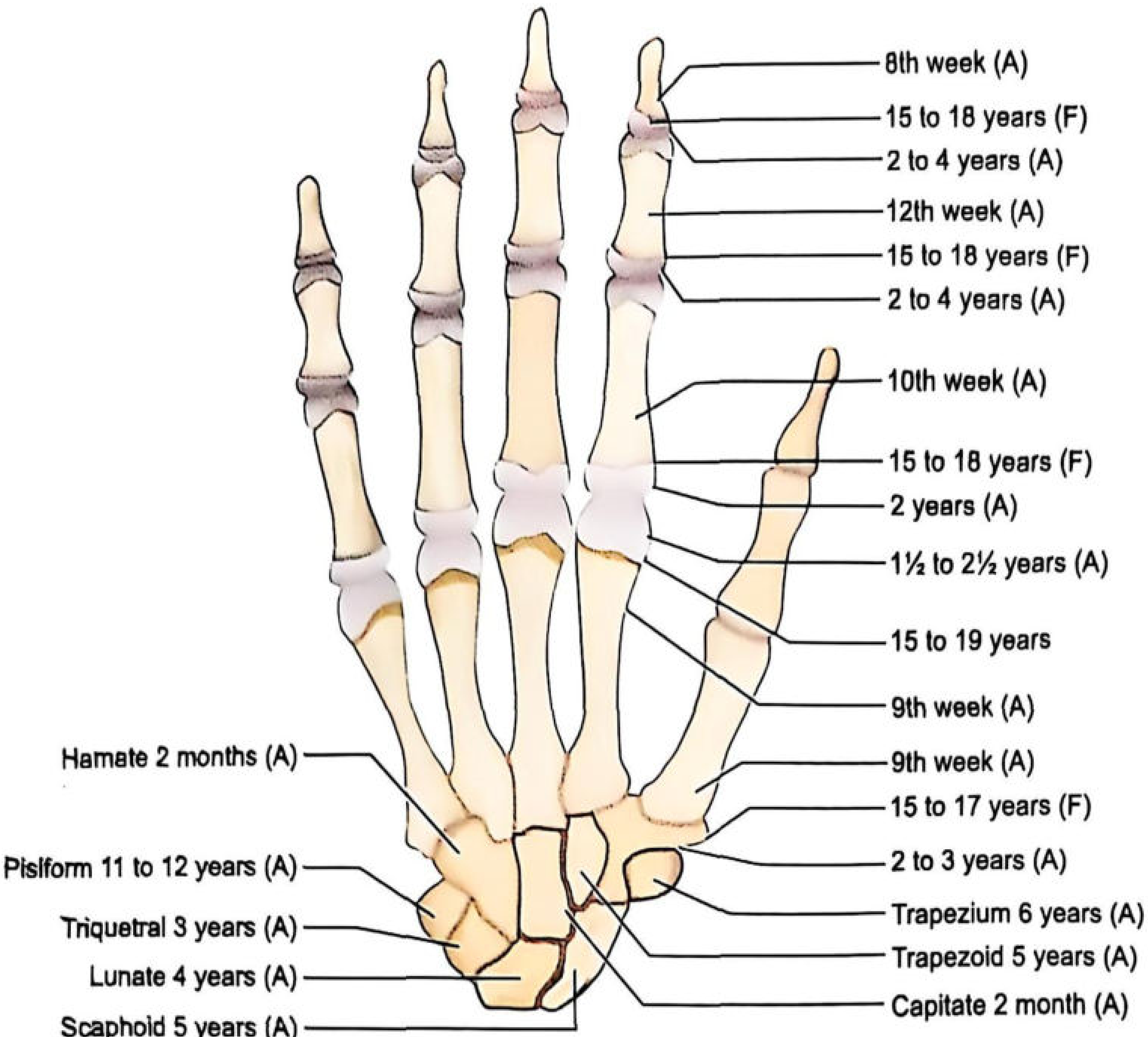
CARPAL BONES (useful 2-6 years):
| Bone | Appears |
|---|---|
| Capitate | 2-3 months |
| Hamate | 1st year |
| Triquetrum | 3 years |
| Lunate | 4 years |
| Scaphoid + Trapezoid | 5 years |
| Trapezium | 6 years |
| Pisiform | 11-12 years |
Rule: Between 2-6 years, number of carpal bones visible on X-ray = approximate age in years
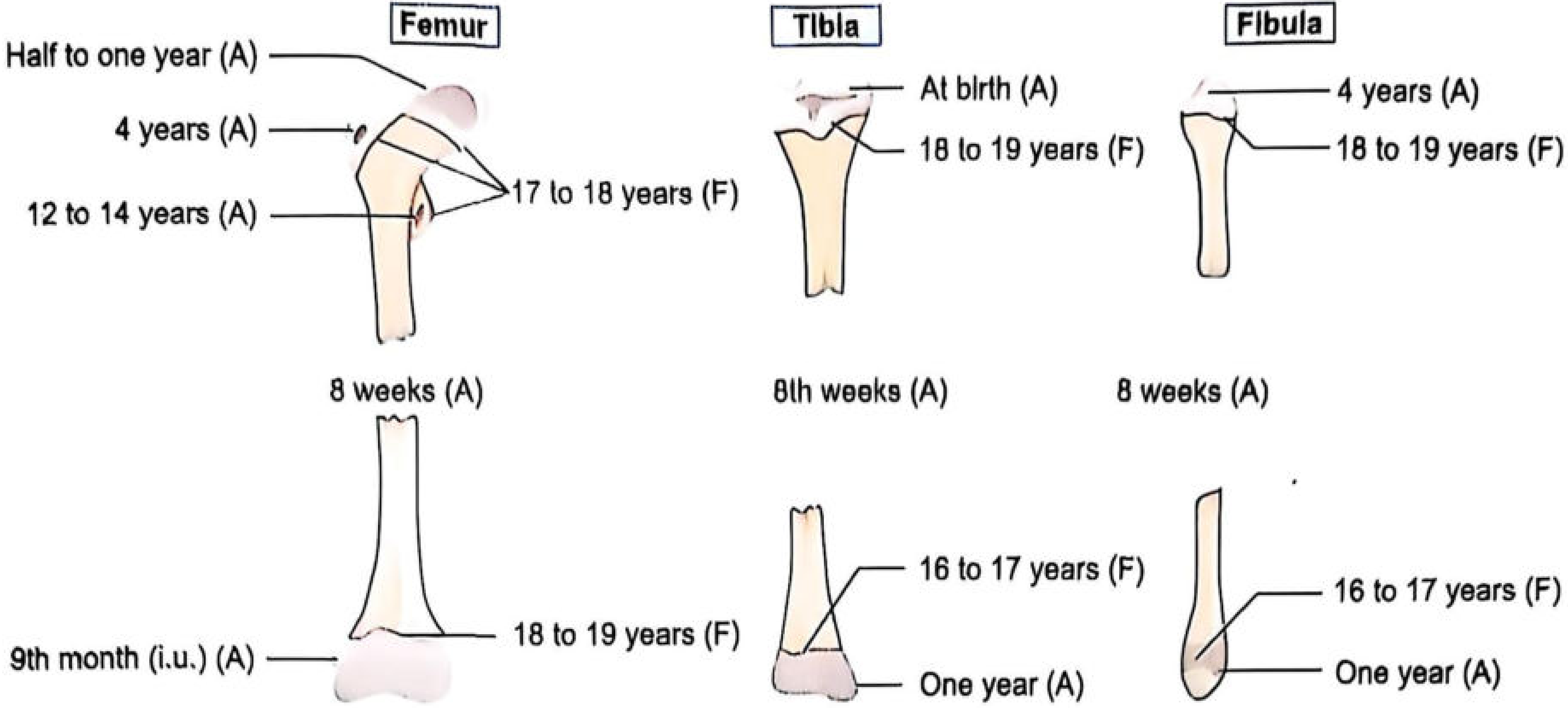
LONG BONES (Lower limb):
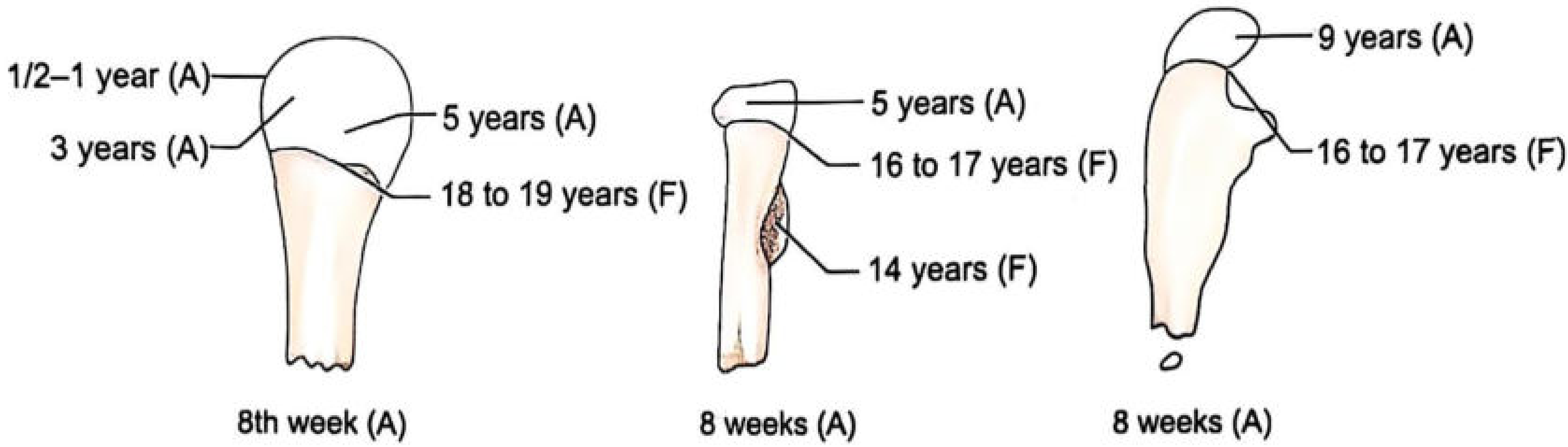
UPPER LIMB - Humerus, Radius, Ulna:
Key Joints (NEET Favourite)
| Joint | Age of Fusion |
|---|---|
| Elbow (medial/lateral epicondyle, olecranon, radial head) | 11-14 years |
| Shoulder (acromion, coracoid, head of humerus) | 15-18 years |
| Greater trochanter of femur | 16-17 years (fuses) |
| Knee (lower femur = at birth; upper tibia = at birth) | Lower femur fuses 18-19 yrs |
| Iliac crest | Appears 14-15 yrs, fuses 21-25 yrs |
| Ischial tuberosity | Fuses ~20-23 years |
Other Bones
- Sternum: 4 pieces fuse 14-25 yrs (bottom to top); xiphoid unites at ~40 yrs; manubrium fuses at ~60 yrs
- Hyoid bone: Greater cornu unites with body at 40-60 years
- Skull sutures: Endocranial closure begins ~22 yrs; complete by 30-40 yrs
Fontanelle Closure
- Lateral + occipital fontanelles: Close within 2 months
- Posterior fontanelle: 6-8 months
- Anterior fontanelle: 1.5-2 years (most important)
SUMMARY FLOWCHART: Identification in Forensic Medicine
UNKNOWN INDIVIDUAL / BODY
          ↓
  PRIMARY METHODS (Most reliable)
  ┌─────────────────────────────────┐
  │  DNA Fingerprinting (gold std.) │
  │  Fingerprints (Dactylography)   │
  └─────────────────────────────────┘
          ↓
  SECONDARY METHODS
  ┌──────────────────────────────────────────┐
  │  Gustafson's Method (age from teeth)     │
  │  Ossification Centres (skeletal age)     │
  │  Superimposition (skull-photo matching)  │
  └──────────────────────────────────────────┘
          ↓
  SUPPORTING METHODS
  ┌─────────────────────────────────┐
  │  Tattoo marks                   │
  │  Scars/deformities              │
  │  Clothing/personal effects      │
  │  Blood group                    │
  └─────────────────────────────────┘
          ↓
  IDENTITY ESTABLISHED or EXCLUDED
RAPID NEET REVISION - One-Liners
| Topic | Key Fact |
|---|---|
| Gustafson's mnemonic | APSRTC (Attrition, Periodontosis, Secondary dentin, Root resorption, Transparency, Cementum) |
| Best single Gustafson criterion | Root transparency |
| Gustafson - error margin | ±4-7 years |
| Gustafson - changes absent below | 15 years |
| DNA fingerprinting inventor | Sir Alec Jeffreys, 1984 |
| DNA identical in | Identical (monozygotic) twins |
| Fingerprint classification (India) | Sir Edward Henry |
| First fingerprint bureau in India | Calcutta, 1897 |
| Most common fingerprint pattern | Ulnar loop (~60-65%) |
| Least common pattern | Plain arch (~5%) |
| Tattoo pigment location | Dermis |
| Superimposition - first case | Ruxton murder case, 1935 |
| Superimposition method | Helmer's video technique |
| Carpal bone rule | No. of carpal bones on X-ray = age (2-6 years) |
| Anterior fontanelle closes | 1.5-2 years |
| Hyoid cornu fuses | 40-60 years |
| Manubrium fuses with sternum | Old age (~60 years) |
| Eruption of temporary teeth (1st) | Lower central incisor: 6-8 months |
| Third molar erupts | 17-25 years |
| Legal age for consent (India) | 18 years |
| Doli incapax (cannot be tried) | Below 7 years |
This is a shared conversation. Sign in to Orris to start your own chat.