I now have rich content from the textbooks. Let me compile a thorough, well-structured study guide for a 1st year MBBS student.
Homeostasis - Complete MBBS 1st Year Study Notes
Based on Medical Physiology (Boron & Boulpaep), Ganong's Review of Medical Physiology (26th Ed.), and Costanzo Physiology (7th Ed.)
1. Definition & Historical Background
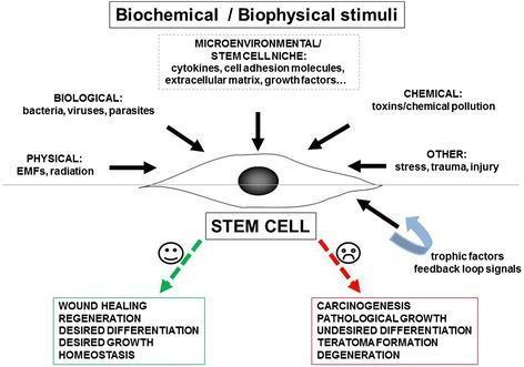
Homeostasis is the control and maintenance of a vital parameter within a narrow, optimal range in the body's internal environment.
- The concept of the milieu intérieur (internal environment) was introduced by the French physiologist Claude Bernard (1813-1878). He observed that living organisms maintain a stable internal environment despite changes in the external world.
- Walter B. Cannon later coined the term "homeostasis" (from Greek: homoios = similar, stasis = standing still) and extended Bernard's work, emphasizing that an organism's ability to survive depends on maintaining this internal constancy.
"The result of many homeostatic systems controlling many vital parameters is a milieu intérieur with a stable composition."
- Medical Physiology (Boron & Boulpaep)
2. What Parameters Are Regulated?
The body maintains homeostasis at two levels:
a) Milieu Intérieur (Extracellular fluid / whole body level)
| Parameter | Normal Range |
|---|
| Body core temperature | 36.5 - 37.5°C |
| Blood glucose | 70-110 mg/dL (fasting) |
| Arterial blood pressure | ~120/80 mmHg |
| Blood osmolality | 275-299 mOsm |
| Plasma O₂ (PaO₂) | 80-100 mmHg |
| Plasma K⁺ | 3.5 - 5.0 mEq/L |
| Plasma Ca²⁺ | 8.5 - 10.5 mg/dL |
| Blood pH (H⁺) | 7.35 - 7.45 |
| Blood volume | ~5 L |
b) Intracellular Level
Single cells also regulate their own volume, Na⁺, Ca²⁺, H⁺, and ATP levels - homeostasis is a universal principle at every level of biological organization.
3. Components of a Homeostatic Feedback System
Every homeostatic mechanism has four essential elements:
Stimulus → Sensor (Receptor) → Control Center → Effector → Response
                    ↑___________ Feedback ___________________↑
| Component | Role | Example (Blood Glucose) |
|---|
| Stimulus | Change in the vital parameter | Rise in blood glucose after a meal |
| Sensor/Receptor | Detects the change | Beta cells of pancreas detect high glucose |
| Control Center | Compares input to set-point; generates error signal | Pancreatic islets |
| Effector | Executes the corrective response | Insulin secretion |
| Set-Point | The ideal "target" value | ~80-100 mg/dL |
| Gain | The proportionality factor that amplifies the error signal | Magnitude of insulin release |
4. Negative Feedback - The Master Mechanism
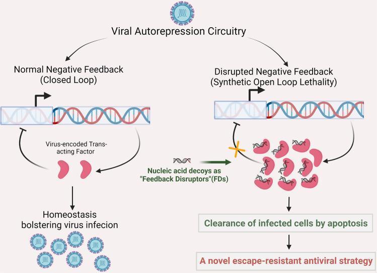
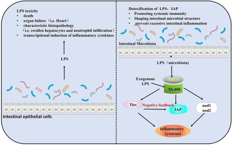
Negative feedback is the most common and most important homeostatic mechanism. It opposes (negates) the initial stimulus, returning the parameter to its set-point.
How it works:
- A vital parameter deviates from the set-point
- Sensors detect the deviation
- Control center generates a corrective output
- Effectors act opposite to the direction of the deviation
- The parameter returns toward the set-point
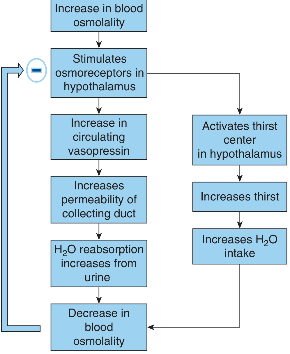
Classic Example: Blood Osmolality Regulation
Ganong's Review of Medical Physiology, 26th Ed.
Step-by-step:
- Dehydration → blood osmolality rises above 299 mOsm
- Osmoreceptors in the hypothalamus are activated
- Two parallel responses occur:
- Vasopressin (ADH) released from posterior pituitary → acts on renal collecting duct → inserts aquaporins → increases water reabsorption from urine
- Thirst center activated → increases water intake
- Blood osmolality falls back to normal
- Negative feedback switches off vasopressin release
Other Key Examples of Negative Feedback:
| System | Sensor | Control Center | Effector | Response |
|---|
| Temperature | Thermoreceptors | Hypothalamus | Sweat glands, muscles | Sweating / shivering |
| Blood glucose ↑ | Beta cells (pancreas) | Islets of Langerhans | Pancreas | Insulin secretion |
| Blood glucose ↓ | Alpha cells (pancreas) | Islets of Langerhans | Pancreas | Glucagon secretion |
| Blood pressure ↑ | Baroreceptors (carotid sinus, aortic arch) | Cardiovascular centre (medulla) | Heart, vessels | ↓HR, vasodilation |
| Thyroid hormone | Hypothalamus, ant. pituitary | HPT axis | Thyroid gland | Feedback inhibition of TRH/TSH |
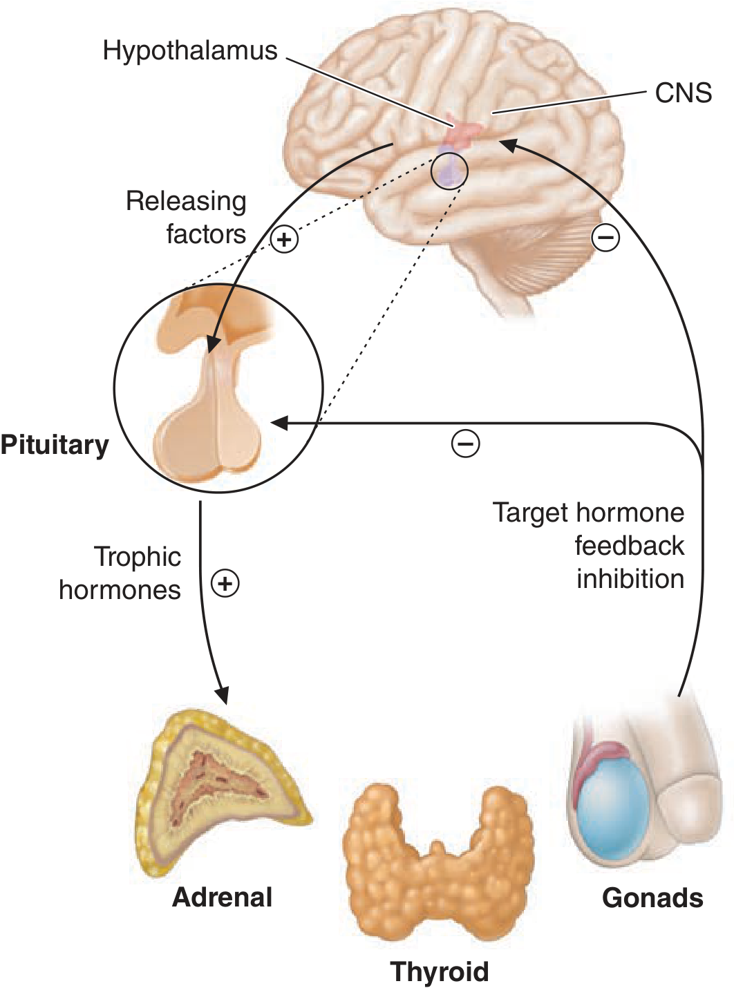
5. Endocrine Feedback Axes
The neuroendocrine system is a classic example of layered negative feedback:
Ganong's Review of Medical Physiology, 26th Ed.
- CNS/Hypothalamus releases releasing factors (+) → stimulates Anterior Pituitary
- Anterior Pituitary releases trophic hormones (+) → stimulates target glands (Adrenal, Thyroid, Gonads)
- Target hormone feeds back (-) to inhibit both the Hypothalamus and Pituitary
- This creates a hierarchical, cascading feedback system for precise control
6. Positive Feedback - The Exception
Positive feedback amplifies the original stimulus instead of opposing it. These loops are inherently unstable - once triggered, they keep escalating until an endpoint is reached.
When is positive feedback used?
Positive feedback is only useful in situations that need to build momentum to reach a definitive outcome:
| Example | How it works |
|---|
| Parturition (childbirth) | Baby's head → pressure on cervix → oxytocin release → stronger contractions → more pressure → more oxytocin (until baby is delivered) |
| Blood clotting (coagulation cascade) | Platelet activation recruits more platelets; clotting factors amplify each step |
| Action potential generation | Na⁺ influx → depolarization → more Na⁺ channels open → more influx (until threshold is passed) |
| LH surge (ovulation) | Rising estrogen (mid-cycle) triggers a massive LH surge → ovulation |
Key rule: Positive feedback always has a natural "off switch" - the endpoint itself terminates the loop (e.g., delivery of the baby, formation of the clot).
7. Feed-Forward (Anticipatory) Control
This is a proactive control mechanism - the body responds to an anticipated disturbance before the vital parameter actually changes.
Example: When you start exercising:
- Heart rate increases even before O₂ levels drop
- This is triggered by motor cortex signals and muscle mechanoreceptors, not by hypoxia
Example: Smell of food → cephalic phase → gastric acid and insulin secretion begins before food even reaches the stomach
Feed-forward + negative feedback together provide faster and more precise homeostatic control than negative feedback alone.
8. Homeostasis: Steady State vs. Equilibrium
This is a commonly confused concept:
| Equilibrium | Steady State |
|---|
| Definition | No net change because processes have stopped | No net change because equal and opposite processes are ongoing |
| Energy required | No | Yes |
| In living organisms | Only in dead tissue | The normal state of all living cells |
| Example | Na⁺ freely diffusing across a dead cell membrane | Na⁺ kept out of live cell by Na⁺/K⁺-ATPase pump |
"When a vital parameter (e.g., blood glucose) is well regulated, that parameter is not in equilibrium... it is in a steady state. The body carefully matches actions that lower the parameter with actions that raise it."
- Medical Physiology (Boron & Boulpaep)
Homeostasis costs energy - this is why metabolism never stops even at rest.
9. Redundancy in Homeostasis
The more vital a parameter is, the more backup systems the body has to regulate it. This explains why:
- Knocking out a single gene sometimes causes no obvious phenotype (another system compensates)
- Multiple hormones (insulin, glucagon, cortisol, epinephrine, GH) all regulate blood glucose
- Blood pressure is controlled by the nervous system, kidneys (RAAS), hormones (ADH, ANP), and local vascular mechanisms
10. Feedback Loops - Network Interactions
A single feedback loop never operates in complete isolation. Multiple loops interact:
- Synergistic loops: Cortisol and epinephrine both raise blood glucose (working together against insulin)
- Antagonistic loops: Insulin lowers blood glucose; glucagon, cortisol, epinephrine raise it
- Hierarchical loops: Hypothalamus → Pituitary → Target gland → Hormone → Feedback (each level modulates the next)
11. Disruption of Homeostasis = Disease
Failure of homeostatic mechanisms is the basis of most diseases:
| Disruption | Disease |
|---|
| Failure of blood glucose regulation | Diabetes mellitus |
| Failure of blood pressure regulation | Hypertension / shock |
| Failure of temperature regulation | Heat stroke / hypothermia |
| Failure of acid-base balance | Acidosis / alkalosis |
| Failure of fluid/electrolyte balance | Hypo/hypernatremia, edema |
| Uncontrolled positive feedback in cells | Cancer (loss of growth homeostasis) |
12. Quick Revision Summary
| Concept | Key Point |
|---|
| Homeostasis | Maintenance of a vital parameter within a normal range |
| Milieu intérieur | Claude Bernard's term for the internal environment |
| Negative feedback | Opposes the deviation; most common type; restores set-point |
| Positive feedback | Amplifies the change; used for definitive outcomes (parturition, clotting, AP) |
| Feed-forward | Anticipatory; acts before the disturbance occurs |
| Steady state | Constant value maintained by ongoing energy-consuming processes |
| Redundancy | Multiple overlapping systems for the most vital parameters |
| Disease | Failure of homeostatic mechanisms |
Exam Tips for MBBS 1st Year
- Always draw the feedback loop diagram - Stimulus → Sensor → Control Center → Effector → Response → Feedback
- Know the 4 elements of negative feedback: sensor, set-point comparison, gain, effector
- Remember positive feedback examples with their natural endpoints
- Distinguish steady state from equilibrium - examiners love this
- Link every clinical condition back to a failure of homeostasis
- The HPT/HPA/HPG axes are the classic hormonal homeostasis examples - learn them as hierarchical negative feedback loops
Sources: Medical Physiology (Boron & Boulpaep) | Ganong's Review of Medical Physiology, 26th Ed. | Costanzo Physiology, 7th Ed.