
Apart from this add photos to the content provided. Add clinical pearls. Also add mechanism of nail change in disease especially psoriasis
cd /home/daytona/workspace && python3 -c " from pypdf import PdfReader reader = …
nail psoriasis mechanism pathophysiology IL-17 TNF enthesitis clinical photos 2024
mkdir -p /home/daytona/workspace/nails-pptx && ls /home/daytona/workspace/nails-…
node /home/daytona/skills/shared/scripts/fetch_images.js \ "https://upload.wik…
curl -s --max-time 10 "https://upload.wikimedia.org/wikipedia/commons/thumb/9/9a…
curl -s --max-time 10 "https://upload.wikimedia.org/wikipedia/commons/thumb/9/90…
ls -la /home/daytona/workspace/nails-pptx/
Ready?
ls /home/daytona/workspace/nails-pptx/ && ls /home/daytona/workspace/attachments…
file /home/daytona/workspace/nails-pptx/nail_anatomy.png /home/daytona/workspace…
# Try fetching actual medical images from open access sources cd /home/daytona/…
python3 << 'EOF' from PIL import Image, ImageDraw, ImageFont import os out = "/…
~/nails-pptx/build_nails.js
const pptxgen = require("pptxgenjs");
const fs = require("fs");
const path = require("path");

const pres = new pptxgen();
pres.layout = "LAYOUT_WIDE"; // 13.3 x 7.5 inches
pres.title = "Nail Anatomy & Disorders — Dermatology Resident Study";
pres.author = "Dermatology Study";

// ── COLOUR PALETTE ──────────────────────────────────────────
const C = {
  navy:    "1A2F5A",
  teal:    "1ABC9C",
  gold:    "F0A500",
  white:   "FFFFFF",
  light:   "F2F6FC",
  dark:    "1C2833",
  red:     "C0392B",
  purple:  "6C3483",
  green:   "1E8449",
  orange:  "A04000",
  gray:    "566573",
  lblue:   "2E86C1",
  pearl:   "FFF8E7",
};

// ── HELPER: image as base64 ──────────────────────────────────
function imgData(filename) {
  const p = path.join("/home/daytona/workspace/nails-pptx/", filename);
  if (!fs.existsSync(p)) return null;
  const buf = fs.readFileSync(p);
  const ext = path.extname(filename).slice(1).toLowerCase();
  const mime = ext === "jpg" || ext === "jpeg" ? "image/jpeg" : "image/png";
  return `${mime};base64,${buf.toString("base64")}`;
}

// ── HELPER: section header slide ────────────────────────────
function sectionSlide(title, subtitle, bgColor) {
  const sl = pres.addSlide();
  sl.addShape(pres.shapes.RECTANGLE, { x:0, y:0, w:13.3, h:7.5, fill:{color: bgColor || C.navy} });
  sl.addShape(pres.shapes.RECTANGLE, { x:0, y:0, w:0.15, h:7.5, fill:{color: C.teal} });
  sl.addShape(pres.shapes.RECTANGLE, { x:0, y:7.2, w:13.3, h:0.3, fill:{color: C.gold} });
  sl.addText(title, { x:0.5, y:2.5, w:12.3, h:1.2, fontSize:40, bold:true, color:C.white, align:"center" });
  if (subtitle) {
    sl.addText(subtitle, { x:0.5, y:3.9, w:12.3, h:0.8, fontSize:20, color:C.teal, align:"center", italic:true });
  }
  return sl;
}

// ── HELPER: content slide with image on right ───────────────
function contentSlide(opts) {
  // opts: {title, bullets, img, imgX, imgY, imgW, imgH, accentColor, pearl}
  const sl = pres.addSlide();
  const ac = opts.accentColor || C.navy;

  // background
  sl.addShape(pres.shapes.RECTANGLE, { x:0, y:0, w:13.3, h:7.5, fill:{color: C.light} });
  // top bar
  sl.addShape(pres.shapes.RECTANGLE, { x:0, y:0, w:13.3, h:0.75, fill:{color: ac} });
  // accent strip left
  sl.addShape(pres.shapes.RECTANGLE, { x:0, y:0.75, w:0.08, h:6.75, fill:{color: C.gold} });
  // bottom bar
  sl.addShape(pres.shapes.RECTANGLE, { x:0, y:7.2, w:13.3, h:0.3, fill:{color: ac} });

  // title
  sl.addText(opts.title, {
    x:0.2, y:0.1, w:13, h:0.58,
    fontSize:22, bold:true, color:C.white, align:"left", valign:"middle", margin:6
  });

  // image
  if (opts.img) {
    const d = imgData(opts.img);
    if (d) {
      sl.addImage({
        data: d,
        x: opts.imgX || 7.8, y: opts.imgY || 0.85,
        w: opts.imgW || 5.2, h: opts.imgH || 5.5,
      });
      // image border
      sl.addShape(pres.shapes.RECTANGLE, {
        x: opts.imgX || 7.8, y: opts.imgY || 0.85,
        w: opts.imgW || 5.2, h: opts.imgH || 5.5,
        fill:{type:"none"}, line:{color: ac, width:1.5}
      });
    }
  }

  // bullets
  const bw = opts.img ? 7.4 : 12.8;
  if (opts.bullets && opts.bullets.length) {
    const items = opts.bullets.map(b => {
      if (b.type === "heading") {
        return [
          { text: b.text, options:{ bold:true, color: ac, fontSize:13, breakLine:true } }
        ];
      } else if (b.type === "sub") {
        return [
          { text: "    " + b.text, options:{ color: C.gray, fontSize:11, breakLine:true, indent:20 } }
        ];
      } else {
        return [
          { text: "• ", options:{ color: ac, bold:true, fontSize:12 } },
          { text: b.text, options:{ color: C.dark, fontSize:12, breakLine:true } }
        ];
      }
    }).flat();
    sl.addText(items, {
      x:0.25, y:0.88, w:bw, h:6.1,
      fontSize:12, fontFace:"Calibri", valign:"top", margin:8,
      wrap:true
    });
  }

  // clinical pearl box
  if (opts.pearl) {
    const py = opts.pearlY || 6.15;
    sl.addShape(pres.shapes.ROUNDED_RECTANGLE, {
      x:0.25, y:py, w:bw, h:1.0,
      fill:{color: C.pearl}, line:{color: C.gold, width:1.5}, rectRadius:0.08,
      shadow:{type:"outer", color:"000000", blur:4, offset:2, angle:135, opacity:0.1}
    });
    sl.addText([
      { text:"💎 Clinical Pearl: ", options:{bold:true, color: C.orange, fontSize:11} },
      { text: opts.pearl, options:{color: C.dark, fontSize:11} }
    ], { x:0.4, y:py+0.05, w:bw-0.3, h:0.9, valign:"middle", wrap:true });
  }

  return sl;
}

// ── HELPER: two-column content slide ────────────────────────
function twoColSlide(opts) {
  const sl = pres.addSlide();
  const ac = opts.accentColor || C.navy;
  sl.addShape(pres.shapes.RECTANGLE, { x:0, y:0, w:13.3, h:7.5, fill:{color: C.light} });
  sl.addShape(pres.shapes.RECTANGLE, { x:0, y:0, w:13.3, h:0.75, fill:{color: ac} });
  sl.addShape(pres.shapes.RECTANGLE, { x:0, y:0.75, w:0.08, h:6.75, fill:{color: C.gold} });
  sl.addShape(pres.shapes.RECTANGLE, { x:0, y:7.2, w:13.3, h:0.3, fill:{color: ac} });
  sl.addText(opts.title, { x:0.2, y:0.1, w:13, h:0.58, fontSize:22, bold:true, color:C.white, align:"left", valign:"middle", margin:6 });

  // col divider
  sl.addShape(pres.shapes.LINE, { x:6.75, y:0.9, w:0, h:6.2, line:{color:ac, width:1, dashType:"dash"} });

  // left col
  if (opts.left && opts.left.length) {
    const items = opts.left.map(b => {
      if (b.type==="heading") return [{text:b.text, options:{bold:true, color:ac, fontSize:13, breakLine:true}}];
      if (b.type==="sub") return [{text:"    "+b.text, options:{color:C.gray, fontSize:11, breakLine:true}}];
      return [{text:"• ", options:{color:ac, bold:true, fontSize:12}},{text:b.text, options:{color:C.dark, fontSize:12, breakLine:true}}];
    }).flat();
    sl.addText(items, { x:0.25, y:0.9, w:6.3, h:6.0, fontSize:12, fontFace:"Calibri", valign:"top", margin:8, wrap:true });
  }
  // right col
  if (opts.right && opts.right.length) {
    const items = opts.right.map(b => {
      if (b.type==="heading") return [{text:b.text, options:{bold:true, color:ac, fontSize:13, breakLine:true}}];
      if (b.type==="sub") return [{text:"    "+b.text, options:{color:C.gray, fontSize:11, breakLine:true}}];
      return [{text:"• ", options:{color:ac, bold:true, fontSize:12}},{text:b.text, options:{color:C.dark, fontSize:12, breakLine:true}}];
    }).flat();
    sl.addText(items, { x:6.9, y:0.9, w:6.2, h:6.0, fontSize:12, fontFace:"Calibri", valign:"top", margin:8, wrap:true });
  }
  return sl;
}

// ── HELPER: full-image diagram slide ─────────────────────────
function diagramSlide(title, imgFile, caption, accentColor) {
  const sl = pres.addSlide();
  const ac = accentColor || C.navy;
  sl.addShape(pres.shapes.RECTANGLE, { x:0, y:0, w:13.3, h:7.5, fill:{color: C.dark} });
  sl.addShape(pres.shapes.RECTANGLE, { x:0, y:0, w:13.3, h:0.7, fill:{color: ac} });
  sl.addShape(pres.shapes.RECTANGLE, { x:0, y:7.2, w:13.3, h:0.3, fill:{color: C.gold} });
  sl.addText(title, { x:0.2, y:0.1, w:13, h:0.55, fontSize:20, bold:true, color:C.white, align:"center", valign:"middle", margin:4 });
  const d = imgData(imgFile);
  if (d) {
    sl.addImage({ data:d, x:0.5, y:0.8, w:12.3, h:6.15 });
  }
  if (caption) {
    sl.addText(caption, { x:0.5, y:7.05, w:12.3, h:0.4, fontSize:10, color:C.teal, align:"center", italic:true });
  }
  return sl;
}

// ── HELPER: mechanism flow slide ─────────────────────────────
function mechanismSlide() {
  const sl = pres.addSlide();
  sl.addShape(pres.shapes.RECTANGLE, { x:0, y:0, w:13.3, h:7.5, fill:{color:"1C1C2E"} });
  sl.addShape(pres.shapes.RECTANGLE, { x:0, y:0, w:13.3, h:0.7, fill:{color: C.purple} });
  sl.addShape(pres.shapes.RECTANGLE, { x:0, y:7.2, w:13.3, h:0.3, fill:{color: C.gold} });
  sl.addText("MECHANISM OF NAIL CHANGES IN PSORIASIS", {
    x:0.2, y:0.1, w:13, h:0.55, fontSize:21, bold:true, color:C.white, align:"center", valign:"middle"
  });

  const steps = [
    { x:0.3,  y:0.85, w:3.7, h:1.2, bg:"6C3483", text:"① TRIGGER\nGenetic susceptibility\n(HLA-Cw6 NEGATIVE)\n+ Microtrauma / Koebner" },
    { x:4.5,  y:0.85, w:3.7, h:1.2, bg:"1A5276", text:"② DC ACTIVATION\nmDC release IL-23\nStressed keratinocytes\nrelease LL-37, hβD-2/3" },
    { x:8.7,  y:0.85, w:4.3, h:1.2, bg:"1E8449", text:"③ Th17 DIFFERENTIATION\nIL-17A, IL-17F, IL-22\nTNF-α, NF-κB activation\nMast cell recruitment" },
    { x:0.3,  y:2.4,  w:3.7, h:1.3, bg:"B7950B", text:"④ MATRIX INFLAMMATION\nParakeratosis in\nproximal matrix\n→ PITTING" },
    { x:4.5,  y:2.4,  w:3.7, h:1.3, bg:"922B21", text:"⑤ NAIL BED PSORIASIS\nPsoriasiform hyperplasia\nMicrovascular changes\n→ OIL-SPOT / ONYCHOLYSIS" },
    { x:8.7,  y:2.4,  w:4.3, h:1.3, bg:"1F618D", text:"⑥ ENTHESITIS @ DIP\nCytokine spread to\nsynovio-entheseal complex\n→ PSORIATIC ARTHRITIS risk" },
  ];

  steps.forEach(s => {
    sl.addShape(pres.shapes.ROUNDED_RECTANGLE, {
      x:s.x, y:s.y, w:s.w, h:s.h,
      fill:{color:s.bg}, line:{color:C.gold, width:1}, rectRadius:0.1,
      shadow:{type:"outer",color:"000000",blur:8,offset:3,angle:135,opacity:0.3}
    });
    sl.addText(s.text, {
      x:s.x+0.1, y:s.y+0.05, w:s.w-0.2, h:s.h-0.1,
      fontSize:11, bold:false, color:C.white, align:"center", valign:"middle", wrap:true, margin:4
    });
  });

  // Arrows between boxes (row 1)
  [[4.0,1.45,0.4,0.15],[8.2,1.45,0.4,0.15]].forEach(([x,y,w,h])=>{
    sl.addShape(pres.shapes.RIGHT_ARROW, {x,y,w,h,fill:{color:C.gold},line:{color:C.gold}});
  });

  // summary box
  sl.addShape(pres.shapes.ROUNDED_RECTANGLE, {
    x:0.3, y:4.05, w:12.7, h:1.3,
    fill:{color:"2D2D4E"}, line:{color:C.teal, width:1.5}, rectRadius:0.12
  });
  sl.addText([
    {text:"KEY CYTOKINE AXIS: ", options:{bold:true, color:C.gold, fontSize:13}},
    {text:"IL-23 → Th17 → IL-17A/F + TNF-α → Nail matrix & nail bed inflammation\n", options:{color:C.white, fontSize:12}},
    {text:"VEGF ↑ ", options:{bold:true, color:C.teal, fontSize:12}},
    {text:"→ dilated tortuous capillaries (onychoscopy finding: 90%)\n", options:{color:"#BBBBBB", fontSize:12}},
    {text:"Onychodermal band breach ", options:{bold:true, color:C.gold, fontSize:12}},
    {text:"→ progressive onycholysis (difficult to reverse)", options:{color:"#BBBBBB", fontSize:12}},
  ], {x:0.5, y:4.1, w:12.3, h:1.2, valign:"middle", wrap:true, margin:6});

  // nail changes legend
  sl.addShape(pres.shapes.RECTANGLE, {x:0.3, y:5.55, w:12.7, h:1.55, fill:{color:"252540"}, line:{color:C.purple, width:1}});
  sl.addText("ZAIAS CLASSIFICATION — Nail Changes by Site", {
    x:0.4, y:5.58, w:12.5, h:0.35, fontSize:12, bold:true, color:C.gold, align:"center"
  });
  const zaias = [
    ["PROXIMAL MATRIX","Pitting (parakeratosis foci)\nTransverse furrows\nLeukonychia (rough surface)"],
    ["DISTAL MATRIX / LUNULA","Red spots in lunula\nFocal onycholysis\nErythema of lunula"],
    ["NAIL BED","Salmon/oil-drop patch\nSubungual hyperkeratosis\nSplinter haemorrhages\nOnycholysis (irregular margin)"],
    ["NAIL FOLD","Periungual psoriasis\nCutaneous psoriasis\n(PsA association ↑↑)"],
  ];
  const cols = [0.4, 3.7, 7.0, 10.3];
  zaias.forEach((z,i) => {
    sl.addShape(pres.shapes.ROUNDED_RECTANGLE, {
      x:cols[i], y:5.98, w:3.1, h:1.0,
      fill:{color:"3A3A5E"}, line:{color:C.teal, width:0.8}, rectRadius:0.07
    });
    sl.addText([
      {text:z[0]+"\n", options:{bold:true, color:C.teal, fontSize:9}},
      {text:z[1], options:{color:C.white, fontSize:9}}
    ], {x:cols[i]+0.05, y:5.99, w:3.0, h:0.95, valign:"top", wrap:true, margin:3});
  });

  return sl;
}

// ── HELPER: quiz slide ───────────────────────────────────────
function quizSlide(qas, title, accentColor) {
  const sl = pres.addSlide();
  const ac = accentColor || C.navy;
  sl.addShape(pres.shapes.RECTANGLE, { x:0, y:0, w:13.3, h:7.5, fill:{color: C.light} });
  sl.addShape(pres.shapes.RECTANGLE, { x:0, y:0, w:13.3, h:0.7, fill:{color: ac} });
  sl.addShape(pres.shapes.RECTANGLE, { x:0, y:7.2, w:13.3, h:0.3, fill:{color: C.gold} });
  sl.addText(title, { x:0.2, y:0.1, w:13, h:0.55, fontSize:20, bold:true, color:C.white, align:"center", valign:"middle" });

  // table header
  sl.addShape(pres.shapes.RECTANGLE, {x:0.3, y:0.78, w:12.7, h:0.4, fill:{color:ac}});
  sl.addText("QUESTION", {x:0.35, y:0.78, w:7.5, h:0.4, fontSize:11, bold:true, color:C.white, valign:"middle"});
  sl.addText("ANSWER", {x:8.0, y:0.78, w:4.8, h:0.4, fontSize:11, bold:true, color:C.white, valign:"middle"});

  qas.forEach((qa, i) => {
    const yy = 1.22 + i * 0.78;
    const bg = i % 2 === 0 ? "FFFFFF" : "EBF5FB";
    sl.addShape(pres.shapes.RECTANGLE, {x:0.3, y:yy, w:12.7, h:0.75, fill:{color:bg}});
    sl.addShape(pres.shapes.LINE, {x:0.3, y:yy+0.75, w:12.7, h:0, line:{color:"CCCCCC", width:0.5}});
    sl.addShape(pres.shapes.LINE, {x:8.0, y:yy, w:0, h:0.75, line:{color:"CCCCCC", width:0.5}});
    sl.addText(qa[0], {x:0.4, y:yy+0.05, w:7.5, h:0.65, fontSize:10, color:C.dark, valign:"middle", wrap:true});
    sl.addText(qa[1], {x:8.1, y:yy+0.05, w:4.8, h:0.65, fontSize:10, color:ac, bold:true, valign:"middle", wrap:true});
  });
  return sl;
}

// ────────────────────────────────────────────────────────────
//  BUILD ALL SLIDES
// ────────────────────────────────────────────────────────────

// ── TITLE SLIDE ──────────────────────────────────────────────
{
  const sl = pres.addSlide();
  sl.addShape(pres.shapes.RECTANGLE, { x:0, y:0, w:13.3, h:7.5, fill:{color: C.navy} });
  sl.addShape(pres.shapes.RECTANGLE, { x:0, y:5.8, w:13.3, h:1.7, fill:{color:"0F1F3D"} });
  sl.addShape(pres.shapes.RECTANGLE, { x:0, y:0, w:0.25, h:7.5, fill:{color: C.teal} });
  sl.addShape(pres.shapes.RECTANGLE, { x:0, y:5.6, w:13.3, h:0.12, fill:{color: C.gold} });
  sl.addText("NAIL ANATOMY & DISORDERS", {
    x:0.5, y:1.2, w:12.3, h:1.2, fontSize:44, bold:true, color:C.white, align:"center",
    shadow:{type:"outer", color:"000000", blur:8, offset:3, angle:135, opacity:0.4}
  });
  sl.addText("Dermatology Resident Study Note", {
    x:0.5, y:2.6, w:12.3, h:0.7, fontSize:24, color:C.teal, align:"center", italic:true
  });
  sl.addText("Nail Unit • Nail Signs • Psoriasis • Onychomycosis • Lichen Planus • Tumours • Clinical Pearls", {
    x:0.5, y:3.4, w:12.3, h:0.55, fontSize:14, color:"AABBCC", align:"center"
  });
  sl.addText([
    {text:"Sources: ", options:{bold:true, color:C.gold, fontSize:12}},
    {text:"Rook's Dermatology Ch.93 • Fitzpatrick's Ch.91 • Bolivnia 5e • Andrews' Ch.33 • JAAD 2024/2025", options:{color:"AABBCC", fontSize:12}}
  ], { x:0.5, y:6.05, w:12.3, h:0.5, align:"center" });
  sl.addText("With Enhanced Clinical Pearls • Mechanism of Nail Changes • Psoriasis Pathway", {
    x:0.5, y:6.65, w:12.3, h:0.45, fontSize:11, color:C.teal, align:"center", italic:true
  });
}

// ── SECTION 1 HEADER ────────────────────────────────────────
sectionSlide("PART 1 — ANATOMY OF THE NAIL UNIT", "Gross anatomy • Microscopy • Blood supply • Growth • Immunology", C.navy);

// ── ANATOMY OVERVIEW ─────────────────────────────────────────
contentSlide({
  title: "1.1 — NAIL UNIT COMPONENTS",
  img: "img_anatomy.png", imgX:7.5, imgY:0.85, imgW:5.5, imgH:5.5,
  accentColor: C.navy,
  bullets: [
    {type:"heading", text:"NAIL PLATE"},
    {text:"Trilaminar: dorsal (from ventral PNF), intermediate (germinal matrix), ventral (nail bed ~21%)"},
    {text:"Hard hair-type keratins in sulfur-rich proteins (high cysteine, glycine, tyrosine)"},
    {type:"heading", text:"NAIL MATRIX (Germinal)"},
    {text:"Proximal matrix → dorsal 80% of nail plate"},
    {text:"Distal matrix (lunula) → ventral 20% of nail plate"},
    {text:"Keratinization along oblique upward-distal axis"},
    {type:"heading", text:"NAIL BED (Sterile Matrix)"},
    {text:"Epidermis 2-3 cells thick, NO granular layer in health"},
    {text:"Granular layer = PATHOLOGICAL (psoriasis, onychomycosis)"},
    {text:"Collagen tightly apposed to periosteum → rapid osteomyelitis risk"},
    {type:"heading", text:"HYPONYCHIUM & ONYCHODERMAL BAND"},
    {text:"Onychodermal band = nail isthmus = most distal nail-bed attachment"},
    {text:"Breach → progressive onycholysis (difficult to reverse)"},
  ],
  pearl: "No granular layer in healthy nail bed. Its appearance signals disease — think psoriasis or onychomycosis when you see parakeratosis on biopsy."
});

// ── KERATINS + GROWTH ────────────────────────────────────────
twoColSlide({
  title: "1.2 — NAIL KERATINS, GROWTH RATES & IMMUNOLOGY",
  accentColor: C.lblue,
  left: [
    {type:"heading", text:"KERATINS"},
    {text:"K5/K14 — basal layer of nail matrix AND nail bed"},
    {text:"K1/K10 — suprabasal nail matrix (absent in HEALTHY nail bed)"},
    {text:"K1/K10 in nail bed = pathological → psoriasis, onychomycosis"},
    {text:"K6/K16 — various subungual locations (development)"},
    {type:"heading", text:"GROWTH RATES"},
    {text:"Fingernails: ~3 mm/month"},
    {text:"Toenails: ~1 mm/month"},
    {type:"heading", text:"FACTORS — FASTER GROWTH"},
    {text:"Daytime, youth, dominant hand, summer, male sex"},
    {text:"Psoriasis, pityriasis rubra pilaris, hyperthyroidism, HIV"},
    {text:"Drugs: levodopa, itraconazole"},
    {type:"heading", text:"FACTORS — SLOWER GROWTH"},
    {text:"Beau lines, yellow nail syndrome, hypothyroidism"},
    {text:"Drugs: methotrexate, azathioprine, targeted therapies"},
  ],
  right: [
    {type:"heading", text:"IMMUNOLOGY OF THE NAIL"},
    {text:"Nail matrix has IMMUNE PRIVILEGE (like hair matrix, eye, testis)"},
    {text:"MHC class I restricted → protects from autoimmune T-cell attack"},
    {text:"Contains LL-37 (kills Candida albicans)"},
    {text:"hβD-2 and hβD-3 (beta-defensins) in matrix"},
    {type:"heading", text:"SIGNALLING PATHWAYS"},
    {text:"Wnt/β-catenin crucial in nail embryogenesis & maintenance"},
    {text:"R-spondin 4 mutations → autosomal recessive anonychia"},
    {text:"Frizzled-6 (Wnt receptor) mutations → inherited nail dysplasia"},
    {text:"BMPs work with Wnt signalling"},
    {type:"heading", text:"BLOOD SUPPLY"},
    {text:"Paired digital arteries (large palmar + small dorsal)"},
    {text:"Glomus bodies (Masson) → regulate capillary circulation"},
    {text:"Glomus bodies DILATE with cold (opposite of arterioles)"},
    {text:"Superficial arcade: supplies nail fold, extensor tendon, matrix"},
  ]
});

// ── SECTION 2 HEADER ────────────────────────────────────────
sectionSlide("PART 2 — NAIL SIGNS & THEIR SIGNIFICANCE", "Shape • Attachment • Surface • Colour • Systemic associations", "0B3D6B");

// ── SHAPE ABNORMALITIES ──────────────────────────────────────
contentSlide({
  title: "2.1 — ABNORMALITIES OF SHAPE: CLUBBING & KOILONYCHIA",
  img: "img_clubbing.png", imgX:7.6, imgY:0.85, imgW:5.4, imgH:5.2,
  accentColor: C.lblue,
  bullets: [
    {type:"heading", text:"CLUBBING (Hippocratic fingers / Acropachy)"},
    {text:"Lovibond angle > 180° (normally < 160°)"},
    {text:"Schamroth window OBLITERATED (normally open diamond)"},
    {text:"Curth angle (DIP joint) < 160°"},
    {text:"Mechanism: ↑VEGF → nail bed angiogenesis + vasodilation"},
    {text:"HPGD / SLCO2A1 mutations → pachydermoperiostosis (primary)"},
    {type:"heading", text:"CAUSES — Secondary Clubbing"},
    {text:"Respiratory: lung Ca, mesothelioma, CF, cryptogenic FA, sarcoid"},
    {text:"Cardiac: cyanotic CHD, infective endocarditis"},
    {text:"GI: IBD, liver disease, oesophageal Ca"},
    {text:"HIV (37%), POEMS syndrome (70%)"},
    {type:"heading", text:"KOILONYCHIA (Spoon nail)"},
    {text:"Concave, spoon-shaped nail plate"},
    {text:"Most common: iron deficiency anaemia, haemochromatosis"},
    {text:"Also: trichothiodystrophy, familial (AD)"},
    {text:"Secondary: psoriasis, LP, dermatophyte infection"},
  ],
  pearl: "Schamroth window test: oppose dorsal surfaces of two homologous fingers. Normal = diamond window. Clubbing = obliteration. Quick bedside sign."
});

// ── ONYCHOLYSIS + PTERYGIUM ───────────────────────────────────
contentSlide({
  title: "2.2 — ONYCHOLYSIS & PTERYGIUM",
  accentColor: C.red,
  bullets: [
    {type:"heading", text:"ONYCHOLYSIS — Separation from nail bed (distal → proximal)"},
    {text:"Detached plate looks WHITE (apparent leukonychia — air underneath)"},
    {text:"Green = Pseudomonas aeruginosa (pyocyanin pigment = chloronychia)"},
    {type:"heading", text:"Dermoscopic Types (4)"},
    {text:"1. Post-traumatic: regular margin, usually lateral"},
    {text:"2. Psoriatic: IRREGULAR margin, whole free edge, erythematous border"},
    {text:"3. Onychomycosis: ragged/spike edge, mycelium visible proximally"},
    {text:"4. Drug-induced: uniform pattern in midline (photo-onycholysis)"},
    {type:"heading", text:"Key Causes"},
    {text:"Psoriasis (30% of psoriatics), fungal, trauma, photo-onycholysis"},
    {text:"Drugs: doxycycline, psoralens, 5-FU, captopril, retinoids"},
    {text:"Systemic: hypothyroidism, hyperthyroidism, yellow nail syndrome"},
    {type:"heading", text:"PTERYGIUM (Dorsal — True)"},
    {text:"Fibrotic band from PNF divides nail in TWO"},
    {text:"Cause: LICHEN PLANUS (most common), trauma, GVHD, leprosy"},
    {type:"heading", text:"PTERYGIUM INVERSUM UNGUIS (Ventral)"},
    {text:"Forward extension of nail bed epithelium"},
    {text:"Causes: systemic sclerosis, Raynaud, SLE, familial, trauma"},
  ],
  pearl: "The onychodermal band is the 'Achilles heel' of nail attachment. Once breached by psoriasis or trauma, onycholysis becomes progressive. Clip, dry, and apply topical antiseptics."
});

// ── SURFACE CHANGES ──────────────────────────────────────────
twoColSlide({
  title: "2.3 — NAIL SURFACE CHANGES",
  accentColor: C.green,
  left: [
    {type:"heading", text:"BEAU LINES (Transverse Grooves)"},
    {text:"Transverse depressions = temporary arrest of proximal matrix mitosis"},
    {text:"Depth = severity of damage; Width = duration of insult"},
    {text:"Migrate distally as nail grows"},
    {text:"All 20 nails simultaneously = SYSTEMIC cause"},
    {text:"1-2 nails only = local trauma / paronychia"},
    {text:"Beau lines → Onychomadesis when full thickness groove"},
    {type:"heading", text:"PITTING"},
    {text:"Shallow depressions from parakeratosis foci in proximal matrix"},
    {text:"Fine pits (<1mm): PSORIASIS (most common)"},
    {text:">10 pits/nail OR >60 pits total supports psoriasis diagnosis"},
    {text:"Coarse pits: ECZEMA"},
    {text:"Geometric/uniform 'Scotch plaid' pattern: ALOPECIA AREATA"},
    {text:"Elkonyxis = deep single large pit (reactive arthritis, psoriasis)"},
    {type:"heading", text:"TRACHYONYCHIA (Twenty-nail dystrophy)"},
    {text:"Rough sandpaper-like opaque surface; all 20 nails"},
    {text:"Associations: alopecia areata (most common), psoriasis, LP, eczema"},
  ],
  right: [
    {type:"heading", text:"ONYCHOSCHIZIA (Lamellar Dystrophy)"},
    {text:"Distal horizontal lamellar splitting"},
    {text:"Causes: repeated wet-dry cycles, chemical exposure"},
    {text:"Common with aging; NOT a sign of systemic disease"},
    {type:"heading", text:"LONGITUDINAL RIDGING / BEADING"},
    {text:"Normal aging change; NOT clinically significant alone"},
    {type:"heading", text:"MEDIAN CANALIFORM DYSTROPHY OF HELLER"},
    {text:"Midline fir-tree groove; thumbs most commonly"},
    {text:"Distinct from habit-tic (washboard nails — disturbed cuticle)"},
    {text:"Often self-resolving"},
    {type:"heading", text:"PINCER NAIL (Trumpet nail)"},
    {text:"Transverse overcurvature increasing toward distal tip"},
    {text:"3 patterns: psoriasis (thumbs/big toes), inherited, isolated"},
    {text:"Treatment: braces, partial matricectomy, Z-plasty"},
    {type:"heading", text:"PACHYONYCHIA"},
    {text:"Thickened plate, yellow, horseshoe free edge, extreme hardness"},
    {text:"Jadassohn-Lewandowsky syndrome (pachyonychia congenita)"},
    {text:"Acquired: psoriasis, PRP, chronic eczema, onychomycosis"},
  ]
});

// ── COLOUR CHANGES ───────────────────────────────────────────
contentSlide({
  title: "2.4 — NAIL COLOUR CHANGES (CHROMONYCHIA)",
  accentColor: C.purple,
  bullets: [
    {type:"heading", text:"LEUKONYCHIA"},
    {text:"True leukonychia (nail plate): parakeratosis in plate; Mees' lines = arsenic / systemic insult"},
    {text:"Apparent leukonychia (nail BED — disappear with pressure):"},
    {text:"  → Muehrcke's lines (paired transverse bands) = hypoalbuminaemia"},
    {text:"  → Terry's nails (white proximally, normal 1-2mm distally) = hepatic cirrhosis"},
    {text:"  → Half-and-half / Lindsay's nails (white proximal, red-brown distal) = chronic renal failure"},
    {text:"  → Azure lunula = Wilson's disease / argyria"},
    {type:"heading", text:"MELANONYCHIA"},
    {text:"Brown/black: nail matrix melanocytes, subungual haematoma"},
    {text:"ABCDEF rule for nail melanoma:"},
    {text:"  A = Age (40-70), African/Asian/Native American; B = Brown/Black >3mm, Blurred borders"},
    {text:"  C = Change in morphology; D = Digit (thumb > hallux > index)"},
    {text:"  E = Extension to nail fold (Hutchinson's sign); F = Family/personal Hx melanoma"},
    {type:"heading", text:"OTHER COLOURS"},
    {text:"Yellow: yellow nail syndrome, psoriasis (subungual hyperkeratosis), fungal"},
    {text:"Green: Pseudomonas aeruginosa (chloronychia)"},
    {text:"Red lunula: psoriasis, CO poisoning, cardiac failure, alopecia areata"},
    {text:"Splinter haemorrhages: psoriasis, endocarditis, vasculitis, trauma"},
  ],
  pearl: "Pseudo-Hutchinson's sign = apparent periungual pigmentation through a TRANSPARENT cuticle (racial melanonychia, benign nevi). Real Hutchinson's = actual melanin in nail fold skin. Dermoscopy distinguishes both."
});

// ── SYSTEMIC NAIL SIGNS ──────────────────────────────────────
diagramSlide(
  "2.5 — NAIL SIGNS IN SYSTEMIC DISEASE (Quick Reference)",
  "img_systemic.png",
  "Source: Rook's Ch.93, Bolivnia 5e, Fitzpatrick's Ch.91, Andrews' Ch.33",
  C.purple
);

// ── SECTION 3 HEADER ────────────────────────────────────────
sectionSlide("PART 3 — NAIL DISORDERS", "Psoriasis • Lichen Planus • Onychomycosis • Paronychia • Darier Disease • Eczema • Yellow Nail • Tumours", C.red);

// ── NAIL PSORIASIS CLINICAL ──────────────────────────────────
contentSlide({
  title: "3.1a — NAIL PSORIASIS — EPIDEMIOLOGY & CLINICAL FEATURES",
  img: "img_psoriasis_clinical.png", imgX:7.5, imgY:0.85, imgW:5.5, imgH:5.0,
  accentColor: C.red,
  bullets: [
    {type:"heading", text:"EPIDEMIOLOGY"},
    {text:">50% prevalence in plaque psoriasis; 80-90% lifetime incidence"},
    {text:"5-10% have nail psoriasis WITHOUT skin/joint disease"},
    {text:"Annual PsA risk with nail involvement: 0.55-2.55%"},
    {text:"HLA: Cw6 NEGATIVE (cutaneous psoriasis is HLA-Cw6 positive)"},
    {type:"heading", text:"MATRIX SIGNS"},
    {text:"Pitting — proximal matrix parakeratosis (most common, fine pits)"},
    {text:"Transverse furrows — proximal matrix extension"},
    {text:"Crumbling nail — entire matrix; prolonged disease"},
    {text:"Leukonychia with rough surface"},
    {type:"heading", text:"NAIL BED SIGNS"},
    {text:"Salmon / Oil-drop patch — NEARLY PATHOGNOMONIC (yellow-red translucent zone)"},
    {text:"Onycholysis — irregular margin involving whole free edge; erythematous border"},
    {text:"Subungual hyperkeratosis — prolonged; hard keratinous material"},
    {text:"Splinter haemorrhages — short episodes; capillary rupture"},
    {text:"Yellow-green discolouration — secondary yeast / Pseudomonas"},
  ],
  pearl: "Salmon patch (oil-drop sign) = nearly pathognomonic for psoriasis. Unlike pitting (also in AA, eczema), an oil-spot is nearly exclusive to psoriasis. Confirm with onychoscopy: dilated tortuous capillaries (90%)."
});

// ── PSORIASIS MECHANISM SLIDE ────────────────────────────────
mechanismSlide();

// ── PSORIASIS TREATMENT ──────────────────────────────────────
contentSlide({
  title: "3.1b — NAIL PSORIASIS — TREATMENT ALGORITHM",
  accentColor: C.red,
  bullets: [
    {type:"heading", text:"≤3 NAILS — MATRIX INVOLVEMENT"},
    {text:"1st line: Intralesional triamcinolone acetonide (after digital nerve block)"},
    {text:"2nd line: Topical vitamin D analogue (calcipotriol) ± topical steroid"},
    {type:"heading", text:"≤3 NAILS — NAIL BED INVOLVEMENT"},
    {text:"1st line: Topical steroids OR topical vitamin D analogue"},
    {type:"heading", text:"> 3 NAILS INVOLVED"},
    {text:"Intralesional steroids ± topical vitamin D + steroids — 1st line"},
    {text:"SYSTEMIC: Methotrexate (≤15mg/week), Ciclosporin (3-5mg/kg short-term)"},
    {text:"Acitretin (0.2-0.4 mg/kg × 6 months)"},
    {type:"heading", text:"BIOLOGICS"},
    {text:"Adalimumab — ONLY FDA-approved specifically for nail psoriasis"},
    {text:"IL-17i: Secukinumab, Ixekizumab, Brodalumab — excellent nail clearance"},
    {text:"IL-23i: Risankizumab, Guselkumab, Ustekinumab"},
    {text:"Anti-TNF: Infliximab, Etanercept, Certolizumab, Golimumab"},
    {type:"heading", text:"SMALL MOLECULES"},
    {text:"Apremilast (PDE4 inhibitor) — especially for mild-moderate nail psoriasis"},
    {text:"Tofacitinib (JAK inhibitor) — emerging evidence"},
    {type:"heading", text:"SCORING: NAPSI"},
    {text:"0-8 per nail (matrix 0-4 + nail bed 0-4); total max = 160 (all 20 nails)"},
  ],
  pearl: "Nail psoriasis is HLA-Cw6 NEGATIVE — the opposite of cutaneous plaque psoriasis. This genetically distinct subset has stronger PsA association. Always screen for joint disease."
});

// ── LICHEN PLANUS ────────────────────────────────────────────
contentSlide({
  title: "3.2 — LICHEN PLANUS OF THE NAILS",
  img: "img_lichen_planus.png", imgX:7.6, imgY:0.85, imgW:5.4, imgH:5.2,
  accentColor: C.gold,
  bullets: [
    {type:"heading", text:"EPIDEMIOLOGY & FEATURES"},
    {text:"5-10% of LP patients; 5th-6th decade; may precede skin LP"},
    {type:"heading", text:"CLINICAL SIGNS"},
    {text:"Dorsal pterygium — MOST CHARACTERISTIC; fibrotic PNF-matrix fusion"},
    {text:"Onychorrhexis — irregular longitudinal ridging and grooving"},
    {text:"Thinning of nail plate → atrophy → nail shedding"},
    {text:"Trachyonychia — 20-nail dystrophy (shiny variant)"},
    {text:"Erythronychia — red longitudinal streaks"},
    {text:"Subungual hyperpigmentation and keratosis"},
    {text:"Idiopathic atrophy of nails = most severe variant (rapid irreversible destruction)"},
    {type:"heading", text:"HISTOLOGY"},
    {text:"Interface dermatitis with band-like lymphocytic infiltrate"},
    {text:"Basal cell liquefaction (identical to cutaneous LP)"},
    {type:"heading", text:"TREATMENT"},
    {text:"Intralesional triamcinolone acetonide (digital nerve block first)"},
    {text:"Oral prednisone 0.5-1 mg/kg × 3 weeks"},
    {text:"IM triamcinolone 0.5-1mg/kg/month × 3-6 months (children)"},
    {text:"Combination: oral retinoids + topical steroids"},
  ],
  pearl: "Dorsal pterygium = lichen planus until proven otherwise. It is irreversible scarring — early aggressive treatment is necessary. LP pterygium splits the nail; psoriasis does NOT cause pterygium."
});

// ── ONYCHOMYCOSIS ────────────────────────────────────────────
contentSlide({
  title: "3.3 — ONYCHOMYCOSIS — CLASSIFICATION & TREATMENT",
  img: "img_onychomycosis.png", imgX:7.6, imgY:0.85, imgW:5.4, imgH:5.2,
  accentColor: C.green,
  bullets: [
    {type:"heading", text:"TYPES & ORGANISMS"},
    {text:"DLSO (most common): T. rubrum (70-80%) — free edge/lateral; hyperkeratosis"},
    {text:"PSO (proximal subungual): T. rubrum — lunula leukonychia — HIV!"},
    {text:"SWO (superficial white): T. interdigitale, moulds — dorsal powdery"},
    {text:"Endonyx: T. soudanense/violaceum — milky white, no hyperkeratosis"},
    {text:"Candida (CDLSO): chronic mucocutaneous candidiasis"},
    {type:"heading", text:"DIFFERENTIAL FROM PSORIASIS"},
    {text:"Onychomycosis: KOH = filaments; ragged/spike dermoscopy edge"},
    {text:"Psoriasis: fine pitting, salmon patch, KOH usually negative"},
    {text:"Up to 20% of psoriatics also have co-infection — treat both!"},
    {type:"heading", text:"DIAGNOSIS"},
    {text:"KOH direct microscopy; Fungal culture (gold standard)"},
    {text:"PAS histology of nail clippings; PCR (faster, more sensitive)"},
    {type:"heading", text:"TREATMENT"},
    {text:"Terbinafine oral 250mg/day × 6 wks (fingernails), 12 wks (toenails)"},
    {text:"Itraconazole: 200mg/day continuous OR pulse (400mg/day × 1 wk/month)"},
    {text:"Topical: ciclopirox 8%, efinaconazole, tavaborole (adjunct)"},
  ],
  pearl: "PSO (proximal subungual onychomycosis) in a young patient = HIV testing MANDATORY. PSO entering via the nail fold is rare in immunocompetent patients. Always get KOH/culture before treating — misdiagnosis rate is high."
});

// ── PARONYCHIA ───────────────────────────────────────────────
twoColSlide({
  title: "3.4 — PARONYCHIA (Acute & Chronic)",
  accentColor: C.orange,
  left: [
    {type:"heading", text:"ACUTE PARONYCHIA"},
    {text:"Rapid onset pain, swelling, erythema of nail folds"},
    {text:"Most common cause: Staphylococcus aureus"},
    {text:"Also: Streptococcus, herpes simplex (herpetic whitlow), orf"},
    {text:"Neutropenic patients: Fusarium, Aspergillus"},
    {text:"3× more common in females; index finger + thumb most common"},
    {type:"heading", text:"TREATMENT — Acute"},
    {text:"Superficial abscess: drain with pointed scalpel (no anaesthesia)"},
    {text:"Deep: penicillinase-resistant antibiotic + surgery if no response 48h"},
    {text:"Herpetic whitlow: oral aciclovir/famciclovir (topical has NO benefit)"},
    {text:"Artificial nails harbour bacteria inaccessible to hand hygiene"},
  ],
  right: [
    {type:"heading", text:"CHRONIC PARONYCHIA"},
    {text:"Insidious; inflammatory dermatosis of nail folds"},
    {text:"Predominantly: domestic/catering workers (wet work), females"},
    {text:"First 3 fingers of dominant hand most affected"},
    {type:"heading", text:"PATHOPHYSIOLOGY (Critical!)"},
    {text:"Candida is NOT the primary pathogen"},
    {text:"Primary cause = IRRITANT REACTION / contact allergy / food hypersensitivity"},
    {text:"Candida = secondary coloniser only"},
    {text:"Systemic antifungals are NOT useful"},
    {type:"heading", text:"CLINICAL FEATURES"},
    {text:"Loss of cuticle (pathognomonic), Beau lines, cross-ridging"},
    {text:"Dark discolouration at lateral edges"},
    {type:"heading", text:"TREATMENT — Chronic"},
    {text:"Avoid wet work; topical imidazoles (for Candida colonisation)"},
    {text:"Topical / intralesional steroids — primary treatment"},
    {text:"Surgical: crescent-shaped excision of PNF (refractory cases)"},
    {text:"Heals by secondary intention in < 2 weeks"},
  ]
});

// ── DARIER + ECZEMA ──────────────────────────────────────────
twoColSlide({
  title: "3.5 & 3.6 — DARIER DISEASE & ECZEMATOUS NAIL DYSTROPHY",
  accentColor: C.purple,
  left: [
    {type:"heading", text:"DARIER DISEASE OF NAILS"},
    {text:"Candy-cane appearance: red (erythronychia) + white (leukonychia) longitudinal bands"},
    {text:"PATHOGNOMONIC — can diagnose Darier disease from nails alone"},
    {text:"Free margin: V-shaped NOTCH and fissure (fragility)"},
    {text:"Multiple nails affected simultaneously"},
    {type:"heading", text:"HISTOLOGY"},
    {text:"Acantholysis + multinucleate giant cells"},
    {text:"Epithelial hyperplasia in nail bed"},
    {text:"Similar to skin Darier disease (ATP2A2 mutation)"},
    {type:"heading", text:"CLINICAL PEARL"},
    {text:"Red+white longitudinal bands + V-notch at free edge = Darier disease"},
    {text:"Even if skin Darier is not clinically apparent"},
  ],
  right: [
    {type:"heading", text:"ECZEMATOUS NAIL DYSTROPHY"},
    {text:"Prevalence: almost ALL hand eczema patients; ~16% atopic dermatitis"},
    {type:"heading", text:"FEATURES"},
    {text:"Thickened, pitted, rough, discoloured nails"},
    {text:"Beau lines (very common)"},
    {text:"COARSE pits (distinguish from fine pits of psoriasis!)"},
    {text:"Loss of cuticle → Candida/bacterial superinfection"},
    {type:"heading", text:"PHOTO-ONYCHOLYSIS"},
    {text:"Tetracyclines, 5-FU, capecitabine → phototoxic onycholysis"},
    {text:"Pattern: matches shape of proximal nail fold"},
    {type:"heading", text:"YELLOW NAIL SYNDROME"},
    {text:"TRIAD: Yellow thickened overcurved nails + Lymphoedema + Pleural effusion"},
    {text:"Also: bronchiectasis, rhinosinusitis, chronic cough"},
    {text:"Absent lunula, loss of cuticle"},
    {text:"Growth < 0.1mm/week (extreme slowdown)"},
    {text:"Treatment: vitamin E (topical/systemic), triazoles"},
  ]
});

// ── NAIL TUMOURS ─────────────────────────────────────────────
twoColSlide({
  title: "3.7 — NAIL TUMOURS (Benign & Malignant)",
  accentColor: C.dark,
  left: [
    {type:"heading", text:"GLOMUS TUMOUR"},
    {text:"1-2% of hand tumours; predominantly women, 4th-5th decade"},
    {text:"TRIAD: Love test (point tenderness) + Hildreth's sign (tourniquet relief) + Cold sensitivity"},
    {text:"Small reddish/bluish spot under nail OR longitudinal erythronychia + distal notch"},
    {text:"Investigation: MRI (gold standard); ultrasound + colour Doppler"},
    {text:"Bone erosion in 50% of cases"},
    {text:"Treatment: surgical excision (transungual approach preferred)"},
    {type:"heading", text:"SUBUNGUAL EXOSTOSIS"},
    {text:"Benign osteochondral outgrowth from distal phalanx"},
    {text:"75% great toenail; porcelain white with telangiectasias"},
    {text:"X-ray: cup-shaped bone outgrowth (diagnostic)"},
    {type:"heading", text:"ONYCHOMATRICOMA"},
    {text:"Classic: longitudinal xanthopachyonychia + woodworm cavities at free edge"},
    {text:"Sea-anemone matrix tumour on nail avulsion"},
  ],
  right: [
    {type:"heading", text:"SUBUNGUAL MELANOMA (Nail Apparatus Melanoma)"},
    {text:"0.18-2.8% of all cutaneous melanomas"},
    {text:"~25% of melanomas in Japanese and African Americans"},
    {text:"Most arise from MATRIX (75%) → longitudinal melanonychia"},
    {text:"Subtype: acral lentiginous melanoma"},
    {text:"BRAF mutations LOW; KIT and NRAS mutations HIGH"},
    {text:"Treatment: in situ → en bloc 5-10mm margins; invasive → amputation"},
    {text:"Sentinel LN biopsy for melanoma > 1mm thickness"},
    {type:"heading", text:"SQUAMOUS CELL CARCINOMA (Most Common Malignant)"},
    {text:"Mean age 60; 75% male"},
    {text:"HPV-associated (genito-digital transmission, up to 60%)"},
    {text:"Right index / middle finger most common"},
    {text:"Mean diagnosis DELAY = 6 years"},
    {text:"Bowen disease = in situ SCC"},
    {text:"Treatment: Mohs surgery; amputation only if bone involved"},
    {type:"heading", text:"DIGITAL MYXOID PSEUDOCYST"},
    {text:"Type B: in PNF → longitudinal nail groove"},
    {text:"MRI confirms DIP joint capsule breach (> 85%)"},
  ]
});

// ── SECTION 4: PSORIASIS MECHANISM (diagram) ─────────────────
sectionSlide("MECHANISM OF NAIL CHANGES IN PSORIASIS", "The IL-23 → Th17 → IL-17/TNF pathway explained step-by-step", C.purple);

// Already added mechanismSlide() above — add another dedicated one here with detail
{
  const sl = pres.addSlide();
  const ac = C.purple;
  sl.addShape(pres.shapes.RECTANGLE, { x:0, y:0, w:13.3, h:7.5, fill:{color: C.light} });
  sl.addShape(pres.shapes.RECTANGLE, { x:0, y:0, w:13.3, h:0.7, fill:{color: ac} });
  sl.addShape(pres.shapes.RECTANGLE, { x:0, y:0.75, w:0.08, h:6.75, fill:{color: C.gold} });
  sl.addShape(pres.shapes.RECTANGLE, { x:0, y:7.2, w:13.3, h:0.3, fill:{color: C.gold} });
  sl.addText("PSORIASIS NAIL MECHANISM — DETAILED CYTOKINE PATHWAY", {
    x:0.2, y:0.1, w:13, h:0.55, fontSize:19, bold:true, color:C.white, align:"center", valign:"middle"
  });

  const rows = [
    {heading:"GENETIC BASIS", text:"HLA-Cw6 NEGATIVE (distinct from cutaneous psoriasis which is HLA-Cw6 positive). Asian patients: HLA-B46, HLA-A*02:07. This suggests nail psoriasis is a genetically distinct entity with stronger PsA association."},
    {heading:"ANATOMICAL TRIGGER — ENTHESITIS", text:"The extensor tendon inserts adjacent to the nail root at the DIP joint. Microtrauma (Koebner phenomenon) triggers enthesitis at this synovio-entheseal complex. Nail matrix is anatomically contiguous with the DIP enthesis — inflammation spreads both ways."},
    {heading:"INNATE IMMUNITY (Step 1)", text:"Stressed keratinocytes release antimicrobial peptides (LL-37, hβD-2, hβD-3). These activate plasmacytoid dendritic cells (pDCs), stimulate myeloid DCs (mDCs), and breach the immune privilege of the nail matrix. NF-κB pathway activated."},
    {heading:"IL-23/Th17 AXIS (Step 2-3)", text:"mDCs release IL-23 → differentiates naïve T cells into Th17 cells. Th17 cells produce IL-17A, IL-17F, IL-22 and induce TNF-α from mDCs and mast cells. VEGF upregulation → angiogenesis → dilated tortuous capillaries visible on onychoscopy (90% of cases)."},
    {heading:"MATRIX EFFECTS → PITTING (Step 4)", text:"IL-17/TNF-α in proximal matrix → defective keratinization → parakeratosis foci (clusters of nucleated cells) in the dorsal nail plate surface → these shed as tiny depressions = PITS. Severity correlates with inflammation duration."},
    {heading:"NAIL BED EFFECTS → OIL-SPOT + ONYCHOLYSIS (Step 5)", text:"Psoriasiform hyperplasia of nail bed + parakeratosis + microvascular changes + neutrophil trapping → translucent yellow-red oil-drop/salmon patch. Onychodermal band breach → progressive onycholysis. Subungual hyperkeratosis = nail bed hyperkeratosis."},
    {heading:"PsA RISK (Step 6)", text:"Pro-inflammatory cytokines (IL-17, IL-22, TNF-α) from nail unit spread to adjacent DIP joint synovio-entheseal complex → sensitisation → psoriatic arthritis. Annual PsA risk: 0.55-2.55% with nail involvement vs 0.26-1.14% without."},
  ];

  rows.forEach((r, i) => {
    const yy = 0.82 + i * 0.91;
    const bg = i % 2 === 0 ? "FFFFFF" : "F0ECF9";
    sl.addShape(pres.shapes.RECTANGLE, {x:0.25, y:yy, w:12.8, h:0.88, fill:{color:bg}, line:{color:"DDDDDD", width:0.5}});
    sl.addText(r.heading, {x:0.35, y:yy+0.05, w:2.8, h:0.75, fontSize:10, bold:true, color:ac, valign:"middle", wrap:true});
    sl.addShape(pres.shapes.LINE, {x:3.2, y:yy, w:0, h:0.88, line:{color:ac, width:0.8}});
    sl.addText(r.text, {x:3.3, y:yy+0.05, w:9.6, h:0.75, fontSize:10, color:C.dark, valign:"middle", wrap:true});
  });
}

// ── CONNECTIVE TISSUE & DRUGS ─────────────────────────────────
twoColSlide({
  title: "3.8 — NAILS IN CONNECTIVE TISSUE DISEASES & DRUG-INDUCED CHANGES",
  accentColor: C.teal,
  left: [
    {type:"heading", text:"CONNECTIVE TISSUE DISEASES"},
    {text:"Systemic sclerosis: periungual telangiectasias; megacapillaries + avascular areas (4 stages on capillaroscopy); pterygium inversum unguis; acro-osteolysis"},
    {text:"SLE: periungual erythema, splinter haemorrhages, onycholysis"},
    {text:"Dermatomyositis: dilated irregular nail fold capillaries (PATHOGNOMONIC); ragged cuticles (Gottron's equivalent)"},
    {text:"Raynaud: capillaroscopy — normal (primary); decreased + dropout (secondary)"},
    {text:"RA: tortuous/ramified capillary loops; 'fish shoal' pattern"},
    {type:"heading", text:"CAPILLAROSCOPY PEARLS"},
    {text:"Megacapillaries + avascular areas + hazy background = SYSTEMIC SCLEROSIS"},
    {text:"Use nail fold capillaroscopy to distinguish primary vs secondary Raynaud"},
  ],
  right: [
    {type:"heading", text:"DRUG-INDUCED NAIL CHANGES"},
    {text:"Retinoids: onycholysis, paronychia, ingrown toenails, fragility"},
    {text:"EGFR inhibitors (cetuximab, gefitinib): multiple pyogenic granulomas, paronychia"},
    {text:"Taxanes (docetaxel, paclitaxel): multiple PGs, onycholysis, subungual haematoma"},
    {text:"Capecitabine: onycholysis, pigmentation"},
    {text:"Antiretrovirals (indinavir): paronychia, pyogenic granuloma"},
    {text:"Hydroxyurea: melanonychia (drug-induced)"},
    {text:"β-blockers: pincer nails, digital necrosis"},
    {text:"ACE inhibitors (captopril): onycholysis"},
    {text:"Methotrexate: slower nail growth"},
    {text:"Doxycycline / psoralens: photo-onycholysis"},
    {text:"Ciclosporin: ingrown toenails, pyogenic granuloma"},
  ]
});

// ── NAIL SURGERY ─────────────────────────────────────────────
contentSlide({
  title: "PART 4 — NAIL SURGERY OVERVIEW",
  accentColor: C.dark,
  bullets: [
    {type:"heading", text:"ANAESTHESIA"},
    {text:"Distal digital block (preferred over ring block)"},
    {text:"1 cm proximal/lateral to PNF-lateral nail fold junction"},
    {text:"0.5 mL dorsal + 0.5 mL palmar branches × BOTH sides"},
    {text:"Addresses dual innervation (ring block misses dorsal branches)"},
    {type:"heading", text:"KEY PROCEDURES"},
    {text:"Nail avulsion (partial preferred; total avulsion causes nail bed shrinkage + distal pulp expansion)"},
    {text:"Punch biopsy 3mm — for small pigmented lesion < 3mm in distal matrix"},
    {text:"Longitudinal ellipse excision — for longitudinal pigmented bands"},
    {text:"Tangential excision — for wide pigmented areas"},
    {text:"Lateral longitudinal biopsy (sigmoid-shaped) — includes all lateral nail unit structures"},
    {text:"Crescent-shaped PNF excision — chronic paronychia; heals 2° intention in < 2 weeks"},
    {text:"Chemical cautery 88% phenol × 4 min — ingrowing toenail; tourniquet mandatory"},
    {text:"Phenol oozing lasts up to 5 weeks; < 3% recurrence"},
    {type:"heading", text:"IMAGING"},
    {text:"MRI: gold standard for glomus tumour, soft tissue masses, melanoma extent"},
    {text:"High-res ultrasound (>15 MHz hockey-stick): glomus tumour, psoriatic enthesitis"},
    {text:"X-ray: subungual exostosis, acro-osteolysis, SCC bone involvement"},
    {text:"Onychoscopy/dermoscopy: pitting patterns, capillaries, melanonychia"},
  ],
  pearl: "Distal digital block — NOT a ring block — is the correct anaesthesia for nail surgery. Nail has dual innervation (dorsal + palmar). A classic ring block at the base of the digit can miss dorsal branches."
});

// ── CLINICAL PEARLS SLIDE ────────────────────────────────────
diagramSlide(
  "CLINICAL PEARLS — HIGH-YIELD SUMMARY",
  "img_pearls.png",
  "Sources: Rook's Ch.93 • Fitzpatrick's Ch.91 • Andrews' Ch.33 • Bolivnia Dermatology 5e",
  C.orange
);

// ── CLINICAL PEARLS EXPANDED ──────────────────────────────────
{
  const sl = pres.addSlide();
  const ac = C.orange;
  sl.addShape(pres.shapes.RECTANGLE, { x:0, y:0, w:13.3, h:7.5, fill:{color: C.light} });
  sl.addShape(pres.shapes.RECTANGLE, { x:0, y:0, w:13.3, h:0.7, fill:{color: ac} });
  sl.addShape(pres.shapes.RECTANGLE, { x:0, y:0.75, w:0.08, h:6.75, fill:{color: C.gold} });
  sl.addShape(pres.shapes.RECTANGLE, { x:0, y:7.2, w:13.3, h:0.3, fill:{color: ac} });
  sl.addText("CLINICAL PEARLS — EXPANDED (High-Yield for Exams & Viva)", {
    x:0.2, y:0.1, w:13, h:0.55, fontSize:19, bold:true, color:C.white, align:"center", valign:"middle"
  });

  const pearls = [
    ["1","Onychodermal band = Achilles heel","Once breached (psoriasis/trauma), onycholysis becomes PROGRESSIVE and hard to reverse. Clip, dry, topical antiseptics."],
    ["2","Chronic paronychia ≠ Candida","Candida is a SECONDARY coloniser. Primary = irritant/allergic dermatitis. Systemic antifungals DO NOT HELP. Treat inflammation first."],
    ["3","PSO + young patient = HIV","Proximal Subungual Onychomycosis in an immunocompetent-appearing patient should always trigger HIV testing."],
    ["4","Nail psoriasis = HLA-Cw6 NEGATIVE","Skin psoriasis = HLA-Cw6 positive. Nail psoriasis patients are MORE frequently HLA-Cw6 NEGATIVE — genetically distinct, stronger PsA link."],
    ["5","Salmon patch = nearly pathognomonic","Unlike pitting, the oil-drop/salmon patch is nearly exclusive to psoriasis. Confirm with onychoscopy: dilated tortuous capillaries (90%)."],
    ["6","Glomus tumour triad","Love test (point tenderness) + Hildreth's sign (tourniquet relieves pain) + cold sensitivity. MRI is gold standard. Bone erosion in 50%."],
    ["7","Darier disease candy-cane nails","Red + white longitudinal bands + V-notch at free edge = PATHOGNOMONIC. Can diagnose Darier disease from nails alone."],
    ["8","Dorsal pterygium = LP until proven otherwise","Lichen planus causes irreversible scarring pterygium. Unlike psoriasis which causes pitting and onycholysis, LP causes pterygium."],
    ["9","SCC > melanoma (malignant nail tumour)","SCC is the MOST COMMON malignant nail tumour (not melanoma). HPV-associated; mean diagnostic delay = 6 years. Right index finger."],
    ["10","Hutchinson vs Pseudo-Hutchinson","Real Hutchinson = melanin pigment IN nail fold skin = melanoma. Pseudo = transparency of cuticle reveals nail pigment = benign (racial melanonychia)."],
  ];

  pearls.forEach((p, i) => {
    const row = Math.floor(i / 2);
    const col = i % 2;
    const x = col === 0 ? 0.2 : 6.85;
    const y = 0.82 + row * 1.25;
    sl.addShape(pres.shapes.ROUNDED_RECTANGLE, {
      x, y, w:6.3, h:1.18,
      fill:{color:C.pearl}, line:{color:ac, width:1}, rectRadius:0.09,
      shadow:{type:"outer", color:"000000", blur:4, offset:2, angle:135, opacity:0.1}
    });
    sl.addText([
      {text:`💎 #${p[0]} — ${p[1]}\n`, options:{bold:true, color:ac, fontSize:10.5}},
      {text:p[2], options:{color:C.dark, fontSize:10}}
    ], {x:x+0.1, y:y+0.06, w:6.1, h:1.05, valign:"top", wrap:true, margin:4});
  });
}

// ── QUIZ SLIDE 1 ─────────────────────────────────────────────
quizSlide([
  ["Normal fingernail growth rate?", "~3 mm/month"],
  ["Which matrix zone produces ventral nail plate?", "Distal matrix (lunula)"],
  ["Onychodermal band is also called?", "Nail isthmus"],
  ["Lovibond angle in clubbing exceeds?", "> 180°"],
  ["Koilonychia most commonly = which deficiency?", "Iron deficiency anaemia"],
  ["K1/K10 in nail bed indicates?", "Pathological (psoriasis/onychomycosis)"],
  ["Yellow nails + lymphoedema + pleural effusion = ?", "Yellow nail syndrome"],
  ["Primary pathogen in chronic paronychia?", "NONE — irritant/allergic dermatitis"],
], "QUIZ PART 1 — ANATOMY, GROWTH & NAIL SIGNS", C.navy);

quizSlide([
  ["Most common organism in DLSO?", "Trichophyton rubrum"],
  ["PSO in young patient → test for?", "HIV"],
  ["Candy-cane nails (red+white + V-notch) = ?", "Darier disease"],
  ["Pterygium of nail most commonly caused by?", "Lichen planus"],
  ["Koenen tumours develop in which syndrome?", "Tuberous sclerosis (around puberty)"],
  ["Triangular lunulae = pathognomonic of?", "Nail-patella syndrome (LMX1B mutation)"],
  ["Most common MALIGNANT nail tumour?", "Squamous cell carcinoma (not melanoma)"],
  ["FDA-approved biologic for nail psoriasis?", "Adalimumab (anti-TNF)"],
  ["NAPSI maximum total score?", "160 (80 fingernails + 80 toenails)"],
  ["Green discolouration under onycholytic nail?", "Pseudomonas aeruginosa (pyocyanin)"],
], "QUIZ PART 2 — NAIL DISORDERS & TREATMENT", C.red);

// ── VIVA Q&A SLIDE ───────────────────────────────────────────
contentSlide({
  title: "VIVA QUESTIONS — PSORIASIS NAIL MECHANISM",
  accentColor: C.purple,
  bullets: [
    {type:"heading", text:"Q: Describe the mechanism of nail pitting in psoriasis."},
    {text:"Proximal matrix is affected by psoriatic inflammation → IL-17/TNF-α → defective keratinization → parakeratosis foci (clusters of nucleated cells without normal cornification) in dorsal nail plate surface → these foci are shed as nail plate grows distally, leaving tiny depressions = PITS. Fine pits (<1mm) = psoriasis. >10 pits/nail OR >60 total supports diagnosis."},
    {type:"heading", text:"Q: Why does nail psoriasis cause psoriatic arthritis?"},
    {text:"The nail matrix is anatomically contiguous with the DIP joint extensor tendon enthesis. Pro-inflammatory cytokines (IL-23, IL-17A/F, TNF-α) from the psoriatic nail unit spread to the adjacent synovio-entheseal complex. This sensitises the DIP joint to trauma-triggered inflammation (Koebner). Annual PsA risk doubles with nail involvement."},
    {type:"heading", text:"Q: What is the Zaias classification of nail psoriasis?"},
    {text:"Based on anatomical site: Proximal matrix → pitting, transverse furrows, leukonychia, crumbling. Distal matrix/lunula → erythema of lunula, focal onycholysis. Nail bed → salmon/oil-drop patch, subungual hyperkeratosis, splinter haemorrhages, onycholysis. Nail fold → periungual psoriasis (PsA association)."},
    {type:"heading", text:"Q: What makes the oil-drop/salmon patch pathognomonic?"},
    {text:"It results from psoriasiform hyperplasia + parakeratosis + microvascular changes + neutrophil trapping in the nail bed — a combination specific to psoriasis. Unlike pitting (also in AA, eczema, LP), oil-spotting is nearly exclusive to psoriasis."},
    {type:"heading", text:"Q: NAPSI scoring?"},
    {text:"0-8 per nail: matrix component (0-4) + nail bed component (0-4). Each scored 0=none, 1=¼ nail, 2=½ nail, 3=¾ nail, 4=entire nail affected. Max total = 160 (all 20 nails × 8)."},
  ],
});

// ── THANK YOU SLIDE ───────────────────────────────────────────
{
  const sl = pres.addSlide();
  sl.addShape(pres.shapes.RECTANGLE, { x:0, y:0, w:13.3, h:7.5, fill:{color: C.navy} });
  sl.addShape(pres.shapes.RECTANGLE, { x:0, y:0, w:13.3, h:0.12, fill:{color: C.teal} });
  sl.addShape(pres.shapes.RECTANGLE, { x:0, y:7.38, w:13.3, h:0.12, fill:{color: C.gold} });
  sl.addText("NAIL ANATOMY & DISORDERS", {
    x:0.5, y:2.0, w:12.3, h:1.0, fontSize:36, bold:true, color:C.white, align:"center"
  });
  sl.addText("Dermatology Resident Study Note — Complete Reference", {
    x:0.5, y:3.1, w:12.3, h:0.6, fontSize:20, color:C.teal, align:"center", italic:true
  });
  sl.addText([
    {text:"Sources: ", options:{bold:true, color:C.gold, fontSize:13}},
    {text:"Rook's Dermatology 9e Ch.93 • Fitzpatrick's Dermatology Ch.91 • Bolivnia Dermatology 5e Ch.71 • Andrews' Diseases of the Skin Ch.33\nGoodman & Gilman's Pharmacology • Goldman-Cecil Medicine • JAAD 2024 (Hwang JK et al. PMID 38007038) • JAAD 2025 (Ricardo JW et al. PMID 40023404)", options:{color:"AABBCC", fontSize:12}}
  ], { x:0.5, y:4.2, w:12.3, h:1.2, align:"center", wrap:true });
  sl.addText("With Enhanced Clinical Pearls | Mechanism of Nail Changes in Psoriasis | Psoriasis IL-23/Th17/IL-17 Pathway", {
    x:0.5, y:5.6, w:12.3, h:0.6, fontSize:13, color:C.teal, align:"center", italic:true
  });
}

// ── WRITE FILE ───────────────────────────────────────────────
pres.writeFile({ fileName: "/home/daytona/workspace/nails-pptx/Nails_Enhanced.pptx" })
  .then(() => console.log("SUCCESS: Nails_Enhanced.pptx written"))
  .catch(e => { console.error("ERROR:", e); process.exit(1); });
cd /home/daytona/workspace/nails-pptx && node build_nails.js 2>&1
ls -lh /home/daytona/workspace/nails-pptx/Nails_Enhanced.pptx && python3 -m mark…
python3 -m markitdown /home/daytona/workspace/nails-pptx/Nails_Enhanced.pptx 2>/…
python3 -c " from pypdf import PdfReader import zipfile, os # check PPTX is vali…
Nails Enhanced
Presentation · PPTX
What's in the 29-slide deck
| Slide(s) | Content |
|---|---|
| 1 | Title slide — styled, with source list |
| 2 | Section header — Anatomy |
| 3 | Nail unit components — with illustrated anatomy diagram (image panel) |
| 4 | Keratins, growth rates & immunology — two-column layout |
| 5 | Section header — Nail Signs |
| 6 | Clubbing & Koilonychia — with illustrated image + Schamroth pearl |
| 7 | Onycholysis & Pterygium — all 4 dermoscopic types |
| 8 | Nail surface changes — Beau lines, pitting, trachyonychia |
| 9 | Chromonychia — leukonychia, melanonychia, ABCDEF rule |
| 10 | Systemic nail signs — full diagram panel |
| 11 | Section header — Nail Disorders |
| 12 | Nail psoriasis — clinical — with illustrated image + salmon patch pearl |
| 13 | 🔬 MECHANISM OF NAIL CHANGES IN PSORIASIS — step-by-step coloured flow boxes: trigger → DC activation → Th17 → IL-17/TNF-α → matrix effects (pitting) → nail bed effects (oil-spot) → PsA risk; plus Zaias classification table |
| 14 | Psoriasis detailed cytokine pathway — 7-row table: genetic basis, enthesitis, innate immunity, IL-23/Th17 axis, matrix pitting mechanism, nail bed oil-spot mechanism, PsA risk |
| 15 | Nail psoriasis treatment algorithm — NAPSI, biologics, small molecules |
| 16 | Lichen planus — dorsal pterygium, histology, treatment |
| 17 | Onychomycosis — types, KOH, treatment |
| 18 | Paronychia (acute vs chronic) — two-column with Candida myth pearl |
| 19 | Darier disease & Eczema / Yellow nail — two-column |
| 20 | Nail tumours — glomus, exostosis, SCC, melanoma |
| 21 | CTD & Drug-induced — capillaroscopy, drug table |
| 22 | Nail surgery — anaesthesia, procedures, imaging |
| 23 | Clinical Pearls diagram |
| 24 | Clinical Pearls expanded — 10 boxed pearls, each with title + explanation |
| 25–26 | Quiz Part 1 & 2 — 18 Q&A in table format |
| 27 | Viva Q&A — Psoriasis mechanism — 5 structured viva questions |
| 28 | Thank you / References |
Just give me in text
NAIL ANATOMY & DISORDERS — DERMATOLOGY RESIDENT STUDY NOTE
PART 1 — ANATOMY OF THE NAIL UNIT
1.1 Gross Anatomy
| Component | Description | Function |
|---|---|---|
| Nail plate | Trilaminar: dorsal (from ventral PNF), intermediate/main (germinal matrix), ventral (nail bed ~21%) | Protection, fine touch, grooming |
| Proximal matrix | Ventral aspect of PNF | Produces dorsal 80% of nail plate |
| Distal matrix (lunula) | Visible white half-moon | Produces ventral 20% of nail plate |
| Nail bed (sterile matrix) | Epidermis 2-3 cells thick; NO granular layer in health | Plate adherence |
| Proximal nail fold (PNF) | Double-layered fold overlying proximal nail plate | Protects matrix from trauma/infection |
| Cuticle (eponychium) | Thin strip of cornified epithelium | Seal against ingress |
| Hyponychium | Junction nail bed/digit tip | Distal barrier |
| Onychodermal band | Most distal attachment of cornified epithelium to nail undersurface | Critical for nail-bed adhesion |
- Hard hair-type keratins embedded in sulfur-rich, high-cysteine and high-glycine/tyrosine proteins
- No granular layer in health; granular layer = PATHOLOGICAL (psoriasis, onychomycosis)
- Dermis tightly apposed to periosteum — bacterial infections spread rapidly to bone → osteomyelitis risk
- Keratinization along oblique upward-distal axis
- MIB-1 (Ki-67) labels proliferating matrix cells
- Contains dormant melanocytes (distal matrix has active compartment)
💎 Clinical Pearl: No granular layer in healthy nail bed. Its appearance on biopsy signals disease — think psoriasis or onychomycosis.
1.2 Microscopic Anatomy — Keratins
| Keratin Pair | Location |
|---|---|
| K5/K14 | Basal layer of nail matrix AND nail bed |
| K1/K10 | Suprabasal nail matrix — ABSENT in healthy nail bed; appears in disease (psoriasis, onychomycosis) |
| K6/K16 | Various subungual locations during development |
💎 Clinical Pearl: K1/K10 expression in the nail bed = pathological granular layer. This is one histological hallmark distinguishing diseased from healthy nail.
1.3 Nail Growth
| Factor | Faster | Slower |
|---|---|---|
| Rate | Fingernails ~3 mm/month | Toenails ~1 mm/month |
| Time of day | Daytime | Night |
| Dominance | Dominant hand | Non-dominant |
| Age | Youth | Old age |
| Season | Summer | Winter/cold |
| Sex | Male | Female |
| Disease (faster) | Psoriasis, PRP, hyperthyroidism, HIV, AV shunts | — |
| Disease (slower) | — | Beau lines, yellow nail syndrome, hypothyroidism, CHF, onychomycosis |
| Drugs (faster) | Levodopa, itraconazole | — |
| Drugs (slower) | — | Methotrexate, azathioprine, targeted therapies |
1.4 Blood Supply
- Paired digital arteries (large palmar + small dorsal on each side)
- Superficial arcade: distal to DIP joint → supplies nail fold, extensor tendon, germinal matrix
- Glomus bodies (Masson): specialized neuromyoarterial structures; regulate capillary circulation; DILATE with cold (opposite of arterioles) to maintain acral circulation
1.5 Nerve Supply
- Dual innervation: dorsal + palmar branches
- Ring block misses dorsal branches → distal digital block is preferred for nail surgery
1.6 Immunology of the Nail
- Nail matrix has immune privilege (like hair matrix, eye, testis)
- MHC class I molecules restricted → protects from autoimmune T-cell attack
- Contains LL-37 (kills Candida albicans), hβD-2 and hβD-3 (beta-defensins)
1.7 Signalling Pathways
- Wnt/β-catenin: crucial in nail embryogenesis and adult maintenance
- R-spondin 4 mutations → autosomal recessive anonychia
- Frizzled-6 (Wnt receptor) mutations → inherited nail dysplasia
PART 2 — NAIL SIGNS AND THEIR SIGNIFICANCE
2.1 Abnormalities of Shape
Clubbing (Hippocratic fingers / Acropachy / Watch-glass nails)
| Measurement | Normal | Clubbing |
|---|---|---|
| Lovibond's angle (nail plate - PNF junction) | < 160° | > 180° |
| Curth's angle (DIP joint) | ~180° | < 160° |
| Schamroth's window | Open diamond window | Obliterated |
- Respiratory: thoracic carcinoma, mesothelioma, asbestosis, cryptogenic fibrosing alveolitis, sarcoidosis, cystic fibrosis, pulmonary AVM, TB
- Cardiac: cyanotic heart disease (PDA, septal defect), infective endocarditis, hepatopulmonary syndrome
- GI: IBD, oesophageal carcinoma, liver disease
- Endocrine: thyroid disease (thyroid acropachy)
- Other: HIV (37%), POEMS syndrome (70%), subungual tumour, lupus
💎 Clinical Pearl: Schamroth window test — oppose dorsal surfaces of two homologous fingers from opposite hands. Normal = open diamond window. In clubbing, the window is obliterated as Lovibond's angle exceeds 180°. Quick bedside sign requiring no instruments.
Koilonychia (Spoon nail)
- Concave, spoon-shaped nail plate
- Most common systemic associations: iron deficiency anaemia, haemochromatosis
- Also: trichothiodystrophy, familial (AD) in adults
- Secondary: psoriasis, lichen planus, dermatophyte infection
Pincer Nail (Trumpet nail / Arched nail)
- Transverse overcurvature increasing toward distal tip, pinching soft tissues
- Three patterns: (1) associated with psoriasis (thumbs and big toes), (2) inherited, (3) isolated
- Treatment: orthopaedic braces, nail plate ablation/partial matricectomy, Z-plasty
Pachyonychia
- Thickened nail plate, yellowish, horseshoe free edge, extreme hardness
- Congenital: Jadassohn-Lewandowsky syndrome (pachyonychia congenita)
- Acquired: psoriasis, PRP, chronic eczema, onychomycosis
Brachyonychia (Racquet nails)
- Width of nail plate > length; usually thumb; AD with higher prevalence in females
- Premature ossification of epiphysis of distal phalanx (age 7-10 years)
Anonychia / Nail-Patella Syndrome
- Anonychia: R-spondin 4, Frizzled-6 or Wnt10a gene mutations
- Nail-Patella syndrome (Fong syndrome): triangular lunulae are PATHOGNOMONIC; LMX1B gene mutation; absent/hypoplastic patella, posterior iliac horns, glomerulonephritis (20% develop renal failure), "Lester iris"
2.2 Abnormalities of Nail Attachment
Onycholysis
- Separation of nail plate from nail bed starting DISTALLY at onychodermal band, moving proximally
- Detached plate looks white (apparent leukonychia — air underneath)
- Green discolouration = Pseudomonas aeruginosa
- Post-traumatic: regular margin, usually lateral
- Psoriatic: irregular margin, involves whole free edge, erythematous border
- Onychomycosis: ragged/spike edge, mycelium visible proximally
- Drug-induced: uniform pattern in midline (photo-onycholysis)
💎 Clinical Pearl: The onychodermal band is the "Achilles heel" of nail plate attachment. Once breached — in psoriasis, trauma, or infection — onycholysis becomes progressive and is difficult to reverse. Clipping, drying, and topical antiseptics are the cornerstone of management.
Pterygium
- Dorsal (true) pterygium: fibrotic band from PNF divides nail in two; LICHEN PLANUS (most common), trauma, GVHD, leprosy
- Ventral pterygium (pterygium inversum unguis): forward extension of nail bed epithelium; systemic sclerosis, Raynaud, lupus, familial, trauma
2.3 Nail Surface Changes
Beau Lines (Transverse Grooves)
- Transverse depressions = temporary interruption of proximal nail matrix mitotic activity
- Depth = extent of matrix damage; Width = duration of insult
- Migrate distally with nail growth
- All 20 nails simultaneously = SYSTEMIC cause (cytotoxics, severe febrile illness, erythroderma)
- 1-2 nails only = local trauma or paronychia
- Beau lines → onychomadesis when full-thickness groove
💎 Clinical Pearl: Beau lines at the same level in all 20 nails simultaneously = systemic cause. Beau lines on only 1-2 nails = local trauma or paronychia.
Pitting
- Shallow depressions from foci of parakeratosis in proximal matrix
- Fine pits (<1mm): PSORIASIS — most common; >10 pits/nail OR >60 total supports diagnosis
- Coarse pits: ECZEMA
- Geometric "Scotch plaid" / tartan pattern: ALOPECIA AREATA
- Elkonyxis = deep full-thickness single large pit (reactive arthritis, psoriasis)
Trachyonychia (Twenty-nail dystrophy)
- Rough, sandpaper-like, opaque surface of all 20 nails; also shiny variant
- Associations: alopecia areata (most common), psoriasis, lichen planus, eczema, vitiligo, idiopathic
- Histology: spongiosis and lymphocytic infiltrate of nail matrix
Onychoschizia (Lamellar Dystrophy)
- Distal horizontal lamellar splitting
- Causes: repeated wet-dry cycles, chemical exposure
- Common with aging; NOT a sign of systemic disease
Median Canaliform Dystrophy of Heller
- Midline longitudinal split with fir-tree appearance; thumbs most commonly
- Distinct from habit-tic (washboard nails: disturbed cuticle)
- Often self-resolving
2.4 Nail Colour Changes (Chromonychia)
| Colour | Cause |
|---|---|
| White — true leukonychia (nail plate) | Parakeratosis in plate; Mees' lines = arsenic/systemic insult |
| White — apparent leukonychia (nail BED, disappear with pressure) | Muehrcke's lines = hypoalbuminaemia; Terry's nails = liver disease; half-and-half nails = renal disease |
| Yellow | Yellow nail syndrome, psoriasis (subungual hyperkeratosis), fungal |
| Green | Pseudomonas aeruginosa (chloronychia, pyocyanin pigment) |
| Brown/black | Melanonychia (matrix melanocytes), subungual haematoma |
| Red lunula | Psoriasis, cardiac failure, CO poisoning, alopecia areata |
| Splinter haemorrhages | Longitudinal capillary haemorrhages; psoriasis, endocarditis, trauma |
- Muehrcke's lines (paired transverse white bands, disappear with pressure) = hypoalbuminaemia (liver disease, nephrotic syndrome)
- Terry's nails (white proximally, normal 1-2mm distally) = hepatic cirrhosis
- Half-and-half / Lindsay's nails (proximal white, distal red-brown) = chronic renal failure
- Azure lunula = Wilson's disease / argyria
💎 Clinical Pearl: In ALL apparent leukonychia, changes are in the nail BED (not plate) and disappear with pressure — because the vascular nail bed changes are compressed. This distinguishes them from true leukonychia (nail plate change — does NOT disappear with pressure).
Melanonychia — ABCDEF Rule for Nail Melanoma
| Letter | Stands For |
|---|---|
| A | Age (40-70), African/Asian/Native American ancestry |
| B | Brown/Black band >3mm, Blurred borders |
| C | Change in morphology/colour |
| D | Digit (thumb > hallux > index; single digit) |
| E | Extension of pigment to nail fold (Hutchinson's sign) |
| F | Family/personal history of melanoma |
💎 Clinical Pearl: Pseudo-Hutchinson's sign = periungual pigmentation that simulates Hutchinson's sign but is actually visible nail plate pigmentation through a TRANSPARENT cuticle. Seen in racial melanonychia, benign nevi, Laugier-Hunziker syndrome. Real Hutchinson's sign = actual extension of melanin pigment INTO the nail fold skin. Dermoscopy distinguishes the two.
2.5 Nail Signs in Systemic Disease
| Sign | Disease |
|---|---|
| Clubbing | Cardiopulmonary, GI, liver, HIV, POEMS |
| Koilonychia | Iron deficiency anaemia, haemochromatosis |
| Splinter haemorrhages | Endocarditis, vasculitis, psoriasis, trauma |
| Muehrcke's lines | Hypoalbuminaemia (liver disease, nephrotic syndrome) |
| Terry's nails | Hepatic cirrhosis |
| Half-and-half nails (Lindsay's) | Chronic renal failure |
| Yellow nail syndrome | Lymphoedema, pleural effusion, bronchiectasis |
| Onycholysis | Hypothyroidism, hyperthyroidism |
| Periungual telangiectasia | Dermatomyositis (most specific), SLE, systemic sclerosis |
| Ragged cuticles | Dermatomyositis |
| Triangular lunula | Nail-patella syndrome |
| Red lunula | Psoriasis, CO poisoning, cardiac failure |
| Beau lines (all nails) | Systemic illness, cytotoxics, erythroderma |
| Periungual fibromas (Koenen tumours) | Tuberous sclerosis (develop around puberty) |
PART 3 — NAIL DISORDERS
3.1 Nail Psoriasis
Epidemiology
-
50% prevalence in plaque psoriasis; 80-90% lifetime incidence
- 5-10% nail psoriasis WITHOUT skin/joint disease
- Major risk factor for psoriatic arthritis: annual PsA risk 0.55-2.55% with nail involvement vs 0.26-1.14% without
- Genetics: HLA-Cw6 NEGATIVE (contrast with skin psoriasis which is HLA-Cw6 positive)
- Asian patients: HLA-B46, HLA-A*02:07 association
Clinical Features — Zaias Classification
| Clinical Feature | Site of Disease | Duration |
|---|---|---|
| MATRIX SIGNS | ||
| Pitting | Proximal matrix (parakeratosis foci) | Episodic (short) |
| Transverse furrows | Proximal matrix + distal extension | 1-2 weeks |
| Crumbling nail plate | Entire matrix | Prolonged |
| Leukonychia with rough surface | Proximal (± distal) matrix | Variable |
| NAIL BED SIGNS | ||
| Splinter haemorrhages | Nail bed dermal ridge haemorrhage | Short |
| Oil spot / Salmon patch | Nail bed psoriasis | Prolonged |
| Onycholysis | Nail bed psoriasis | Prolonged |
| Subungual hyperkeratosis | Nail bed psoriasis | Prolonged |
| Yellow/green discolouration | Secondary yeast/Pseudomonas | Prolonged |
- Salmon (oil-drop) patch: characteristic yellowish-red translucent zone at leading edge of onycholysis — NEARLY PATHOGNOMONIC
- Acrodermatitis continua of Hallopeau (ACH): sterile pustular form; severe destruction
- POPP: psoriatic onychopachydermoperiostitis — nail thickening, periosteal reaction, bone erosion without joint involvement
- Co-infection with onychomycosis: more common in psoriatics; must treat with antifungals alongside psoriasis treatment (Koebner effect)
💎 Clinical Pearl: Salmon patch (oil-drop sign) = NEARLY PATHOGNOMONIC for psoriasis. Unlike pitting (also seen in AA, eczema, LP), oil-spotting is nearly exclusive to psoriasis. Confirm with onychoscopy: dilated tortuous capillaries in 90% of nail psoriasis cases.
═══ MECHANISM OF NAIL CHANGES IN PSORIASIS ═══
Step 1 — Genetic Basis & Trigger
- HLA-Cw6 NEGATIVE — genetically distinct from cutaneous plaque psoriasis
- Asian patients: HLA-B46, HLA-A*02:07
- Trigger: microtrauma at nail unit → Koebner phenomenon
- The extensor tendon inserts adjacent to the nail root at the DIP joint — the nail matrix is anatomically contiguous with the synovio-entheseal complex
Step 2 — Innate Immune Activation
- Stressed keratinocytes release antimicrobial peptides (LL-37, hβD-2, hβD-3)
- These activate plasmacytoid dendritic cells (pDCs) and myeloid DCs (mDCs)
- This breaches the immune privilege of the nail matrix
- NF-κB pathway activated; mast cells recruited
Step 3 — IL-23 / Th17 Axis
- mDCs release IL-23 → differentiates naïve T cells into Th17 cells
- Th17 cells produce: IL-17A, IL-17F, IL-22, TNF-α
- TNF-α also released directly by mDCs and stressed keratinocytes
- VEGF upregulated → angiogenesis → dilated tortuous capillaries (visible on onychoscopy in 90% of nail psoriasis)
Step 4 — Matrix Inflammation → PITTING
- IL-17/TNF-α in proximal nail matrix → defective keratinization
- Parakeratosis foci form in the dorsal nail plate surface (clusters of nucleated cells without normal cornification)
- These foci are shed as the nail plate grows distally, leaving tiny depressions = PITS
- Fine pits (<1mm) = psoriasis; >10 pits/nail OR >60 total supports diagnosis
- More severe/prolonged matrix inflammation → transverse furrows → crumbling nail → leukonychia
Step 5 — Nail Bed Inflammation → OIL-SPOT / ONYCHOLYSIS
- Psoriasiform hyperplasia of nail bed + parakeratosis + microvascular changes + neutrophil trapping
- → Salmon/oil-drop patch (translucent yellow-red zone at leading edge of onycholysis) — nearly pathognomonic
- Onychodermal band breach → progressive onycholysis (difficult to reverse)
- Nail bed hyperkeratosis → subungual hyperkeratosis
- Dermal ridge capillary rupture → splinter haemorrhages
Step 6 — Enthesitis → Psoriatic Arthritis
- Pro-inflammatory cytokines (IL-23, IL-17A/F, IL-22, TNF-α) from the nail unit spread to the adjacent DIP joint synovio-entheseal complex
- This sensitises the DIP joint to trauma-triggered inflammation
- Dysbiosis of nail microbiome + immunogenic antimicrobial peptides from the nail matrix → amplify local cytokine release
- Result: psoriatic arthritis (PsA) — annual risk doubles with nail involvement
- This explains why nail psoriasis is the strongest clinical predictor of PsA development
Summary Cytokine Axis:
Microtrauma/Koebner
        ↓
Keratinocyte stress → LL-37, hβD-2/3
        ↓
mDC activation → IL-23
        ↓
Th17 differentiation → IL-17A/F + IL-22 + TNF-α
        ↓
     ┌──────────────────┬────────────────────┐
     ↓                  ↓                    ↓
Proximal matrix    Nail bed            DIP enthesis
Parakeratosis     Hyperplasia +        Enthesitis
→ PITTING         microvascular →      → PsA
                  OIL-SPOT +
                  ONYCHOLYSIS
💎 Clinical Pearl: Nail psoriasis is HLA-Cw6 NEGATIVE — the exact opposite of cutaneous plaque psoriasis. This is a genetically distinct subset with a far stronger association with psoriatic arthritis. Always screen nail psoriasis patients for joint disease.
Nail Psoriasis — Treatment Algorithm
- Intralesional triamcinolone acetonide (digital nerve block first) — 1st line
- Topical vitamin D analogue ± topical steroids — 2nd line
- Topical steroids OR topical vitamin D — 1st line
- Intralesional steroids ± topical vitamin D + steroids — 1st line
- Systemic: Methotrexate (≤15mg/week), Ciclosporin (3-5mg/kg short-term), Acitretin (0.2-0.4mg/kg × 6 months)
- Biologics:
- Adalimumab — ONLY FDA-approved specifically for nail psoriasis
- IL-17 inhibitors: secukinumab, ixekizumab, brodalumab (excellent nail clearance)
- IL-23 inhibitors: risankizumab, guselkumab, ustekinumab
- Anti-TNF: infliximab, etanercept, certolizumab, golimumab
- Small molecules: apremilast (PDE4 inhibitor), tofacitinib (JAK inhibitor)
- 0-8 per nail: matrix component (0-4) + nail bed component (0-4)
- Each scored 0=none, 1=¼ nail affected, 2=½, 3=¾, 4=entire nail
- Maximum total score = 160 (all 20 nails)
3.2 Lichen Planus of the Nails (NLP)
- Dorsal pterygium — MOST CHARACTERISTIC: fibrotic band from PNF divides nail in two (irreversible scarring)
- Onychorrhexis — irregular longitudinal ridging and grooving
- Thinning of nail plate → atrophy → nail shedding
- Trachyonychia — 20-nail dystrophy (shiny variant)
- Erythronychia — red longitudinal streaks
- Subungual hyperpigmentation and keratosis
- Idiopathic atrophy of nails = most severe variant (acute onset, rapid irreversible destruction)
- Intralesional triamcinolone acetonide (digital nerve block first)
- Oral prednisone 0.5-1 mg/kg × 3 weeks
- IM triamcinolone 0.5-1mg/kg/month × 3-6 months (children)
- Combination: oral retinoids + topical corticosteroids
💎 Clinical Pearl: Dorsal pterygium = lichen planus until proven otherwise. It represents irreversible scarring — aggressive early treatment is essential. LP pterygium splits the nail plate in two; psoriasis does NOT cause pterygium (it causes pitting and onycholysis).
3.3 Onychomycosis
Classification
| Type | Description | Common Organism |
|---|---|---|
| DLSO (most common) | Starts at free edge/lateral margins; hyperkeratosis, onycholysis | T. rubrum (70-80%) |
| PSO (Proximal Subungual) | Via nail fold; leukonychia at LUNULA; HIV patients | T. rubrum |
| SWO (Superficial White) | Powdery white patches on dorsal surface | T. interdigitale, moulds |
| Endonyx | Milky white, no subungual hyperkeratosis | T. soudanense, T. violaceum |
| CDLSO | Chronic mucocutaneous candidiasis | Candida spp. |
| Total dystrophic | Advanced end stage | Any |
Differential from Psoriasis
| Feature | Onychomycosis | Psoriasis |
|---|---|---|
| Pitting | Infrequent | Often, fine |
| Onycholysis | Frequent | Frequent |
| Dermoscopy edge | Ragged/spike | Irregular, erythematous border |
| KOH | Filaments abundant | Up to 20% also have co-infection |
Treatment
- Terbinafine oral 250mg/day × 6 weeks (fingernails), 12 weeks (toenails) — most effective single agent
- Itraconazole: 200mg/day continuous OR pulse (400mg/day × 1 week/month × 2-3 months)
- Fluconazole: 150-450mg weekly × 3-6 months
- Topical: ciclopirox 8%, efinaconazole, tavaborole (adjunct or mild SWO)
💎 Clinical Pearl: PSO (proximal subungual onychomycosis) in a young patient = HIV testing MANDATORY. PSO entering via the nail fold is rare in immunocompetent patients. Always get KOH/culture before treating — misdiagnosis rate is significant.
3.4 Paronychia
Acute Paronychia
- Rapid onset: pain, swelling, erythema of nail folds ± abscess
- Most common cause: Staphylococcus aureus; also Streptococcus, herpes simplex (herpetic whitlow), orf
- Neutropenic patients: Fusarium, Aspergillus
- 3× more common in females; index finger + thumb most affected
- Superficial abscess: drain with pointed scalpel (no anaesthesia needed)
- Deep: penicillinase-resistant antibiotic + surgery if no response at 48h
- Herpetic whitlow: oral aciclovir/famciclovir (topical has NO benefit)
Chronic Paronychia
- Insidious; inflammatory dermatosis of nail folds; loss of cuticle
- Predominantly: domestic/catering workers (wet work), females
- Candida is NOT the primary pathogen
- Primary aetiology = irritant reaction / contact allergy / food hypersensitivity
- Candida = secondary coloniser only
- Systemic antifungals are NOT useful
- Avoid wet work (protective gloves)
- Topical imidazoles (for Candida colonisation)
- Topical / intralesional steroids — primary treatment
- Surgical: crescent-shaped excision of PNF (refractory cases) — heals by secondary intention in < 2 weeks
💎 Clinical Pearl: Chronic paronychia ≠ Candida infection. Candida is a secondary coloniser. The primary aetiology is irritant/allergic dermatitis. Systemic antifungals DO NOT HELP. Treat the inflammation with topical steroids and eliminate wet-work exposure first.
3.5 Darier Disease of the Nails
- Candy-cane appearance: red (erythronychia) + white (leukonychia) longitudinal bands — PATHOGNOMONIC
- Free margin: V-shaped notch and fissure (fragility)
- Histology: acantholysis + multinucleate giant cells + epithelial hyperplasia in nail bed
💎 Clinical Pearl: Candy-cane nails (red + white longitudinal bands + V-notch at free edge) = Darier disease. This combination is pathognomonic and can be used to diagnose Darier disease even when the nail is the only clinical manifestation.
3.6 Eczematous Nail Dystrophy
- Prevalence: almost all hand eczema patients; ~16% in atopic dermatitis
- Features: thickened/pitted/rough/discoloured nails; Beau lines (very common); loss of cuticle → Candida/bacterial superinfection
- Coarse pits — distinguish from fine pits of psoriasis
- Photo-onycholysis (tetracyclines, 5-FU, capecitabine): matches shape of proximal nail fold
3.7 Yellow Nail Syndrome
- Yellow, thickened, overcurved nails with absent lunula and loss of cuticle
- Lymphoedema (legs)
- Pleural effusion
- Growth arrest: < 0.1 mm/week
- Treatment: vitamin E (topical or systemic), triazoles; treat underlying condition
3.8 Nail Tumours
Glomus Tumour
- 1-2% of all hand tumours; predominantly women (up to 90%); 4th-5th decade
- Triad: Love test (point tenderness) + Hildreth's sign (tourniquet relieves pain) + cold sensitivity
- Presentations: small reddish/bluish spot under nail plate OR longitudinal erythronychia with distal notching/fissure
- Investigation: MRI (gold standard — highest sensitivity, best extent assessment); high-resolution ultrasound + colour Doppler
- Bone erosion in 50% of cases
- Treatment: surgical excision (transungual approach preferred)
💎 Clinical Pearl: Glomus tumour triad = Love test + Hildreth's sign + cold sensitivity. Affects predominantly women under age 40. If there is a mismatch between severity of pain and clinical exam findings, think glomus tumour.
Subungual Exostosis
- Benign osteochondral outgrowth from distal phalanx; 75% great toenail
- Porcelain white hue with surface telangiectasias
- X-ray: cup-shaped bone outgrowth (diagnostic)
- Treatment: surgical excision
Onychomatricoma
- Classic features: longitudinal xanthopachyonychia, woodworm cavities at free edge, transverse + longitudinal overcurvature
- Nail avulsion reveals sea-anemone matrix tumour
Subungual Melanoma (Nail Apparatus Melanoma)
- 0.18-2.8% of all cutaneous melanomas
- ~25% of melanomas in Japanese and African Americans
- Most arise from MATRIX (75%) → longitudinal melanonychia
- Histological subtype: acral lentiginous melanoma
- BRAF mutations LOW; KIT and NRAS mutations HIGH
- Treatment: in situ → en bloc 5-10mm margins; invasive → amputation
- Sentinel LN biopsy for melanoma > 1mm thickness
Squamous Cell Carcinoma (Most Common Malignant Nail Tumour)
- Mean age 60; 75% male
- HPV-associated (up to 60%; genito-digital transmission); right index/middle finger
- Presents: subungual hyperkeratosis, onycholysis, oozing, nail plate destruction
- Mean diagnosis delay = 6 years
- Bowen disease = in situ SCC
- Treatment: Mohs surgery first line; amputation only if bone involved
💎 Clinical Pearl: SCC is the MOST COMMON malignant nail tumour — NOT melanoma. HPV-associated; genito-digital transmission; right index finger most common. Mean diagnosis delay = 6 years. Treat any chronic subungual hyperkeratosis with persistent oozing as SCC until proven otherwise.
3.9 Nail Changes in Connective Tissue Diseases
| Disease | Nail Findings |
|---|---|
| Systemic sclerosis | Periungual telangiectasias; megacapillaries + avascular areas on capillaroscopy (4 stages); pterygium inversum unguis; acro-osteolysis |
| SLE | Periungual erythema; splinter haemorrhages; onycholysis |
| Dermatomyositis | Dilated irregular nail fold capillaries (PATHOGNOMONIC); ragged cuticles |
| Raynaud | Capillaroscopy: normal architecture (primary); decreased + avascular (secondary) |
| Rheumatoid arthritis | Tortuous/ramified capillary loops; "fish shoal" pattern |
💎 Clinical Pearl: Nail-fold capillaroscopy distinguishes primary Raynaud from secondary Raynaud. Megacapillaries + avascular areas + hazy background = secondary (scleroderma pattern). Normal capillary architecture = primary Raynaud. This is a non-invasive bedside investigation.
3.10 Drug-Induced Nail Disorders
| Drug | Nail Change |
|---|---|
| Retinoids | Onycholysis, paronychia, ingrown toenails, fragility |
| EGFR inhibitors (cetuximab, gefitinib) | Multiple pyogenic granulomas, paronychia |
| Taxanes (docetaxel, paclitaxel) | Multiple PGs, onycholysis, subungual haematoma, pain |
| Capecitabine | Onycholysis, pigmentation |
| Antiretrovirals (indinavir) | Paronychia, pyogenic granuloma |
| Hydroxyurea | Melanonychia |
| β-blockers | Pincer nails, digital necrosis |
| ACE inhibitors (captopril) | Onycholysis |
| Methotrexate | Slower nail growth |
| Doxycycline / psoralens | Photo-onycholysis |
| Ciclosporin | Ingrown toenails, pyogenic granuloma |
PART 4 — IMAGING OF THE NAIL
| Modality | Key Uses |
|---|---|
| X-ray | Subungual exostosis, acro-osteolysis, bone involvement in SCC |
| Ultrasound (>15 MHz, hockey-stick probe) | Real-time blood flow; solid vs cystic; glomus tumour; psoriatic enthesitis/dactylitis |
| MRI | Gold standard for glomus tumour; myxoid cysts; melanoma extent; 1mm resolution |
| Dermoscopy (onychoscopy) | Pitting patterns, onycholysis types, capillaries, melanonychia |
| Capillaroscopy | CTD (4-stage SSc pattern); dermatomyositis megacapillaries |
| OCT | Very high resolution (5-10 μm); onychomycosis monitoring |
PART 5 — NAIL SURGERY OVERVIEW
- 1 cm proximal/lateral to PNF-lateral nail fold junction
- 0.5 mL dorsal + 0.5 mL palmar branches × BOTH sides
- Addresses dual innervation (ring block misses dorsal branches)
- Nail avulsion (partial preferred; total avulsion causes nail bed shrinkage + distal pulp expansion)
- Punch biopsy 3mm — small pigmented lesion < 3mm in distal matrix
- Longitudinal ellipse excision — longitudinal pigmented bands
- Tangential excision — wide pigmented areas
- Lateral longitudinal biopsy (sigmoid-shaped) — includes all lateral nail unit structures
- Crescent-shaped PNF excision — chronic paronychia; heals 2° intention in < 2 weeks
- Chemical cautery 88% phenol × 4 min — ingrowing toenail; tourniquet mandatory; < 3% recurrence; oozing lasts up to 5 weeks
💎 Clinical Pearl: Distal digital block — NOT a ring block — is the correct anaesthesia for nail surgery. The nail has dual innervation (dorsal and palmar branches). A classic ring block at the base of the digit can miss dorsal branches, leaving inadequate anaesthesia.
PART 6 — CLINICAL PEARLS (Complete List)
-
Onychodermal band = Achilles heel of nail attachment. Once breached (psoriasis/trauma), onycholysis is progressive and hard to reverse. Clip, dry, topical antiseptics.
-
Chronic paronychia ≠ Candida. Candida is a secondary coloniser. Primary = irritant/allergic dermatitis. Systemic antifungals DO NOT HELP. Treat inflammation first.
-
PSO + young patient = HIV. Proximal Subungual Onychomycosis in an apparently immunocompetent patient should always trigger HIV testing.
-
Nail-fold capillaroscopy distinguishes primary from secondary Raynaud. Megacapillaries + avascular areas = scleroderma pattern (secondary). Normal = primary Raynaud.
-
Pseudo-Hutchinson's sign = periungual pigmentation through a transparent cuticle (racial melanonychia, benign nevi). Real Hutchinson's = actual melanin extension into nail fold skin = melanoma concern.
-
Glomus tumour triad = Love test + Hildreth's sign + cold sensitivity. Predominantly women < 40. Pain disproportionate to clinical findings = think glomus tumour.
-
Darier disease candy-cane nails = red + white longitudinal bands + V-notch at free edge — PATHOGNOMONIC. Can diagnose Darier disease from nails alone.
-
Nail psoriasis is HLA-Cw6 NEGATIVE — opposite of cutaneous plaque psoriasis. Genetically distinct subset with stronger PsA association.
-
Koenen tumours (periungual fibromas of tuberous sclerosis) are histologically identical to acquired ungual fibrokeratomas — but LACK the hyperkeratotic tip.
-
Nail melanoma in dark-skinned individuals: ~25% of melanomas in Japanese and African Americans are nail apparatus melanomas. Proportionately nails matter far more in these populations.
-
Pigment location in free edge: pigment in upper portion → proximal/dorsal matrix origin (higher risk of dystrophy post-biopsy); pigment in lower portion → distal matrix (safer to biopsy).
-
SCC > melanoma as most common malignant nail tumour. HPV-associated (genito-digital route); right index finger most commonly affected. Mean diagnosis delay = 6 years.
-
Onychomycosis in toenails does NOT cure with topical treatment alone. Systemic terbinafine is the most effective single agent. Always get KOH/culture before treating.
-
Beau lines at same level in all 20 nails = systemic cause (cytotoxics, severe illness). Beau lines on 1-2 nails = local trauma or paronychia.
-
Distal digital block (NOT ring block) for nail surgery — dual innervation requires addressing both dorsal and palmar branches at the lateral digit.
PART 7 — VIVA QUESTIONS & ANSWERS
PART 8 — QUIZ (Q & A)
| # | Question | Answer |
|---|---|---|
| 1 | Normal fingernail growth rate? | ~3 mm/month |
| 2 | Which matrix zone produces ventral nail plate? | Distal matrix (lunula) |
| 3 | Onychodermal band = ? | Nail isthmus |
| 4 | Lovibond's angle in clubbing exceeds? | 180° |
| 5 | Koilonychia most commonly = which deficiency? | Iron deficiency anaemia |
| 6 | K1/K10 in nail bed indicates? | Pathological (psoriasis/onychomycosis) |
| 7 | Yellow nails + lymphoedema + pleural effusion = ? | Yellow nail syndrome |
| 8 | Primary pathogen in chronic paronychia? | NONE — irritant/allergic dermatitis; Candida = secondary |
| 9 | Most common organism in DLSO? | Trichophyton rubrum |
| 10 | PSO in young patient → test for? | HIV |
| 11 | Candy-cane nails (red + white + V-notch) = ? | Darier disease |
| 12 | Pterygium of nail most commonly caused by? | Lichen planus |
| 13 | Koenen tumours develop in which syndrome? | Tuberous sclerosis (around puberty) |
| 14 | Triangular lunulae = pathognomonic of? | Nail-patella syndrome (LMX1B mutation) |
| 15 | Nail fold capillaroscopy: megacapillaries + avascular areas = ? | Systemic sclerosis |
| 16 | Most common MALIGNANT nail tumour? | Squamous cell carcinoma (not melanoma) |
| 17 | FDA-approved biologic specifically for nail psoriasis? | Adalimumab (anti-TNF) |
| 18 | NAPSI maximum total score? | 160 |
| 19 | Gold standard imaging for glomus tumour? | MRI |
| 20 | Green discolouration under onycholytic nail = ? | Pseudomonas aeruginosa |
| 21 | Hutchinson's sign is defined as? | Extension of melanin pigment INTO periungual skin (nail fold) |
| 22 | Nail pitting histologically = ? | Foci of parakeratosis in proximal nail matrix |
| 23 | Most effective drug for toenail onychomycosis? | Oral terbinafine 250mg/day × 12 weeks |
| 24 | Chemical cautery for ingrowing toenail = ? | 88% phenol × 4 min with tourniquet; <3% recurrence |
| 25 | HLA in nail psoriasis vs skin psoriasis? | Nail = HLA-Cw6 NEGATIVE; Skin = HLA-Cw6 positive |
| 26 | Crescent-shaped PNF excision used for? | Chronic paronychia (refractory) |
| 27 | Salmon/oil-drop patch = ? | NEARLY PATHOGNOMONIC for nail psoriasis |
| 28 | Nail psoriasis annual PsA risk? | 0.55-2.55% (vs 0.26-1.14% without nail involvement) |
| 29 | SCC of nail — mean diagnosis delay? | 6 years |
| 30 | Key cytokine responsible for pitting in nail psoriasis? | IL-17A/TNF-α → parakeratosis in proximal matrix |
Just give me this content along with photos in text
"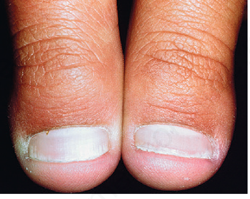
| Measurement | Normal | Clubbing |
|---|---|---|
| Lovibond's angle | < 160° | > 180° |
| Schamroth's window | Open diamond | Obliterated |
| Curth's angle (DIP) | ~180° | < 160° |
- Respiratory: lung Ca, mesothelioma, CF, cryptogenic fibrosing alveolitis, TB
- Cardiac: cyanotic CHD, infective endocarditis
- GI: IBD, liver disease, oesophageal Ca
- HIV (37%), POEMS syndrome (70%)
💎 Clinical Pearl: Schamroth window test — oppose dorsal surfaces of two homologous fingers. Normal = open diamond window of light. Clubbing = window obliterated. Quick, no-instrument bedside sign.
2.2 Koilonychia (Spoon Nail)
- Concave, spoon-shaped nail plate
- Most common: iron deficiency anaemia, haemochromatosis
- Also: trichothiodystrophy, familial (AD)
- Secondary: psoriasis, lichen planus, dermatophyte infection
2.3 Nail-Patella Syndrome
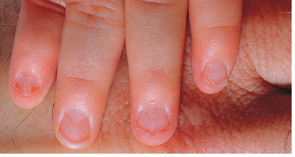
- Triangular lunulae — PATHOGNOMONIC
- LMX1B gene mutation (autosomal dominant)
- Associated: absent/hypoplastic patella, posterior iliac horns, glomerulonephritis (20% → renal failure), "Lester iris"
2.4 Onycholysis
- Separation from nail bed distally → proximally; plate looks WHITE (air underneath)
- Green discolouration = Pseudomonas aeruginosa
- Post-traumatic: regular margin, lateral
- Psoriatic: irregular margin, whole free edge, erythematous border
- Onychomycosis: ragged/spike edge, mycelium proximally
- Drug-induced: uniform midline (photo-onycholysis)
💎 Clinical Pearl: The onychodermal band is the "Achilles heel" of nail plate attachment. Once breached — psoriasis, trauma, infection — onycholysis becomes progressive and is difficult to reverse. Clip, dry, topical antiseptics are the cornerstone.
2.5 Beau Lines
- Transverse depressions = temporary interruption of proximal matrix mitosis
- All 20 nails simultaneously = SYSTEMIC cause (cytotoxics, severe illness, erythroderma)
- 1-2 nails only = local trauma or paronychia
- Beau lines → onychomadesis when full-thickness groove
💎 Clinical Pearl: Beau lines at the same level in all 20 nails = systemic cause. Beau lines on only 1-2 nails = local trauma or paronychia.
2.6 Pitting
- Fine pits (<1mm): PSORIASIS — >10 pits/nail OR >60 total supports diagnosis
- Coarse pits: ECZEMA
- Geometric "Scotch plaid" / tartan pattern: ALOPECIA AREATA
- Elkonyxis = deep single large pit (reactive arthritis, psoriasis)
2.7 Nail Colour Changes
| Colour | Cause |
|---|---|
| True leukonychia (nail plate) | Parakeratosis; Mees' lines = arsenic/systemic insult |
| Apparent leukonychia (nail BED — disappear with pressure) | Muehrcke's = hypoalbuminaemia; Terry's = liver disease; Half-and-half = renal failure |
| Yellow | Yellow nail syndrome, psoriasis, fungal |
| Green | Pseudomonas aeruginosa |
| Red lunula | Psoriasis, CO poisoning, cardiac failure, AA |
| Splinter haemorrhages | Psoriasis, endocarditis, vasculitis, trauma |
💎 Clinical Pearl: All apparent leukonychia (Muehrcke's, Terry's, half-and-half) involves the nail BED not the plate, and disappears with pressure. True leukonychia (nail plate) does NOT disappear with pressure.
2.8 Melanonychia — ABCDEF Rule
| Letter | Meaning |
|---|---|
| A | Age (40-70); African/Asian/Native American |
| B | Brown/Black band >3mm, Blurred borders |
| C | Change in morphology |
| D | Digit (thumb > hallux > index) |
| E | Extension to nail fold (Hutchinson's sign) |
| F | Family/personal history of melanoma |
💎 Clinical Pearl: Pseudo-Hutchinson's sign = apparent periungual pigmentation through a transparent cuticle (racial melanonychia, benign nevi). Real Hutchinson's = actual melanin extension INTO the nail fold skin. Dermoscopy distinguishes the two.
2.9 Nail Signs in Systemic Disease
| Sign | Disease |
|---|---|
| Clubbing | Cardiopulmonary, GI, liver, HIV, POEMS |
| Koilonychia | Iron deficiency anaemia, haemochromatosis |
| Splinter haemorrhages | Endocarditis, vasculitis, psoriasis, trauma |
| Muehrcke's lines | Hypoalbuminaemia |
| Terry's nails | Hepatic cirrhosis |
| Half-and-half nails (Lindsay's) | Chronic renal failure |
| Yellow nail syndrome | Lymphoedema, pleural effusion |
| Periungual telangiectasia | Dermatomyositis (most specific), SLE, SSc |
| Ragged cuticles | Dermatomyositis |
| Triangular lunula | Nail-patella syndrome |
| Beau lines (all 20 nails) | Systemic illness, cytotoxics, erythroderma |
| Koenen tumours (periungual fibromas) | Tuberous sclerosis |
PART 3 — NAIL DISORDERS
3.1 Nail Psoriasis
-
50% prevalence in plaque psoriasis; 80-90% lifetime incidence
- 5-10% nail psoriasis without skin/joint disease
- Annual PsA risk with nail involvement: 0.55-2.55%
- HLA-Cw6 NEGATIVE (skin psoriasis = HLA-Cw6 positive)
| Clinical Feature | Site | Duration |
|---|---|---|
| Pitting | Proximal matrix (parakeratosis foci) | Episodic |
| Transverse furrows | Proximal matrix | 1-2 weeks |
| Crumbling nail | Entire matrix | Prolonged |
| Leukonychia with rough surface | Proximal matrix | Variable |
| Splinter haemorrhages | Nail bed capillaries | Short |
| Oil spot / Salmon patch | Nail bed | Prolonged — PATHOGNOMONIC |
| Onycholysis | Nail bed | Prolonged |
| Subungual hyperkeratosis | Nail bed | Prolonged |
💎 Clinical Pearl: Salmon patch (oil-drop sign) = NEARLY PATHOGNOMONIC for psoriasis. Unlike pitting (also in AA, eczema), oil-spotting is nearly exclusive to psoriasis. Confirm with onychoscopy: dilated tortuous capillaries in 90% of cases.
═══ MECHANISM OF NAIL CHANGES IN PSORIASIS ═══
Step 1 — Genetic Basis & Trigger
- HLA-Cw6 NEGATIVE — genetically distinct from cutaneous psoriasis
- Trigger: microtrauma at nail unit → Koebner phenomenon
- Extensor tendon inserts adjacent to nail root at DIP joint → nail matrix is anatomically contiguous with the synovio-entheseal complex of DIP
Step 2 — Innate Immune Activation
- Stressed keratinocytes → antimicrobial peptides (LL-37, hβD-2, hβD-3)
- These activate myeloid DCs (mDCs) and plasmacytoid DCs
- Breaches immune privilege of nail matrix
- NF-κB pathway activated; mast cells recruited
Step 3 — IL-23 / Th17 Axis
- mDCs release IL-23 → Th17 cell differentiation
- Th17 → IL-17A, IL-17F, IL-22, TNF-α
- VEGF upregulated → angiogenesis → dilated tortuous capillaries (onychoscopy finding in 90%)
Step 4 — Matrix Inflammation → PITTING
- IL-17/TNF-α in proximal nail matrix → defective keratinization
- Parakeratosis foci (clusters of nucleated cells without normal cornification) form in dorsal nail plate
- These foci shed as nail grows distally → tiny depressions = PITS
- More prolonged matrix inflammation → transverse furrows → crumbling → leukonychia
Step 5 — Nail Bed Inflammation → OIL-SPOT / ONYCHOLYSIS
- Psoriasiform hyperplasia + parakeratosis + microvascular changes + neutrophil trapping in nail bed
- → Salmon/oil-drop patch (translucent yellow-red zone) — NEARLY PATHOGNOMONIC
- Onychodermal band breach → progressive onycholysis
- Nail bed hyperkeratosis → subungual hyperkeratosis
- Dermal ridge capillary rupture → splinter haemorrhages
Step 6 — Enthesitis → Psoriatic Arthritis
- IL-17/IL-22/TNF-α from nail unit spread to adjacent DIP joint synovio-entheseal complex
- Sensitises DIP joint to trauma-triggered inflammation → psoriatic arthritis
- Annual PsA risk doubles with nail involvement
- This is why nail psoriasis is the strongest clinical predictor of PsA
Microtrauma / Koebner
         ↓
Keratinocyte stress → LL-37, hβD-2/3
         ↓
mDC activation → IL-23
         ↓
Th17 → IL-17A/F + IL-22 + TNF-α
         ↓
   ┌─────────────────┬────────────────────┐
   ↓                 ↓                    ↓
Proximal matrix   Nail bed           DIP enthesis
Parakeratosis     Hyperplasia +      Enthesitis
→ PITTING         microvascular      → PsA risk
                  → OIL-SPOT +
                    ONYCHOLYSIS
💎 Clinical Pearl: Nail psoriasis is HLA-Cw6 NEGATIVE — opposite of cutaneous plaque psoriasis. Genetically distinct subset with stronger PsA association. Always screen for joint disease.
💎 Clinical Pearl: Adalimumab is the ONLY FDA-approved biologic specifically for nail psoriasis. IL-17 inhibitors (secukinumab, ixekizumab) show excellent nail clearance in trials.
Nail Psoriasis — Treatment Algorithm
| Extent | First Line | Second Line |
|---|---|---|
| ≤3 nails, matrix | Intralesional triamcinolone acetonide | Topical vitamin D ± steroid |
| ≤3 nails, nail bed | Topical steroid OR vitamin D | — |
| >3 nails | IL steroid ± topical vitamin D | Systemic / Biologics |
3.2 Lichen Planus of the Nails
- 5-10% of LP patients; 5th-6th decade
- Dorsal pterygium — MOST CHARACTERISTIC (fibrotic PNF-matrix fusion, splits nail in 2)
- Onychorrhexis — irregular longitudinal ridging
- Thinning → atrophy → shedding
- Trachyonychia (20-nail dystrophy, shiny variant)
- Erythronychia, subungual hyperpigmentation
- Idiopathic atrophy of nails = most severe variant (rapid irreversible destruction)
- Histology: interface dermatitis, band-like lymphocytes, basal cell liquefaction — identical to cutaneous LP
💎 Clinical Pearl: Dorsal pterygium = lichen planus until proven otherwise. Irreversible scarring — early aggressive treatment is essential. LP causes pterygium; psoriasis causes pitting — never confuse the two.
3.3 Onychomycosis
| Type | Description | Organism |
|---|---|---|
| DLSO (most common) | Free edge/lateral; hyperkeratosis, onycholysis | T. rubrum (70-80%) |
| PSO | Via nail fold; leukonychia at lunula; HIV! | T. rubrum |
| SWO | Powdery white patches on dorsal surface | T. interdigitale, moulds |
| Endonyx | Milky white, no hyperkeratosis | T. soudanense/violaceum |
| CDLSO | Chronic mucocutaneous candidiasis | Candida spp. |
| Feature | Psoriasis | Onychomycosis |
|---|---|---|
| Pitting | Fine, irregular | Rare |
| Dermoscopy edge | Irregular, erythematous border | Ragged/spike |
| KOH | Usually negative | Filaments abundant |
| Salmon patch | Present | Absent |
- Terbinafine oral 250mg/day × 6 wks (fingernails), 12 wks (toenails) — most effective
- Itraconazole 200mg/day OR pulse (400mg/day × 1 wk/month × 2-3 cycles)
- Topical: ciclopirox 8%, efinaconazole (adjunct only)
💎 Clinical Pearl: PSO (proximal subungual onychomycosis) in a young patient = HIV testing MANDATORY. Always get KOH/culture before treating — misdiagnosis rate is high. Topical treatment alone rarely cures toenail onychomycosis.
3.4 Paronychia
Acute
- Staphylococcus aureus (most common); herpes simplex (herpetic whitlow); orf
- Treatment: drain abscess; penicillinase-resistant antibiotic; oral aciclovir for herpetic whitlow (topical = no benefit)
Chronic
- Predominantly wet-work workers, females
- Candida is NOT the primary pathogen — it is a secondary coloniser
- Primary = irritant/allergic dermatitis; systemic antifungals are useless
- Treatment: avoid wet work; topical steroids (primary); imidazoles (for Candida); crescent PNF excision if refractory (heals 2° intention < 2 weeks)
💎 Clinical Pearl: Chronic paronychia ≠ Candida infection. Candida is a secondary coloniser. Primary = irritant/allergic dermatitis. Systemic antifungals DO NOT HELP.
3.5 Darier Disease of the Nails
- Candy-cane appearance: red + white longitudinal bands — PATHOGNOMONIC
- Free margin: V-shaped notch and fissure (fragility)
- Histology: acantholysis + multinucleate giant cells + epithelial hyperplasia
💎 Clinical Pearl: Red + white longitudinal bands + V-notch at free edge = Darier disease. Pathognomonic — can diagnose Darier from nails alone even without skin lesions.
3.6 Eczematous Nail Dystrophy
- Almost all hand eczema patients affected; ~16% in atopic dermatitis
- Coarse pits — distinguish from fine pits of psoriasis
- Beau lines very common; loss of cuticle → Candida/bacterial superinfection
- Photo-onycholysis (tetracyclines, 5-FU, capecitabine): matches shape of proximal nail fold
3.7 Yellow Nail Syndrome
- Also: bronchiectasis, rhinosinusitis; growth < 0.1mm/week
- Treatment: vitamin E (topical/systemic), triazoles
3.8 Nail Tumours
Glomus Tumour
- Predominantly women, 4th-5th decade
- Triad: Love test (point tenderness) + Hildreth's sign (tourniquet relieves pain) + cold sensitivity
- Presentations: reddish/bluish spot under nail OR longitudinal erythronychia with distal notch
- Investigation: MRI (gold standard); high-res ultrasound + Doppler
- Bone erosion in 50%
- Treatment: surgical excision (transungual approach preferred)
💎 Clinical Pearl: Glomus tumour triad = Love test + Hildreth's sign + cold sensitivity. Pain disproportionate to exam findings = think glomus tumour. MRI is gold standard.
Subungual Exostosis
- Benign osteochondral outgrowth; 75% great toenail
- X-ray: cup-shaped bone outgrowth (diagnostic)
Subungual Melanoma
- ~25% of melanomas in Japanese and African Americans
- Most from matrix (75%) → longitudinal melanonychia; acral lentiginous subtype
- BRAF mutations LOW; KIT/NRAS mutations HIGH
- Treatment: in situ → en bloc 5-10mm; invasive → amputation; SLNB if >1mm
SCC — Most Common Malignant Nail Tumour
- HPV-associated (up to 60%; genito-digital); right index finger
- Mean diagnosis delay = 6 years
- Treatment: Mohs surgery; amputation only if bone involved
💎 Clinical Pearl: SCC is the MOST COMMON malignant nail tumour — not melanoma. HPV-associated; right index finger. Mean diagnosis delay = 6 years. Persistent subungual hyperkeratosis + oozing = SCC until proven otherwise.
3.9 Connective Tissue Diseases
| Disease | Nail Findings |
|---|---|
| Systemic sclerosis | Periungual telangiectasias; megacapillaries + avascular areas (4 stages capillaroscopy); pterygium inversum unguis; acro-osteolysis |
| SLE | Periungual erythema, splinter haemorrhages, onycholysis |
| Dermatomyositis | Dilated irregular nail fold capillaries — PATHOGNOMONIC; ragged cuticles |
| RA | Tortuous/ramified capillary loops; "fish shoal" pattern |
💎 Clinical Pearl: Nail-fold capillaroscopy distinguishes primary from secondary Raynaud. Megacapillaries + avascular areas + hazy background = systemic sclerosis (secondary). Normal architecture = primary Raynaud.
3.10 Drug-Induced Nail Changes
| Drug | Nail Change |
|---|---|
| Retinoids | Onycholysis, paronychia, ingrown toenails, fragility |
| EGFR inhibitors | Multiple pyogenic granulomas, paronychia |
| Taxanes | Multiple PGs, onycholysis, subungual haematoma |
| Hydroxyurea | Melanonychia |
| β-blockers | Pincer nails, digital necrosis |
| ACE inhibitors (captopril) | Onycholysis |
| Methotrexate | Slower nail growth |
| Doxycycline/psoralens | Photo-onycholysis |
PART 4 — NAIL SURGERY OVERVIEW
- 0.5 mL dorsal + 0.5 mL palmar × both sides
- Ring block misses dorsal branches (dual innervation)
- Nail avulsion: partial preferred (total → nail bed shrinkage)
- Punch biopsy 3mm — small pigmented lesion < 3mm
- Longitudinal ellipse — longitudinal pigmented bands
- Lateral longitudinal biopsy (sigmoid-shaped) — all lateral nail structures
- Crescent PNF excision — chronic paronychia; heals 2° intention < 2 weeks
- 88% phenol × 4 min with tourniquet — ingrowing toenail; < 3% recurrence
- MRI: gold standard — glomus tumour, soft tissue masses, melanoma extent
- Ultrasound >15 MHz: glomus tumour, psoriatic enthesitis/dactylitis
- X-ray: subungual exostosis, acro-osteolysis, SCC bone involvement
💎 Clinical Pearl: Distal digital block — NOT ring block — for nail surgery. The nail has dual innervation. A ring block at the digit base misses dorsal branches.
PART 5 — ALL CLINICAL PEARLS
- Onychodermal band = once breached → progressive onycholysis, difficult to reverse. Clip, dry, antiseptics.
- Chronic paronychia ≠ Candida. Candida = secondary. Primary = irritant dermatitis. Antifungals useless.
- PSO + young patient = HIV. Test every time.
- Nail-fold capillaroscopy — megacapillaries + avascular areas = scleroderma; normal = primary Raynaud.
- Pseudo-Hutchinson's = transparent cuticle revealing nail pigment (benign). Real Hutchinson's = melanin IN nail fold skin (melanoma).
- Glomus tumour triad = Love test + Hildreth's sign + cold sensitivity. MRI = gold standard.
- Darier candy-cane nails = red + white bands + V-notch = pathognomonic. Diagnose from nails alone.
- Nail psoriasis HLA-Cw6 NEGATIVE — skin psoriasis is HLA-Cw6 positive. Genetically distinct; stronger PsA link.
- Koenen tumours (tuberous sclerosis) lack the hyperkeratotic tip — histologically identical to ungual fibrokeratomas otherwise.
- Nail melanoma in Japanese/African Americans = ~25% of all their melanomas.
- SCC > melanoma as most common malignant nail tumour. Mean diagnosis delay = 6 years. HPV-associated.
- Toenail onychomycosis does not cure with topical alone. Always get KOH/culture first.
- Beau lines all 20 nails = systemic. 1-2 nails only = local.
- Distal digital block, not ring block, for nail surgery.
- Salmon/oil-drop patch = nearly pathognomonic for psoriasis. Onychoscopy: tortuous capillaries in 90%.
- Adalimumab = only FDA-approved biologic specifically for nail psoriasis.
- Dorsal pterygium = LP. Psoriasis NEVER causes pterygium — it causes pitting and onycholysis.
- NAPSI max = 160 (all 20 nails × 8).
PART 6 — VIVA QUESTIONS & ANSWERS
PART 7 — QUIZ (Q&A)
| # | Question | Answer |
|---|---|---|
| 1 | Normal fingernail growth rate? | ~3 mm/month |
| 2 | Which matrix zone produces ventral nail plate? | Distal matrix (lunula) |
| 3 | Onychodermal band = ? | Nail isthmus |
| 4 | Lovibond's angle in clubbing exceeds? | 180° |
| 5 | Koilonychia — most common systemic cause? | Iron deficiency anaemia |
| 6 | K1/K10 in nail bed = ? | Pathological — psoriasis/onychomycosis |
| 7 | Yellow nails + lymphoedema + pleural effusion = ? | Yellow nail syndrome |
| 8 | Primary pathogen in chronic paronychia? | NONE — irritant dermatitis; Candida = secondary |
| 9 | Most common organism in DLSO? | Trichophyton rubrum |
| 10 | PSO in young patient → test for? | HIV |
| 11 | Candy-cane nails = ? | Darier disease |
| 12 | Pterygium most commonly caused by? | Lichen planus |
| 13 | Koenen tumours → which syndrome? | Tuberous sclerosis |
| 14 | Triangular lunulae → which syndrome? | Nail-patella syndrome (LMX1B) |
| 15 | Megacapillaries + avascular areas on capillaroscopy? | Systemic sclerosis |
| 16 | Most common MALIGNANT nail tumour? | SCC (not melanoma) |
| 17 | FDA-approved biologic for nail psoriasis? | Adalimumab |
| 18 | NAPSI maximum score? | 160 |
| 19 | Gold standard imaging for glomus tumour? | MRI |
| 20 | Green discolouration under onycholytic nail? | Pseudomonas aeruginosa |
| 21 | Hutchinson's sign = ? | Melanin extending INTO periungual skin = melanoma |
| 22 | Nail pitting histologically = ? | Parakeratosis foci in proximal nail matrix |
| 23 | Best drug for toenail onychomycosis? | Oral terbinafine 250mg/day × 12 weeks |
| 24 | Phenol cautery time for ingrowing toenail? | 4 min with tourniquet; < 3% recurrence |
| 25 | HLA in nail psoriasis? | HLA-Cw6 NEGATIVE |
| 26 | Salmon patch = ? | Nearly pathognomonic for psoriasis |
| 27 | Key cytokine pathway in nail psoriasis? | IL-23 → Th17 → IL-17A/F + TNF-α |
| 28 | SCC of nail — mean diagnosis delay? | 6 years |
| 29 | Crescent PNF excision used for? | Chronic paronychia (refractory) |
| 30 | Nail psoriasis annual PsA risk? | 0.55-2.55% (doubles vs no nail involvement) |