
1. Discuss benefits of exercise in patients with coronary artery disease. Write about exercise program for coronary artery disease. 30 M (Summer 2023)
"coronary artery disease" AND "exercise" AND "cardiac rehabilitation"
Exercise in Coronary Artery Disease: Benefits and Exercise Program
1. Overview - Cardiac Rehabilitation as the Framework
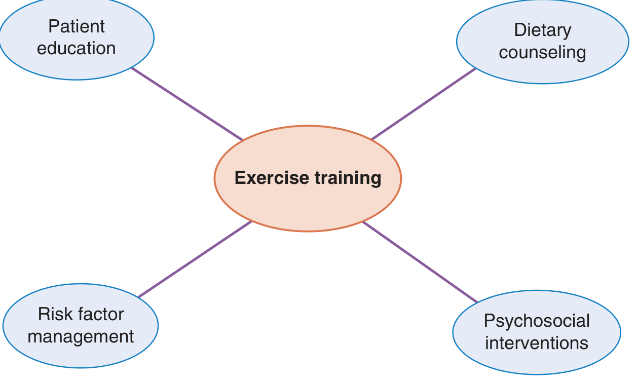
- Fuster and Hurst's The Heart, 15th Ed., p. 714
2. Benefits of Exercise in CAD Patients
A. Physical Fitness Benefits
- Improved exercise functional capacity - patients can exercise at higher intensity and for longer duration over time.
- Improved muscle strength - through resistance (strengthening) exercise.
- Improved tolerance for activities of daily living - previously intolerable tasks become manageable for deconditioned patients.
- Improved physical self-efficacy - patients gain confidence in their physical abilities.
- Improved endothelial function - exercise may be the single most effective method for improving endothelial function among all available options.
- Fuster and Hurst's The Heart, 15th Ed., p. 716; Pfenninger and Fowler, p. 648
B. Reduction in Anginal Symptoms
- Fuster and Hurst's The Heart, 15th Ed., p. 716
C. Reduction in Adverse Cardiac Events (Mortality and Morbidity)
| Outcome | Magnitude of Benefit |
|---|---|
| All-cause mortality | 26% reduction (meta-analysis of 34 trials) |
| Cardiac mortality | 36% reduction |
| Cardiovascular mortality (recent era) | 58% reduction (van Halewijn et al., 2010-2015 trials) |
| Risk of MI | 30% reduction |
| Risk of stroke | 60% reduction |
| Recurrent hospitalizations | Significant reduction |
| Downstream medical costs | Lower than usual care |
- Fuster and Hurst's The Heart, 15th Ed., p. 716-717
- Sabiston Textbook of Surgery, p. 2514
D. Physiological Mechanisms Behind the Benefits
- Anti-atherogenic effects: improves lipid profile (raises HDL, lowers LDL and triglycerides)
- Anti-thrombotic effects: reduces platelet aggregability and fibrinogen levels
- Anti-arrhythmic effects: increases vagal tone, reduces resting heart rate, improves heart rate variability
- Hemodynamic effects: lowers resting blood pressure, reduces cardiac workload at submaximal exercise levels
- Metabolic effects: improves insulin sensitivity, reduces body weight and adiposity
- Endothelial function: increases nitric oxide bioavailability, reduces vascular inflammation
- Autonomic effects: reduces sympathetic nervous system activation
E. Psychological Benefits
- Significant improvement in quality of life
- Reduction in depression symptoms - depression affects up to 20% of post-revascularization patients and is a major independent cardiac risk factor
- Reduction in anxiety
- Improved self-confidence and psychological resilience
- Fuster and Hurst's The Heart, 15th Ed., p. 714
3. Indications for Cardiac Rehabilitation / Exercise Program
- Post-acute myocardial infarction (AMI)
- Post-coronary artery bypass graft surgery (CABG)
- Post-percutaneous coronary intervention (PCI)
- Chronic stable angina pectoris
- Peripheral vascular disease
- Heart failure with reduced ejection fraction
- Post-cardiac transplantation
- Post-valvular repair or replacement
- Sabiston Textbook of Surgery, p. 2514
4. Contraindications to Exercise / Cardiac Rehabilitation
| Contraindication |
|---|
| Inability to exercise (musculoskeletal conditions) |
| Cognitive dysfunction preventing compliance |
| Worsening/unstable chest pain |
| Decompensated heart failure |
| Recent stroke or TIA |
| Atrial arrhythmia with uncontrolled ventricular response |
| Complex ventricular arrhythmia |
| Severe pulmonary arterial hypertension |
| Intracavitary thrombus |
| Recent thrombophlebitis or pulmonary embolism |
| Severe obstructive cardiomyopathy |
| Symptomatic or severe aortic stenosis |
| Acute infection |
5. Structure of the Exercise Program (Cardiac Rehabilitation)
Phases of Cardiac Rehabilitation
- Begins during hospitalization following AMI, CABG, or PCI
- Progressive ambulation initiated within 1 day of acute event ("armchair" ambulation)
- Patient education on risk factor modification ("teachable moment")
- Now largely abbreviated in the US due to short hospital stays
- Team: cardiologists, physiotherapists, occupational therapists, social workers
- Standard: 36 sessions, conducted 2-3 times per week over 12-18 weeks
- Globally, median duration is 24 sessions
- Continuous ECG monitoring during all sessions
- Components: warm-up, monitored aerobic exercise, resistance training, cool-down, education
- Optional extended exercise maintenance program
- May or may not be medically supervised
- Generally no continuous ECG monitoring
- Maintains the gains achieved in Phase 2
Pre-Exercise Assessment
- Symptom-limited or modified exercise tolerance test (ETT) - conducted with patients on their usual medications
- Identifies inducible symptoms, ischemia, or arrhythmia
- Used to formulate target training heart rate range
- If no baseline ETT performed: initial target HR = resting standing HR + 20 beats/min
- Customized exercise prescription based on true maximal heart rate (MHR), aerobic capacity, and test results
- Pfenninger and Fowler, p. 648
Exercise Intensity Prescription
- Target: moderate intensity - equivalent to brisk walking
- Use of Borg Perceived Exertion Scale (RPE 12-14, "somewhat hard")
- Initial training may start at low intensity to build confidence
- Heart rate target range based on ETT results
- Intensity adjusted based on: baseline conditioning, comorbidities, age, motivation
Structure of a Typical Session
- Warm-up (5-10 minutes): stretching exercises and light calisthenics
- Aerobic Exercise Phase: cross-training on:
- Treadmills
- Stationary bicycles
- Elliptical machines
- Other aerobic equipment
- ECG monitored throughout
- Resistance/Strengthening Exercises: upper- and lower-body muscle strengthening incorporated into program
- Cool-down phase
- Education sessions: integrated into supervision time
Types of Exercise
| Type | Description | Benefit |
|---|---|---|
| Aerobic (isotonic) | Walking, cycling, swimming | Cardiorespiratory fitness, weight reduction |
| Resistance (isotonic/isometric) | Weight training | Muscle strength, functional capacity |
| Flexibility | Stretching | Range of motion, injury prevention |
| High-Intensity Interval Training (HIIT) | Alternating high/low intensity | Superior VO2 max gains vs moderate continuous training |
Staffing of Cardiac Rehabilitation
- Medical director (usually cardiologist)
- Nurses for exercise supervision and medication administration
- Exercise physiologists
- Dieticians, psychologists, social workers as needed
- Fuster and Hurst's The Heart, 15th Ed., p. 716
6. Special Populations
- Sabiston Textbook of Surgery, p. 2514
7. Home-Based vs. Centre-Based Programs
- Traditional CR is centre-based with ECG monitoring
- Home-based programs (telerehabilitation) reduce costs and increase access
- Recent meta-analyses (2023) confirm that cardiac telerehabilitation produces similar longer-term benefits in CAD patients (PMID: 37505803)
- The future likely includes hybrid programs combining centre-based and home-based supervised activities
- Currently limited in the US by insurance coverage constraints
- Fuster and Hurst's The Heart, 15th Ed., p. 714
8. Barriers to Participation
- Older adults
- Women
- Individuals with less education
- Patients with fewer socioeconomic resources
- Patients living farther from CR centres (longer drive times)
- Unemployed individuals
- Patients with less social support
Summary Table: Exercise Program in CAD
| Parameter | Recommendation |
|---|---|
| Timing | Before discharge or at first outpatient visit |
| Frequency | 2-3 sessions per week |
| Sessions | 36 sessions total (Phase 2) |
| Duration | 12-18 weeks |
| Intensity | Moderate (Borg RPE 12-14) |
| Mode | Aerobic + resistance + flexibility |
| Monitoring | Continuous ECG (Phase 2) |
| Pre-assessment | Exercise tolerance test |
| Guideline class | Class I, Level A (ACC/AHA 2021) |
- Zhong et al. (2023) - Cardiac telerehabilitation produces equivalent longer-term benefits (PMID: 37505803)
- Gao et al. (2025) - HIIT superior to moderate continuous training for cardiorespiratory capacity in CAD (PMID: 39977401)
- Shi et al. (2025) - Exercise-based CR post-coronary artery surgery: strong RCT-based meta-analytic support (PMID: 39903572)
1. Ergonomic approach to housewife coming from low socio-economic status from urban area while working in kitchen. 10 M (Summer 2023) 2. Define screening tools and write about their role to identify source of symptoms in desk workers. 10 M (Summer 2023) 3. Write a short note on Ergonomic advice for a computer operator. 10 M (Summer 2023) 4. Write down the movement analysis of lifting and carrying activity in a housewife. Write down the instructions w.r.t. correct body mechanics and ergonomic advice to improve the efficiency of household and social activities during participation. 30 M (Summer 2022) 5. Write a note on adaptive devices and ergonomics for transfer and mobility household geriatric dwellers.10 M (Summer 2022) 6. Evaluation & Ergonomic advice for housewife with lateral elbow pain on dominant side. 10 M (Summer 2022) 7. Discuss the Ergonomics for a 55-year-old Bus driver and list out the exercises to be performed for his low back along. 10 M (Summer 2022) 8. Explain in detail about manual therapy approaches for IT software official with radiating pain down to the back of thigh and numbness along the lateral two toes. Discuss kinematics of the same and give ergonomic advice. 30 M(Winter 2022) 9. Discuss ergonomic assessment and management of knee joint in housemaid. 10 M (Winter 2022) 10. Write a note on biomechanical considerations while giving ergonomic advice in lifting and handling of loads. 10 M(Summer 203)
Ergonomics - Complete Answer Set (Summer/Winter 2022-2023)
Q1. Ergonomic Approach to a Housewife from Low Socio-Economic Status (Urban Area) Working in the Kitchen (10 M, Summer 2023)
Definition of Ergonomics
- Park's Textbook of Preventive and Social Medicine
Special Considerations for Low Socio-Economic Urban Housewife
- Small, cramped kitchen space with poor layout
- Non-adjustable, low-height work surfaces (chulas/floor cooking)
- Limited or no kitchen appliances - manual grinding, pounding, kneading
- Extended cooking hours with repetitive tasks
- No ergonomic furniture - working on floor, low platforms
Common Kitchen Tasks and Associated Ergonomic Hazards
| Task | Posture/Risk | Probable Condition |
|---|---|---|
| Floor cooking (chula) | Prolonged squatting/kneeling | Knee OA, patellofemoral pain |
| Grinding/pounding | Repetitive wrist/shoulder motion | Lateral epicondylitis, CTS |
| Chopping/cutting | Prolonged wrist ulnar/radial deviation | De Quervain's tenosynovitis |
| Lifting heavy vessels | Forward flexion of trunk | Low back pain |
| Washing utensils | Stooped posture at low sink | Thoracic and lumbar pain |
| Rolling chapattis | Repetitive shoulder/wrist rotation | Rotator cuff tendinopathy |
| Carrying water/groceries | Asymmetric loading | Scoliosis, shoulder pain |
Ergonomic Assessment - REBA/RULA Approach
- Observe and document postures during all kitchen tasks
- Identify risk factors: awkward postures, repetition, force, static loading, contact stress
- Prioritize high-risk tasks for modification
Ergonomic Advice and Modifications
A. Work Surface Height
- Ideal counter height: elbow height minus 5-10 cm (approximately 85-90 cm for average Indian woman)
- Improvised solution (low cost): use a sturdy wooden platform/raised block to elevate the work surface
- Avoid prolonged floor-level work - use a low stool at minimum
B. Cooking Area (Chula/Stove)
- Raise the chula/stove to tabletop or knee-high level using brickwork or platform
- Avoid prolonged squatting - use a low plastic stool if floor cooking is unavoidable
- Take 5-minute postural breaks every 30 minutes
C. Body Mechanics During Cooking
- Spine: maintain neutral lumbar lordosis; avoid sustained forward flexion
- Knees: avoid deep squatting; if needed, use full-squat with heels on ground rather than half-squat
- Wrists: keep wrists in neutral position during cutting, kneading, grinding
- Shoulders: keep elbows close to body; avoid overhead reaching
D. Grinding and Kneading
- Switch from stone grinder (silbatta) to mechanical mixer-grinder if budget allows
- If manual grinding: sit at appropriate height, alternate hands, take frequent breaks
- Kneading dough: stand rather than sit; use body weight through extended arms
E. Lifting Heavy Pots and Vessels
- Squat-lift technique: bend knees, keep back straight, object close to body
- Use two hands symmetrically
- Avoid twisting while carrying - pivot entire body
- Use smaller, lighter vessels and refill them rather than carrying full heavy loads
F. Storage and Reach
- Store frequently used items at between shoulder and hip height (power zone)
- Avoid deep overhead shelves - use a step stool rather than stretching
- Place heavy items at waist level, light items above
G. Personal Protective Measures
- Wear flat, supportive footwear (not slippers)
- Use padded anti-fatigue mat if standing for prolonged periods
- Use insulated gloves when handling hot vessels to avoid grip strain
H. Work Organization
- Batch similar tasks to reduce repetitive transitions
- Prepare mise en place (pre-arrangement) before cooking
- Schedule rest breaks of 5-10 minutes every 30-45 minutes of continuous work
- Perform gentle wrist circles, shoulder rolls, and trunk extension stretches during breaks
I. Task Rotation
- Alternate between standing tasks (cutting, stirring) and seated tasks
- Avoid >2 hours of continuous same-posture task
Q2. Screening Tools for Desk Workers - Definition and Role in Identifying Source of Symptoms (10 M, Summer 2023)
Definition of Screening Tools
Purpose of Screening Tools in Desk Workers
- Identify the source and location of musculoskeletal symptoms
- Quantify exposure to risk factors (posture, repetition, force, duration)
- Prioritize interventions - determine which workers or workstations need immediate attention
- Provide baseline data for monitoring over time
- Enable comparison between pre- and post-intervention status
Categories of Screening Tools
A. Symptom-Based Tools (Identify WHERE the problem is)
- Most widely used standardized tool for desk workers
- Maps symptoms across 9 body regions: neck, shoulders, upper back, elbows, wrists/hands, lower back, hips/thighs, knees, ankles/feet
- Records prevalence of symptoms in the last 12 months and last 7 days
- Identifies whether symptoms interfered with normal activity
- Role: Identifies which body region is symptomatic; guides targeted assessment
- Quantifies pain intensity (0-10)
- Simple, quick administration
- Used for monitoring progress
- 30-item tool for upper extremity function
- Especially relevant for desk workers with wrist/hand/shoulder symptoms
- Identifies functional disability related to computer use
- Specifically designed for office/computer workers
- Combines body map with discomfort frequency, severity, and interference with work
- Useful for workstation-specific assessment
B. Postural/Risk Factor Assessment Tools (Identify WHY the problem is happening)
- Evaluates upper limb postures: upper arm, lower arm, wrist, neck, trunk, legs
- Generates an action level score (1-4):
- Level 1-2: Acceptable
- Level 3: Investigate and change
- Level 4: Investigate and change immediately
- Role: Identifies postural source of symptoms in desk/computer workers
- Extension of RULA covering the whole body
- Includes trunk, neck, legs, upper arm, lower arm, wrist
- Generates action level 0-4
- Role: Comprehensive postural analysis for varied desk tasks
- Codes work postures of back, arms, and legs
- Classifies into 4 action categories
- Useful for tasks involving varied postures
C. Workstation-Specific Tools
- Evaluates: monitor height and distance, chair adjustments, keyboard/mouse position, document holder, lighting, footrest
- Checks for: monitor at eye level (top of screen at/just below eye level), keyboard at elbow height, forearms parallel to floor, 90-degree elbow angle, lumbar support, feet flat on floor
- Role: Directly maps workstation deficiencies to reported symptoms
- Specific to mouse and keyboard: neutral wrist position, mouse close to keyboard, wrist rest use
- Monitor: 50-70 cm distance, 15-20 degrees below horizontal gaze
- Chair: seat pan depth, armrest height, backrest recline
D. Functional Assessment Tools
- Quantifies exposure to risk factors for distal upper extremity disorders
- Six factors: intensity of exertion, exertion duration, efforts per minute, wrist posture, wrist speed, daily duration
- Role: Identifies whether keyboard/mouse work intensity is a risk factor
Application in Desk Workers - How They Identify Source of Symptoms
| Symptom Reported | Screening Tool | Source Identified |
|---|---|---|
| Neck pain | NMQ + RULA | Forward head posture, monitor too high/low |
| Shoulder pain | NMQ + RULA | Elevated arm rest, mouse too far |
| Wrist pain/numbness | CMDQ + JSI | Non-neutral wrist during typing (CTS risk) |
| Low back pain | NMQ + REBA | Lack of lumbar support, prolonged sitting |
| Eye strain | Office checklist | Monitor glare, improper distance |
| Elbow pain | DASH + JSI | Repetitive clicking, arm unsupported |
Screening Protocol for Desk Workers
- Step 1: Administer NMQ to identify symptomatic body regions
- Step 2: Apply RULA/REBA to assess postural risk at workstation
- Step 3: Use Office Ergonomics Checklist to audit the workstation
- Step 4: Correlate symptom regions with postural and workstation deficiencies
- Step 5: Prioritize and implement targeted interventions
- Step 6: Re-screen at 3 and 6 months to assess outcome
Q3. Ergonomic Advice for a Computer Operator (10 M, Summer 2023)
Introduction
Ergonomic Advice - Workstation Setup
1. Chair Adjustment
- Seat height: Feet flat on floor (or footrest); thighs parallel to floor; knees at 90-100 degrees
- Seat pan depth: 2-4 finger-widths between front edge of seat and back of knees
- Lumbar support: Adjustable backrest supporting lumbar lordosis at L2-L5 level
- Backrest angle: 100-110 degree recline (slight backward lean reduces lumbar disc pressure)
- Armrests: At elbow height; allow shoulders to relax; elbows at 90-100 degrees
- Seat material: Breathable, cushioned; no hard edges
2. Desk and Keyboard
- Desk height: Match elbow height when seated; forearms horizontal or slightly downward
- Keyboard position: Directly in front; close to edge; elbows at 90 degrees
- Wrist position: Neutral (straight); avoid wrist extension during typing
- Wrist rest: Use only during pauses, NOT while actively typing
- Tilt: Keyboard flat or slight negative tilt (away from user) preferred
3. Mouse
- Position: Immediately beside keyboard (same level); no reaching
- Grip: Light grip; use whole-arm movement (not just wrist)
- Type: Consider vertical mouse or trackpad to reduce forearm pronation
4. Monitor
- Distance: 50-70 cm (arm's length) from eyes
- Height: Top of screen at or just below eye level; eyes look slightly downward
- Tilt: Slight backward tilt (10-15 degrees) toward user
- Angle: Perpendicular to windows; avoid glare and reflections
- Dual monitors: Primary monitor directly in front; secondary at same height, angled slightly
5. Documents and Phone
- Document holder: Place between keyboard and monitor, or beside monitor at same height as screen
- Phone: Use headset for prolonged calls; avoid cradling phone between ear and shoulder
6. Lighting
- Avoid direct glare on screen; use indirect or task lighting
- Ambient lighting: 300-500 lux
- Use anti-glare screen filter if needed
Ergonomic Advice - Work Habits and Breaks
The 20-20-20 Rule (Eye Strain)
Break Schedule
- Micro-breaks: 1-2 minutes every 20-30 minutes - stand, stretch, walk
- Mini-breaks: 5-10 minutes every 1-2 hours
- Macro-breaks: Full rest/lunch break
Posture Reminders
- Avoid "tech neck" (forward head posture) - ear over shoulder over hip
- No crossing of legs - maintain bilateral weight bearing
- Do not sit on the edge of the seat
Exercise Programme for Computer Operators
- Lateral cervical flexion (ear to shoulder)
- Cervical rotation
- Chin tuck (cervical retraction)
- Shoulder rolls (forward and backward)
- Chest stretches / doorway stretch (pectoral stretch)
- Scapular retraction (squeeze shoulder blades together)
- Thoracic extension over chair backrest
- Wrist flexion/extension stretch
- Forearm pronation/supination
- Finger tendon glides
- Prayer stretch (bilateral wrist extension)
- Seated lumbar extension
- Knee-to-chest stretch
- Pelvic tilts
Q4. Movement Analysis of Lifting and Carrying in a Housewife + Correct Body Mechanics and Ergonomic Advice (30 M, Summer 2022)
Part A: Movement Analysis of Lifting
Phases of Lifting
- Subject positions feet shoulder-width apart, close to object
- Hip and knee flexion initiated
- Cervical spine moves into slight extension for visual reference
- Core muscles begin to activate (intra-abdominal pressure rises)
- Hip-dominant or knee-dominant strategy:
- Squat lift: Ankle dorsiflexion, knee flexion (>90°), hip flexion, trunk remains upright
- Stoop lift (commonly used, hazardous): Hip and lumbar flexion predominant, knees relatively straight
- Semi-squat: Combination of both
- Gluteus maximus, quadriceps, hamstrings, and spinal extensors eccentrically active
- Increased lumbar compressive forces during stoop lifting
- Object grasped with both hands symmetrically (ideally)
- Object pulled close to the body - moment arm minimized
- Valsalva maneuver increases intra-abdominal pressure (natural spine stabilization)
- Concentric contraction: gluteus maximus, quadriceps, erector spinae
- Spine moves from flexion to neutral/lordotic
- Head lifts first (cervical extension), followed by trunk
- Object remains close to body throughout
- Hip extension drives movement
- Neutral spine maintained
- Object held at or near hip/waist level
- Core co-contraction maintains stability
Carrying Analysis (Asymmetric Load - e.g., Bucket, Grocery Bag)
- Unilateral carrying creates lateral trunk bending
- Contralateral quadratus lumborum and ipsilateral obliques compensate
- Hip Trendelenburg pattern: pelvis drops on unloaded side
- Shoulder girdle asymmetry: elevation on loaded side
- Compensatory cervical lateral flexion toward loaded side
- Increased lumbar disc loading asymmetrically
Carrying Analysis (Head Load - e.g., Water Pot)
- Common in low socio-economic housewives
- Increased cervical axial compression
- Exaggerated lumbar lordosis and anterior pelvic tilt
- Center of mass shifted superiorly - increased spinal compression throughout
Part B: Correct Body Mechanics
For Lifting
- Get close to the object - keep it within 30 cm of the body
- Feet placement: shoulder-width apart, one foot slightly forward for stability (stagger stance)
- Squat down: bend at knees and hips, NOT at the waist
- Back position: maintain neutral lumbar lordosis; avoid forward flexion
- Head position: look forward/slightly up, not down
- Grip: firm two-handed symmetric grip
- Breathe: exhale during the lifting phase (avoid sustained Valsalva for high-risk cardiac patients)
- Rise: straighten legs first, then hips - use leg power
- No twisting: if turning is needed, pivot feet; do not rotate trunk while lifting
- Limit load: use smaller vessels/loads for frequent trips rather than single heavy lifts
For Carrying
- Hold object close to body - at hip/waist height
- Bilateral symmetric carrying is preferred (use two hands, distribute load equally)
- If unilateral: switch sides regularly; do not carry on same arm for >5 minutes
- Head loads: avoid unless habitual; if used, minimize by reducing load weight
- Maintain neutral spine throughout the carrying task
- Look ahead - scan path for obstacles
For Setting Down (Lowering)
- Reverse of lifting: bend knees and hips, NOT waist
- Control the load eccentrically
- Do not drop or release suddenly
Part C: Ergonomic Advice for Household and Social Activities
Kitchen Activities
- Raise cooking platform to elbow height; avoid floor-level cooking
- Use lightweight utensils and divide large loads into smaller batches
- Store heavy items at waist height in lower cabinets
Washing and Cleaning
- Use long-handled mops and brooms (avoid prolonged stooping)
- Kneel rather than squat for low-level tasks; use knee pad
- For floor washing: use bucket on a stool; do not place bucket on the floor while standing
Laundry
- Place washing basin at waist height
- Wring clothes with two hands symmetrically
- Use a washing machine or raised washtub when possible
Carrying Water (Common in Low-Income Households)
- Use a wheeled cart or trolley instead of head/hand carrying
- Reduce pot size - multiple trips preferred over one heavy load
- Use backpack-style water carrier to distribute load symmetrically
Market/Shopping
- Use a wheeled shopping trolley
- Distribute load equally in two bags if carrying by hand
- Avoid carrying loads >10% of body weight unilaterally
Social Activities (Sitting on Floor, Religious Activities, Visiting)
- Use a low back support cushion when sitting on floor
- Avoid Virasana (kneeling) or deep cross-legged sitting for prolonged periods
- Use a folding stool or low chair at social gatherings
Q5. Adaptive Devices and Ergonomics for Transfer and Mobility in Household Geriatric Dwellers (10 M, Summer 2022)
Introduction
A. Transfer Adaptive Devices
Bed Transfers
- Bed rails / grab rails: Fixed to bedframe; allow patient to push/pull to standing
- Transfer board (sliding board): Bridging device for lateral transfers bed-to-chair (for patients with limited standing ability)
- Bed rope ladder: Attached to foot of bed; patient pulls hand-over-hand to sit up
- Adjustable-height bed: Electric or manual adjustment to optimal transfer height (knee height)
- Over-bed trapeze bar: Overhead bar for pulling self up in bed; assists with repositioning
Toilet Transfers
- Raised toilet seat: Adds 5-15 cm to toilet height; reduces hip and knee flexion requirement; critical for patients with hip OA/arthroplasty
- Toilet safety frame / grab bars: Fixed bars beside toilet for push-up assist
- Commode chair: Portable toilet chair beside bed; eliminates need to walk to bathroom at night
Chair/Sofa Transfers
- Chair raiser (leg extensions): Raises chair height to optimize sit-to-stand mechanics
- Ejector cushion (auto-raise seat): Spring-assisted seat that aids rising from chair
- Arm chair with high seat and armrests: Provides pushing surface during sit-to-stand
B. Mobility Adaptive Devices
| Device | Indication | Ergonomic Consideration |
|---|---|---|
| Walking stick (cane) | Mild balance deficit, unilateral hip/knee OA | Handle at wrist crease height; use on contralateral side |
| Forearm crutch (Lofstrand) | Moderate weakness; stair climbing | Forearm trough reduces wrist stress |
| Standard walking frame (Zimmer frame) | Moderate-severe balance deficit | No wheels; stable base; elbow at 20-30° flexion when gripping |
| Wheeled walker (rollator) | Better mobility with balance issues | Has hand brakes; seat for resting; suitable for community use |
| Hemi-walker | Hemiplegia | Provides wider base on affected side |
| Wheelchair (manual) | Severe mobility limitation | Footrests, armrests, anti-tip bars; seat cushion for pressure relief |
| Wheelchair (powered) | Minimal upper limb strength | Joystick control; home must be wheelchair accessible |
C. Home Environmental Ergonomic Modifications
General
- Remove loose rugs and clutter from walkways
- Non-slip flooring or anti-slip mats in bathroom/kitchen
- Adequate lighting at night (night lights in corridor, bathroom)
- Contrast markings on step edges
Bathroom
- Grab bars: Beside toilet (bilateral), inside shower/bath
- Shower stool/bath seat: Allows seated bathing; reduces fall risk
- Hand-held shower head: Reduces reach and balance demands
- Non-slip bath mat
Bedroom
- Bed height: Seat height at knee level for easy transfers
- Bedside lamp with reachable switch
- Telephone/call device within reach
Kitchen
- Pull-out shelves at waist height
- Turntable (lazy susan) inside cabinets to bring items forward
- Lever-type door handles and tap fittings (reduce grip force requirement)
D. Ergonomic Principles for Geriatric Home Dwellers
- Joint protection: Design activities to minimize peak joint loading
- Energy conservation: Pace activities, use adaptive devices to reduce effort
- Fall prevention: Address environmental hazards, improve footwear, balance exercises
- Accessibility: Ensure clear pathway widths (minimum 90 cm for wheelchair)
Q6. Evaluation and Ergonomic Advice for a Housewife with Lateral Elbow Pain on Dominant Side (10 M, Summer 2022)
Probable Diagnosis
Evaluation
History
- Duration, onset (acute vs. gradual)
- Dominant hand involvement
- Specific aggravating tasks (wringing laundry, lifting pots, cutting)
- Previous treatments
- Occupation-specific repetitive tasks
Clinical Assessment
- Mill's test: Passive wrist flexion with elbow extended and forearm pronated - reproduces lateral elbow pain (positive)
- Cozen's test: Resisted wrist extension with elbow in 90° flexion - pain over lateral epicondyle (positive)
- Chair test: Patient lifts chair by gripping with extended elbow and pronated forearm - pain reproduced
- Grip strength measurement (dynamometer): typically reduced on affected side
- DASH questionnaire score
- Observe patient performing kitchen tasks: note wrist position, grip force, forearm rotation
- Identify: sustained wrist extension, high grip force with pronation, repetitive motion frequency
Ergonomic Advice
Task Modification
- Wringing laundry: Use washing machine or wring with bilateral symmetric motion; use gloves to reduce grip force
- Lifting pots and vessels: Use two-handed lift; use lighter vessels; reduce vessel weight by emptying before lifting
- Cutting and chopping: Use sharp knives (less force required); hold knife with power grip; work at counter height; avoid cutting toward the body
- Rolling chapattis: Stand at elbow height; push with open palm rather than tight grip
- Kneading: Use the heels of hands (reduces wrist extension); maintain neutral wrist
Adaptive Devices
- Tennis elbow strap (counterforce brace): Applied 2-3 finger widths below the lateral epicondyle; reduces force transmission to the epicondyle during activity
- Wrist splint: Worn at night (prevents nocturnal wrist flexion) and during prolonged tasks
- Ergonomic grip aids: Larger handle circumference for kitchen tools (pots, brooms, rolling pins) - reduces grip intensity; wrap foam tubing around handles
- Lever taps and door handles: Reduce wrist torque demands
- Anti-vibration gloves: If using vibrating tools
Kitchen Layout Modifications
- Work surface at elbow height - reduces need for elbow extension with raised arms
- Frequently used items at shoulder to hip height - eliminates reaching
- Use lightweight cookware (aluminium, non-stick) instead of heavy iron vessels
Positioning Advice
- Avoid sustained elbow extension with loaded wrist
- Keep tools/work close to body
- Maintain forearm in mid-pronation (neutral rotation) during tasks - avoids end-range pronation loading
Exercise Programme
- Rest from provocative tasks
- Ice 10 minutes, 3-4 times/day
- Gentle wrist flexion ROM
- Eccentric wrist extension exercise (Tyler Twist / Theraband exercise): most evidence-based conservative treatment
- Wrist flexor/extensor stretching
- Grip strengthening (progressive resistance)
- Forearm supination/pronation strengthening
Q7. Ergonomics for a 55-Year-Old Bus Driver + Exercises for Low Back (10 M, Summer 2022)
Occupational Risk Profile of Bus Driver
- Whole body vibration (WBV): Primary risk factor for low back pain; steering wheel transmits upper extremity vibration
- Prolonged sitting: 8-12 hours/day; increases lumbar disc pressure
- Awkward postures: Left-right asymmetric torso rotation, sustained cervical rotation (mirror checking)
- Psychological stress: Traffic, time pressure - increases muscle tension
- Restricted breaks: Limited opportunity for postural change
Ergonomic Advice for Bus Driver
Seat and Driving Position
- Seat height: Hips at or slightly above knee level; thighs fully supported
- Seat depth: Full thigh contact without pressure behind knees
- Lumbar support: Properly adjusted lordotic support at L2-L5 level
- Backrest angle: 100-110 degrees (slight recline) - reduces lumbar disc pressure
- Seat suspension: Air-ride suspension seat with damping setting adjusted to driver's weight - reduces WBV transmission
- Seat-to-pedal distance: Slight knee flexion when pressing pedals (not full extension or full flexion)
Steering Wheel
- Height and reach: Elbows at 90-120 degrees when gripping; no shoulder shrugging
- Position: 9 and 3 o'clock grip (reduces shoulder fatigue compared to 10 and 2)
- Use power steering to reduce grip force
Mirrors
- Adjust all mirrors before beginning journey to minimize cervical rotation
- Position mirrors to eliminate need for extreme neck rotation
- Convex/wide-angle mirrors reduce mirror-checking frequency
Vibration Reduction
- Anti-vibration seat cushion (gel or foam)
- Vehicle maintenance: Regular check of tire pressure, shock absorbers to minimize transmitted vibration
- Route planning: Avoid rough roads where possible
Break Schedule
- Micro-breaks: 2-3 minutes every 30-45 minutes of continuous driving - step off bus, walk, perform lumbar extension
- Major breaks: Every 2 hours - 10-15 minute rest with walking
- Compliance with driving hours regulations: No >4.5 hours continuous driving (EC regulations)
Postural Habits
- Avoid leaning to one side while driving
- Sit symmetrically; both buttocks equally weighted
- No wallet in back pocket during driving (causes pelvic asymmetry)
- Chin slightly tucked; cervical spine neutral
Exercises for Low Back (Bus Driver, 55 Years)
1. McKenzie Extension Protocol (for discogenic pain)
- Prone lying: 10 minutes; allows disc nucleus to migrate anteriorly
- Press-ups (cobra pose): 10 repetitions, 3 sets; lumbar extension; hold 5 sec at top
2. Core Stabilisation Exercises
- Drawing-in maneuver (abdominal hollowing): Gentle transversus abdominis activation; hold 10 sec, 10 repetitions
- Dead bug: Supine, alternate arm-leg extension; core maintained neutral
- Bird-dog: Quadruped alternate arm-leg extension; 10 repetitions each side, 3 sets
- Plank: Progress from modified (knees down) to full; 20-30 second holds
3. Flexibility Exercises
- Knee-to-chest stretch: Supine; pull both knees to chest; hold 30 sec; relieves lumbar compression
- Piriformis/hip external rotator stretch: Figure-4 stretch supine
- Hip flexor stretch (iliopsoas): Kneeling lunge; lean forward; hold 30 sec each side
- Hamstring stretch: Supine with towel around foot; gentle sustained stretch
4. Aerobic Exercise
- Walking: 30 minutes, 5 days/week; best general exercise for back pain
- Swimming: Non-weight-bearing; excellent for older drivers with pain
5. Mid-Shift Exercises (At Rest Stop)
- Stand and perform 10 lumbar extension press-ups
- Hip flexor stretch
- Thoracic rotation stretches (arm across chest, seated)
- Calf raises and ankle circles (improve circulation)
Q8. Manual Therapy Approaches for IT Software Professional with Radiating Pain Down the Back of Thigh and Numbness Along Lateral Two Toes + Kinematics + Ergonomic Advice (30 M, Winter 2022)
Clinical Interpretation
- Radiating pain down the back of thigh: Involvement of the sciatic nerve (posterior thigh = S1 nerve root or L5 nerve root)
- Numbness along the lateral two toes: 4th and 5th toes = S1 dermatomal distribution
- L5-S1 disc herniation with S1 nerve root compression (most common in desk workers)
- Possible piriformis syndrome (sciatic nerve compression at piriformis muscle)
Part A: Kinematics of L5-S1 Disc Herniation in IT Worker
Normal L5-S1 Biomechanics
- L5-S1 bears the highest compressive load in the lumbar spine
- Normal disc: nucleus pulposus centrally located, annulus fibrosus intact
- Extension: disc height maintained; posterior facets approximate; foraminal height maintained
- Flexion: anterior disc compression; posterior annular tension; nucleus migrates posteriorly
Pathomechanics in Prolonged Sitting (IT Worker)
- Sustained lumbar flexion (slumped posture) → posterior annular stress
- Repetitive flexion (leaning forward) → nuclear migration posteriorly
- Disc creep: sustained loading over 8+ hours reduces disc height (up to 18% during the working day)
- Annular fiber fatigue: repetitive micro-trauma leads to radial tear
- Posterolateral herniation (most common direction): nucleus material extrudes → compresses S1 nerve root in lateral recess or at foramen
Neurological Effect of S1 Root Compression
- Pain pathway: Posterior thigh and calf (sciatic/posterior femoral cutaneous distribution)
- Numbness/paresthesia: Lateral two toes (4th and 5th toes) - S1 dermatomal area
- Motor deficit (if severe): Weakness of plantar flexion (gastrocnemius/soleus = S1 myotome); weak big toe extension (L5)
- Reflex: Absent/diminished ankle jerk reflex (S1)
Piriformis Syndrome Kinematics
- Prolonged sitting causes piriformis to shorten and hypertrophy
- Tight piriformis mechanically compresses sciatic nerve as it passes through/below the muscle
- External hip rotation deformity; pain reproduced with internal rotation of hip (FAIR test)
Part B: Manual Therapy Approaches
Assessment Prerequisites
- Neurological screening (ASIA/manual muscle testing, sensory testing, reflexes)
- SLR (straight leg raise): positive at <70° suggests disc herniation
- Cross SLR: highly specific for disc herniation
- Slump test: neural tension test - highly sensitive
- Red flags ruled out: No cauda equina syndrome (bowel/bladder dysfunction), no malignancy, no fracture
1. Mobilization Techniques (Maitland Approach)
- Grade I-II: Pain relief (neurophysiological - gate control)
- Grade III-IV: Stiffness/hypomobility (mechanical)
- Position: Prone; applied over L5-S1 spinous process
- Grading based on symptoms and clinical findings
- For L5-S1 hypomobility
- Patient side-lying; oscillatory lateral flexion of lumbar segments
- Mechanical or manual traction reduces intradiscal pressure
- Distraction widens foramen → reduces nerve root compression
- Indications: nerve root pain with positive SLR, disc herniation confirmed on MRI
2. Spinal Manipulation (Thrust Techniques)
- Patient side-lying; top leg flexed; operator applies thrust to L5-S1 level
- Cavitation opens facet joint, releases intra-articular meniscoid, reduces pain
- Contraindicated in severe disc herniation with neurological deficit, cauda equina syndrome, osteoporosis
3. Neural Mobilisation (Neurodynamic Techniques)
- Slider: Alternate cervical extension + knee flexion with SLR position → slides nerve without tensioning
- Tensioner: SLR position with ankle dorsiflexion and cervical flexion → progressively loads the neural tissue
- Evidence: reduces intraneural edema, improves axoplasmic flow, reduces mechanosensitivity
- Perform: 10-15 repetitions, 2-3 sets; pain-free range only
- Soft tissue technique: sustained pressure to piriformis belly in side-lying hip flexion/internal rotation
- Stretching: FAIR position (flexion, adduction, internal rotation) - hip flexed 60°, adducted, internally rotated
4. McKenzie Method (MDT - Mechanical Diagnosis and Therapy)
- Repeated lumbar extension: Reduces disc herniation (centralizes peripheralised pain)
- If pain centralizes with extension: positive responder; aggressive extension program
- Centralization phenomenon: Pain moves from periphery (toe, calf) toward midline with specific repeated movements - highly prognostically significant
- Extension protocol: prone lying → press-ups → standing extension
5. Soft Tissue Techniques
- Myofascial release: thoracolumbar fascia, gluteal muscles, piriformis
- Trigger point therapy: gluteus medius, piriformis, tensor fasciae latae
- Ischemic compression to piriformis for 90 seconds
6. Stabilisation / Motor Control Rehabilitation
- Transversus abdominis (TrA) activation (drawing-in)
- Multifidus contraction (lumbar multifidus has preferential atrophy in L4-L5 and L5-S1 disc disease)
- Dead bug, bird-dog, planks
- Progress to functional load patterns
Part C: Ergonomic Advice for IT Software Professional
Workstation Setup
- Chair: Lumbar support at L2-L5; backrest 100-110° recline; seat height for 90° hips and knees
- Monitor: Top at eye level; 50-70 cm distance; avoid forward head posture
- Keyboard: At elbow height; wrists neutral; keyboard close to body
- Mouse: Beside keyboard; no reaching; light grip
Postural Training
- Sit tall: Maintain slight lumbar lordosis (NOT flat back); use lumbar roll if needed
- Hip angle: Consider sit-stand desk or saddle chair to increase hip angle to >90° (reduces disc pressure)
- Foot support: Feet flat on floor or footrest
Break Protocol
- Avoid sitting >30 minutes without a 2-3 minute standing/walking break
- 20-20 rule for posture: Every 20 minutes, stand for 20 seconds and perform 5 lumbar extensions
- Use sit-stand workstation to alternate postures
Activity Modifications
- Avoid sustained lumbar flexion (slouching, leaning forward to read)
- When commuting: lumbar roll in car; avoid prolonged forward flexion
- Lifting: proper squat-lift technique
Q9. Ergonomic Assessment and Management of Knee Joint in a Housemaid (10 M, Winter 2022)
Common Knee Conditions in Housemaids
- Patellofemoral pain syndrome (PFPS): Anterior knee pain with prolonged kneeling/squatting/stairs
- Knee osteoarthritis (OA): Progressive articular cartilage degeneration from repetitive loading
- Prepatellar bursitis ("housemaid's knee"): Inflammation of the prepatellar bursa from repetitive kneeling
- Pes anserine bursitis: Medial proximal tibia; seen with OA and overuse
Ergonomic Assessment of the Knee in a Housemaid
Step 1: Occupational History
- Duration and frequency of kneeling, squatting, stair climbing
- Type of floors cleaned (hard vs. carpeted)
- Weight of loads carried (buckets, mops)
- Duration of standing per day
- Use of knee pads or protective gear
Step 2: Posture and Movement Analysis
- Kneeling posture: Full kneeling vs. half-kneeling vs. squatting
- Duration: Time spent in each posture
- Frequency: Number of transitions per hour
- Load: Weight lifted or carried
Step 3: Clinical Assessment
- Inspection: Swelling (prepatellar vs. infrapatellar vs. suprapatellar), alignment (genu valgum/varum), muscle wasting (VMO - vastus medialis oblique)
- Palpation: Joint line tenderness (medial/lateral), patellar tenderness, tibial tuberosity, pes anserine region
- ROM: Flexion/extension; note pain arc
- Special Tests:
- Clarke's test (patellar grind) - PFPS
- McMurray's test - meniscal pathology
- Valgus/varus stress test - ligament laxity
- Lachman's test - ACL integrity
Step 4: REBA/Risk Assessment
- Assess kneeling posture REBA score
- Quantify knee flexion angle during tasks (contact stress peaks at >120° flexion)
Management
A. Ergonomic Modifications
- Floor mopping: Use a long-handled mop (120-140 cm) to eliminate kneeling and stooping; use squeeze mop with lever to avoid excessive wrist/knee demands
- Floor scrubbing: Use scrubbing brush with long handle or mechanized floor scrubber; avoid kneeling on hard floors
- Wiping low surfaces: Use kneeling pad/knee guard if kneeling is unavoidable; perform in half-kneeling (one knee down) rather than both
- Lifting buckets: Keep bucket at waist height (on platform); avoid repeated deep squats to floor-level bucket
- Avoid sustained deep squatting (>90° knee flexion) for >10 minutes continuously
- Prefer half-kneeling over full kneeling (one knee on ground, other foot planted)
- Use a low stool for floor-level tasks rather than kneeling on the floor
- Reduce stair climbing frequency - batch tasks to minimize trips
- Use knee pads (gel-filled) when kneeling is unavoidable
- Place items at height to minimize kneeling (e.g., cleaning supplies on low shelf)
- Rubber-soled, shock-absorbing footwear
B. Physical Therapy Management
- Rest from provocative activities
- Ice 15-20 minutes, 3-4 times/day
- NSAIDs as prescribed
- Offloading: avoid all kneeling
-
Quadriceps strengthening (especially VMO):
- Quad sets (isometric quads, 10 sec holds)
- Straight leg raises
- Terminal knee extension (TKE) with resistance band
- Mini squats (0-45° range to avoid high patellofemoral joint stress)
- Step-ups
-
Hip strengthening (reduces valgus collapse, offloads medial knee):
- Clamshells (hip abduction with resistance band)
- Side-lying hip abduction
- Glute bridges
-
Calf strengthening (reduces knee joint loading by improving shock absorption):
- Calf raises (standing and single-leg)
- Quadriceps stretch
- Hamstring stretch
- IT band foam rolling
- Calf stretch
- Single-leg standing (flat, then wobble board)
- Important for preventing falls in housemaids working on wet floors
- Knee sleeve or patellar stabilizing brace: For PFPS
- Medial wedge insole: For medial compartment OA (reduces medial compartment loading)
- Walking stick: Contralateral hand; reduces knee joint loading by 25-30%
Q10. Biomechanical Considerations While Giving Ergonomic Advice in Lifting and Handling of Loads (10 M, Summer 2023)
Introduction
Key Biomechanical Concepts
1. Moment of Force (Torque)
- The moment arm of a load is the horizontal distance between the load and the lumbar spine (L5-S1)
- Even a light load held far from the body generates very high lumbar torque
- A 10 kg load held at 60 cm from the spine generates: 10 × 9.8 × 0.6 = 58.8 Nm of torque
- Same load at 15 cm: 10 × 9.8 × 0.15 = 14.7 Nm - 4x reduction by holding object close
2. Spinal Compressive Force
- During forward bending with 20 kg load, lumbar compressive force exceeds 6000 N (National Institute of Occupational Safety and Health [NIOSH] action limit: 3400 N; maximum permissible limit: 6400 N)
- Erector spinae and multifidus must contract with high force to counter the flexion moment, dramatically increasing compression
3. Stoop vs. Squat Lift
| Parameter | Stoop Lift | Squat Lift |
|---|---|---|
| Trunk posture | Flexed forward | Upright |
| Lumbar compressive force | High (>6000 N with load) | Lower (4000-5000 N) |
| Shear force on L5-S1 | Higher | Lower |
| Quadriceps demand | Lower | Higher |
| Preferred for light loads on floor | Yes | No |
| Preferred for heavy loads | No | Yes |
| Semi-squat (compromise) | Both knees and hips partially flexed | Most energy efficient for most workers |
4. Intra-Abdominal Pressure (IAP)
- During lifting, the diaphragm and pelvic floor contract, increasing IAP
- IAP creates a hydraulic rigid column within the trunk (pneumatic spine concept)
- This reduces compressive load on the lumbar spine by 10-40%
- IAP is generated naturally during Valsalva maneuver
5. NIOSH Lifting Equation
- LC = Load Constant (23 kg under ideal conditions)
- HM = Horizontal Multiplier (load distance from body)
- VM = Vertical Multiplier (height of lift)
- DM = Distance Multiplier (vertical travel distance)
- AM = Asymmetric Multiplier (twisting angle)
- FM = Frequency Multiplier (lifts/minute)
- CM = Coupling Multiplier (grip quality)
- LI < 1: Low risk
- LI 1-3: Moderate risk (ergonomic intervention recommended)
- LI > 3: High risk (immediate redesign needed)
6. Role of Asymmetric and Twisting Lifts
- Rotation of the trunk during lifting superimposes torsional stress on the disc
- Combined compression + torsion dramatically increases risk of annular tear
- A 45° trunk rotation reduces the maximum permissible lift by 57% (per NIOSH AM = 0.57)
7. Cumulative Loading (Fatigue and Creep)
- Repeated sub-threshold loads produce cumulative disc fatigue
- Spinal creep: Prolonged loading causes viscoelastic deformation of disc and ligaments (water loss, reduced stiffness), making the spine vulnerable
- Greatest risk: first lifts in the morning (disc maximally hydrated, stiffer) and after prolonged sitting (disc already partially loaded)
- Warm-up before heavy lifting sessions
- Avoid heavy lifting immediately after prolonged sitting
- Schedule high-demand tasks mid-shift rather than at start
8. Biomechanical Principles Summary for Ergonomic Advice
| Biomechanical Principle | Ergonomic Rule |
|---|---|
| Minimize moment arm | Hold load close to body (<30 cm) |
| Reduce trunk flexion | Squat or semi-squat lift |
| Avoid asymmetry | No twisting; face the load |
| Optimize lift height | Work in power zone (hip to shoulder height) |
| Reduce frequency | Use mechanical aids; rest between lifts |
| Improve coupling | Better grip; handles on containers |
| Team lift | For loads >25 kg |
| NIOSH RWL | Keep actual load < RWL (LI < 1) |
| Fatigue management | Rotate tasks; warm up before heavy work |
| Intra-abdominal pressure | Train core musculature; breathing technique |
Practical NIOSH Recommendations for Manual Handling
- Single-person lift limit: 23 kg under ideal conditions
- Team lift recommended: When load >25 kg or LI consistently >1
- Mechanical aids: Trolleys, hoists, conveyors - eliminate manual lifting when load >50 kg
- Training: Workers should receive specific manual handling training including correct technique, NIOSH limits, and risk recognition
11. Discuss ergonomics of lifting for a normal labourer. 10 M(Summer 2020) 12. Significance of evaluation of work place and environement in IT industry. 10 M(Summer 2019) 13. Write a short note on correct patient handling and lifting techniques. 10 M(Winter 2019) 14. Workspace ergonomics. 10 M(Summer 2018) 15. Ergonomic considerations for computer operators. 10 M(Winter 2017) 16. Discuss ergonomics for textile mill workers. 10 M(Summer 2023) 17. Explain body mechanics and ergonomic advice for computer operators. 10 M(Winter 2021) 18. Management of msk injuries of lower limb in city bus conductor. 10 M(Summer 2021) 19. Describe ergonomic considerations for msk dysfunction of lower limb. 10 M(Winter 2020) 20. Ergonomics and body mechanics advice for dentists. 10M(Summer 2020) 21. Describe management of msk dysfunction in glass and aluminium industry workers.10M(Summer 2020) 22. Ergonomic evaluation and management guidelines for sitting in a chair for person with hip and knee pain. 10 M(Winter 2018) 23. Ergonomic management post- acute cervical intervertebral disc prolapse for computer professional. 10 M(Winter 2022)
Ergonomics - Complete Answer Set (Questions 11–23)
Q11. Ergonomics of Lifting for a Normal Labourer (10 M, Summer 2020)
Definition
Why Lifting is a High-Risk Activity
- Load weight and size
- Frequency of lifting
- Horizontal distance of load from the spine
- Vertical height of lift
- Degree of trunk rotation and asymmetry
- Floor surface and footing conditions
- Duration of lifting tasks
Biomechanical Basis
- A 20 kg load held 60 cm from the spine produces ~120 Nm of lumbar torque
- Same load at 15 cm produces ~30 Nm - a 4-fold reduction simply by holding the load closer
- NIOSH action limit: 3,400 N at L5-S1
- Maximum permissible limit: 6,400 N
- Stoop lifting a 20 kg load can generate >6,000 N compression
- Squat lifting same load: approximately 4,000-5,000 N
NIOSH Lifting Equation
- LC = 23 kg (load constant under ideal conditions)
- HM = horizontal multiplier (load distance from body)
- VM = vertical multiplier (height of hands from floor)
- DM = distance multiplier (vertical travel distance)
- AM = asymmetric multiplier (trunk rotation angle)
- FM = frequency multiplier (lifts per minute)
- CM = coupling multiplier (quality of grip)
- LI < 1: Low risk (acceptable)
- LI 1-3: Moderate risk - ergonomic review needed
- LI > 3: High risk - immediate redesign required
Correct Lifting Technique for the Labourer
The Squat-Lift Technique (Recommended)
- Stand close to the load (within 30 cm) - reduces horizontal moment arm
- Feet shoulder-width apart with one foot slightly forward (stagger stance for stability)
- Face the load squarely - no oblique approach
- Bend at both the hips AND knees simultaneously
- Maintain natural lumbar lordosis - do NOT flex the lumbar spine
- Head in neutral to slight extension - look forward
- Core muscles pre-activated (drawing-in manoeuvre)
- Firm two-handed symmetrical grip
- Object pulled tight against the body before lifting
- Use handles or handholds where available (improves coupling multiplier)
- Exhale during lifting
- Extend legs first (quadriceps-driven), then hips
- Keep object close to the body throughout the entire lift
- Do NOT jerk - smooth controlled movement
- Object held at approximately hip-to-waist height
- Spine in neutral throughout carrying
- Take small steps; do not twist trunk
- Reverse of lifting technique
- Bend knees and hips; keep back straight
- Controlled eccentric descent - do not drop the load
The Stoop Lift vs. Squat Lift
| Parameter | Stoop Lift | Squat Lift | Semi-Squat |
|---|---|---|---|
| Back posture | Flexed | Upright | Partial flexion |
| Lumbar compression | Very high | Moderate | Moderate |
| Disc shear force | High | Low | Low-moderate |
| Energy cost | Lower | Higher | Moderate |
| Quad demand | Low | Very high | Moderate |
| Recommended for | Light, frequent tasks | Heavy loads | Most common loads |
Administrative and Engineering Controls
Hierarchy of Hazard Controls (NIOSH):
- Elimination: Remove the manual lifting need entirely
- Substitution: Replace heavy materials with lighter alternatives
- Engineering controls:
- Mechanical hoists, forklifts, conveyor belts, pallet jacks
- Adjustable-height platforms to keep loads in the power zone (hip to shoulder height)
- Handles and hand-holds on all containers
- Administrative controls:
- Job rotation to distribute spinal loading
- Team lifting for loads >25 kg
- Limit to <25 kg single-person lift; <12 kg for repetitive lifting
- Rest breaks between lifting sessions
- Personal Protective Equipment:
- Lumbar support belts (limited evidence; used as adjunct only)
- Non-slip footwear
- Gloves for grip and temperature protection
Work Zone Principle (Power Zone)
- Below knee level: >40% increase in lumbar load
- Above shoulder level: compromised stability and vision
-
30 cm horizontal distance: exponential increase in lumbar torque
Specific Ergonomic Guidelines for Labourers
| Task | Guidance |
|---|---|
| Sack/bag carrying | Shift load on back (backpack style) rather than one shoulder |
| Stacking/unstacking | Work between knee and shoulder height; use staging platform |
| Carrying over distance | Use trolley or barrow for >15 m distances |
| Team lifting | Designate one leader; lift in coordinated count |
| Asymmetric loads | Balance load with counterweight in other hand |
| Hot/humid environment | Increase rest periods; hydration |
Q12. Significance of Evaluation of Workplace and Environment in IT Industry (10 M, Summer 2019)
Definition
Why IT Industry is a High-Risk Sector
- Sit for 8-12 hours/day at computer workstations
- Perform repetitive keyboard and mouse operations (up to 10,000-15,000 keystrokes/hour)
- Maintain static, awkward postures for prolonged periods
- Face high psychosocial demands (deadlines, performance pressure)
- Work in open-plan offices with noise and lighting issues
- Often work from home with suboptimal setups (since 2020 onward)
Components of Workplace Evaluation
A. Physical/Workstation Evaluation
- Observation and recording of actual sitting postures
- RULA (Rapid Upper Limb Assessment) - assesses neck, trunk, upper limbs
- REBA (Rapid Entire Body Assessment) - full body posture scoring
- Action levels 1-4 guide urgency of intervention
| Component | Evaluation Criteria |
|---|---|
| Chair | Height, lumbar support, seat depth, armrest height, swivel base |
| Monitor | Height (top at eye level), distance (50-70 cm), angle, glare |
| Keyboard | Height, tilt, distance from edge, wrist rest |
| Mouse | Proximity to keyboard, grip style, surface |
| Desk | Height, surface area, leg room, clutter |
| Documents | Holder placement, proximity to monitor |
| Telephone | Headset availability, proximity |
- Lighting: Adequate ambient light (300-500 lux); avoid glare on screens; natural light orientation
- Temperature and Humidity: 20-24°C; 40-60% relative humidity for comfort and cognitive performance
- Noise: Open offices <55 dB for cognitive tasks; noise masking systems for concentration
- Air Quality: Ventilation rate, CO2 levels (>1000 ppm CO2 causes cognitive impairment), volatile organic compounds from printers/carpets
- Space: Minimum 4.6 m² per workstation; fire exit compliance
B. Psychosocial Evaluation
- Job content (variety, meaning, use of skills)
- Work demands (quantitative overload, emotional demands)
- Work control (autonomy, decision latitude)
- Social support (supervisors and colleagues)
- Job security
- Job Content Questionnaire (JCQ): Demand-control-support model
- Copenhagen Burnout Inventory (CBI): Burnout risk
- Work Ability Index (WAI): Overall functional capacity
- General Health Questionnaire (GHQ-12): Psychological well-being
C. Symptom Prevalence Survey
- Nordic Musculoskeletal Questionnaire (NMQ): Maps symptoms across 9 body regions; reveals population-level hotspots (e.g., 65% neck pain in a given department)
- Cornell Musculoskeletal Discomfort Questionnaire (CMDQ): Office-specific; frequency, severity, and work interference ratings
- These data identify clusters of risk linked to specific workstation setups or job roles
D. Task and Time Analysis
- Keystroke logging / motion capture: Quantifies repetitive hand-wrist movements
- Sampling: Observe percentage of time in each posture
- Time-pressure analysis: Ratio of workload to available hours
- Break compliance: Whether workers take mandated micro-breaks
Significance of Evaluation
1. Injury Prevention
- Early identification of high-risk workstations/postures before symptomatic injury develops
- Reduces incidence of carpal tunnel syndrome, cervical spondylosis, PFPS, and low back disorders
2. Productivity Enhancement
- Poor ergonomics reduces typing speed, accuracy, and cognitive output
- Discomfort distracts workers; chronic pain causes presenteeism (working while unwell)
- Correcting workstation can improve productivity by 10-15%
3. Legal and Regulatory Compliance
- OSHA (US), Health and Safety at Work Act (UK), national labour acts require hazard-free workplaces
- Medical surveillance records are required for documented WRMSDs
- Failure to act on identified risks creates liability
4. Cost Reduction
- WRMSDs are the most expensive occupational health burden globally
- Ergonomic intervention ROI (return on investment): typically 3:1 to 10:1
- Reduces absenteeism, compensation claims, recruitment costs due to attrition
5. Psychosocial Risk Management
- High-demand, low-control work environments predict burnout, anxiety, and depression
- Addressing psychosocial hazards reduces turnover and improves retention of skilled IT workers
Evaluation Protocol for IT Industry
- Walk-through survey: Observe the physical workspace environment
- Workstation audit: Standardised ergonomics checklist for each workstation type
- Symptom survey: Administer NMQ or CMDQ to all workers
- Detailed assessment: RULA/REBA on workers with high symptom scores
- Psychosocial survey: JCQ or equivalent
- Report and prioritise: Risk score each finding; match symptoms to environmental causes
- Intervention: Implement workstation modifications, breaks, training, policy changes
- Follow-up: Re-survey at 6 months; track injury incidence trends
Q13. Correct Patient Handling and Lifting Techniques (10 M, Winter 2019)
Introduction
Assessment Before Patient Handling
- Level of consciousness and cooperation
- Weight and height (BMI)
- Ability to weight-bear, pivot, or assist
- Pain levels and movement restrictions
- Presence of drains, catheters, IV lines, fractures
- Skin integrity (bruising, fragility)
- Space available (clear floor space for turning, pivoting)
- Floor surface (wet/dry/uneven)
- Bed height adjustable?
- Adequate staff available for task?
Principles of Correct Patient Handling
- Plan before acting: Know the technique, available equipment, and patient condition
- Communicate: Inform the patient what will happen; give clear instructions
- Use minimal manual force: Employ mechanical aids first
- Maintain neutral spine: Avoid lumbar flexion at all times; use leg drive
- Work at optimal height: Adjust bed/trolley to hip height before handling
- Keep load close: Patient's body should be close to the handler's body
- Avoid twisting: Pivot with feet; face direction of movement
- Two-person rule: Use two or more handlers for obese patients (>80 kg with no assistance ability)
Specific Patient Handling Techniques
A. Turning Patient in Bed (Lateral Roll)
- Adjust bed to working height (mid-thigh to hip level of handler)
- Raise side rails on far side
- Position patient's arm and leg for roll
- Handler 1 at patient's shoulder; Handler 2 at patient's hip
- Both adopt lunge stance (one foot forward)
- "1-2-3 roll" - simultaneously log-roll patient away from handlers
- Position patient with pillow support
- Use rolled draw sheet as grip points
- Move in segments: shoulder, hip, legs
B. Moving Patient Up in Bed
- Adjust bed to flat position; remove pillows
- Place two slide sheets under patient (facing in opposite directions)
- Two handlers at head end; adopt walk-stance, forward lean with straight back
- "1-2-3 slide" - pull draw sheet toward head of bed
- Smooth, coordinated movement; no jerking
C. Sitting Patient Up from Supine
- Adjust bed height to hip level
- Patient rolls to side-lying (log-roll technique first)
- Patient's legs swing off bed as trunk is raised
- Handler supports patient's shoulder and knee simultaneously
- Patient uses arm push if able
- Check for orthostatic hypotension before standing
D. Bed-to-Chair Transfer
- Chair adjacent to bed (45° angle; casters locked)
- Bed at patient's knee height
- Patient slides to edge of bed; feet flat on floor
- Handler in walk-stance, knees bent, back straight
- Handler grips patient's transfer belt (gait belt) - NOT under arms
- "1-2-3 stand" - patient pushes up with arms while handler assists with knee block and gait belt
- Patient pivots (feet pivot, not slides) to face away from chair
- Handler guides controlled lowering
- Board bridging bed to chair seat
- Patient lifts body slightly and slides across board
- Handler guides and stabilises; does not lift
- Full body hoist with appropriate sling (hammock, toileting, standing sling)
- Position sling under patient using log-roll technique
- Attach sling to hoist spreader bar
- One handler operates hoist; second guides patient
- Safest transfer method; eliminates manual lifting
E. Floor-to-Chair Transfer (Post-Fall)
- Call for additional help
- Assess for injury before moving
- Roll patient to prone → kneeling → standing via a sturdy chair
- Use hoist if injured or too heavy
F. Lifting from Toilet / Bath
- Use grab bars; patient actively assists
- Raised toilet seat reduces hip/knee flexion demand on patient and handler
- Bath hoist for dependent patients
Assistive Equipment
| Equipment | Use | Benefit |
|---|---|---|
| Gait belt/transfer belt | Standing pivot transfers | Safe grip point; avoids axilla injury |
| Slide sheet | Bed repositioning | Reduces friction; eliminates manual lifting |
| Transfer board | Lateral transfers (bed-chair) | Eliminates lifting for partial-weight-bearing patients |
| Stand-assist hoist | Partial weight-bearing patients | Reduces handler load by 60-80% |
| Full body hoist | Completely dependent patients | Eliminates manual lifting |
| Banana board | Toilet transfers | Smooth sliding surface |
| Bed elevator | Elevate bed to handler hip height | Eliminates stooping |
Handler Body Mechanics Summary
- Wide base of support (feet shoulder-width apart; stagger stance for direction of movement)
- Knees bent; work at hip height
- Spine in neutral lordosis - never flex the lumbar spine while handling patients
- Keep patient/load close to your body
- Breathe out during exertion; no prolonged breath-holding
- No solo lifting of dependent patients >40 kg without mechanical aid
Q14. Workspace Ergonomics (10 M, Summer 2018)
Definition
Domains of Workspace Ergonomics
1. Physical Ergonomics
2. Cognitive Ergonomics
3. Organisational Ergonomics
Key Principles of Workspace Design
| Principle | Application |
|---|---|
| Neutral posture | Design tasks so joints work near midrange of motion |
| Work in power zone | All tasks between hip and shoulder height |
| Reduce repetition | Job rotation; automated assists |
| Minimise force | Use mechanical aids; lighter tools; better grips |
| Proper work height | Seated: elbow height; Standing: elbow height -5 cm |
| Reduce static loading | Allow postural change; sit-stand options |
| Provide adequate space | Minimum 4.6 m² per workstation |
Components of Workspace Ergonomics
A. Seating Ergonomics
- Seat height: Adjustable; feet flat on floor; thighs parallel; knees at 90-100°
- Seat depth: 2-4 finger-widths between front edge and popliteal fossa
- Seat width: 5 cm wider than hip width on each side
- Backrest: Adjustable lumbar support at L2-L5; backrest angle 100-110°
- Armrests: At elbow height; width adjustable; allow shoulder relaxation
- Seat material: Breathable upholstery; firm cushion (not too soft)
- Swivel base: 5-castor base; lockable for stability when required
- Headrest: For prolonged computer work; supports cervical spine
- Supine: 25 kg
- Standing: 100 kg
- Sitting without support: 140 kg
- Sitting forward flexed: 185-275 kg
- Standing forward flexed: 150 kg
B. Workstation Height
- Seated work: Desk height = elbow height (typically 68-76 cm)
- Standing work: Counter height = elbow height -5 to -10 cm
- Precision work: Higher surface (forearm support)
- Heavy force work: Lower surface (use body weight advantage)
C. Lighting Ergonomics
- Ambient lighting: 300-500 lux for general office work
- Task lighting: 500-750 lux for reading/detailed work
- Monitor screens: No direct overhead glare; perpendicular to windows
- Contrast: Avoid high contrast between task and surroundings (eye fatigue)
- Colour temperature: 3000-5000 K for office environments
- Emergency lighting: Minimum 1 lux at floor level
D. Thermal Comfort
- Temperature: 20-24°C for sedentary work; 16-20°C for light physical work
- Humidity: 40-60% relative humidity
- Air velocity: <0.1 m/s (avoids draught sensation)
- Radiant heat sources (machinery) must be shielded or ventilated
E. Noise
- Cognitive work: <55 dB(A)
- General office: <60 dB(A)
- Loud noise (>85 dB) causes hearing damage with prolonged exposure
- Engineering controls: sound-absorbing panels, acoustic ceiling tiles, isolated machinery
F. Spatial Layout
- Clearance beneath desk: minimum 60 cm width, 45 cm depth for leg room
- Walkway width: 90 cm minimum; 120 cm for wheelchair accessibility
- Reach zone: frequently used items within 45-50 cm reach without stretching
- Emergency egress: clearly marked, unobstructed
G. Tools and Equipment Design
- Handle diameter: 30-40 mm for power grip; 8-16 mm for precision grip
- Tool weight: <2 kg for one-handed use
- Vibrating tools: anti-vibration handles; limit exposure to <2 m/s² (EU Directive)
- Key travel on keyboards: 2-4 mm (reduces finger fatigue)
Workspace Ergonomics Assessment Process
- Identify hazards: Systematic walk-through; checklist audit
- Measure risk: RULA/REBA/OWAS; NIOSH equation for lifting tasks
- Evaluate exposure: Duration, frequency, intensity
- Implement controls: Engineering → Administrative → PPE
- Evaluate effectiveness: Re-audit, symptom surveys, incident reports
- Monitor continuously: Annual reassessment; change assessment for new equipment
Q15. Ergonomic Considerations for Computer Operators (10 M, Winter 2017)
Introduction
Common WRMSDs in Computer Operators
| Region | Condition | Causative Factors |
|---|---|---|
| Neck | Cervical spondylosis, cervicogenic headache | Forward head posture; monitor too high/low |
| Shoulder | Rotator cuff tendinopathy, TOS | Elevated arms; mouse too far; armrests absent |
| Elbow | Lateral epicondylitis, cubital tunnel | Unsupported forearm; elbow leaning on hard edge |
| Wrist/Hand | Carpal tunnel syndrome, De Quervain's | Non-neutral wrist during typing; repetitive finger flexion |
| Thoracic | Kyphosis, thoracic outlet syndrome | Rounded upper back; poor posture |
| Lumbar | Disc prolapse, lumbar spondylosis | Slumped sitting; no lumbar support; prolonged flexion |
Ergonomic Considerations - Detailed
1. Monitor
- Height: Top edge of screen at or just below horizontal eye level; eyes look slightly downward at 15-20°
- Distance: 50-70 cm (one arm's length); further for larger screens
- Angle: Screen perpendicular to window; slight backward tilt (10-15°) toward user
- Brightness: Adjusted to ambient light (should not appear as a light source in a dim room)
- Anti-glare: Use matte screen or anti-glare filter
- Dual monitors: Primary centred; both at same height
2. Chair
- Seat height, depth, lumbar support, armrests, backrest angle (all as detailed in Q14)
- Dynamic seating: Encourage frequent micro-postural shifts; forward tilt option for active sitting
3. Keyboard and Mouse
- Keyboard at elbow height; wrists in neutral (not extended or flexed)
- Negative tilt keyboard (tilted slightly downward away from user) reduces wrist extension
- Mouse immediately beside keyboard; elbow at side; wrist neutral
- Keyboard shortcuts reduce mouse use (reduces shoulder/wrist exposure)
4. Wrist and Forearm Position
- Forearms parallel to floor or slightly downward
- No pressure on wrist against desk edge (cuts off ulnar nerve/artery)
- Wrist rest used during pauses only; NOT during active typing/mousing
5. Document Holder
- Adjacent to monitor at same viewing distance and height
- Eliminates repeated head turning and focal accommodation changes
6. Footrest
- Used when chair must be raised above foot height; maintains 90° knee angle
7. Headset for Phone
- Eliminates head-tilting (lateral flexion) during phone calls
- Critical for operators who simultaneously type and speak
Work Organisation
- Every 20-30 minutes: 1-2 minute micro-break (stand, stretch)
- Every 1-2 hours: 5-10 minute rest away from screen
- 20-20-20 rule: Every 20 min, look 20 ft away for 20 sec (eye strain)
- Alternate between input tasks and reading/reviewing (reduces repetition)
- Use voice recognition for some input to vary modality
Exercise Programme (integrate with breaks)
- Chin tucks (10 repetitions)
- Shoulder shrugs and rolls
- Wrist circles and finger spreads
- Thoracic extension (lean back over chair)
- Seated lumbar extension
- Walking
- Doorway pectoral stretch
- Calf raises
- Lateral neck stretch
Q16. Ergonomics for Textile Mill Workers (10 M, Summer 2023)
Introduction
Occupational Hazard Profile
| Task | Posture/Risk | Common Condition |
|---|---|---|
| Weaving at loom | Prolonged standing; cervical flexion; shoulder abduction | Cervical spondylosis, shoulder tendinopathy |
| Spinning (charkha/machine) | Repetitive upper limb; seated or standing | Carpal tunnel syndrome, de Quervain's |
| Sewing/stitching | Sustained cervical flexion; wrist deviation | Cervicogenic headache, wrist tendinopathy |
| Dyeing/finishing | Prolonged standing; chemical exposure; wet floors | Varicose veins, foot problems, chemical dermatitis |
| Cutting | Forceful scissor/blade use; standing | Forearm and wrist tendinopathy |
| Packing/loading | Manual handling; lifting; carrying | Low back pain, shoulder injuries |
| Power loom operation | Whole-body vibration; noise >90 dB | Noise-induced hearing loss, lumbar disc disease |
Specific Ergonomic Evaluations
RULA/REBA
- Sewing and weaving tasks typically score REBA level 3-4 (need immediate investigation/change)
- Identify: cervical flexion >30°, shoulder abduction >60°, wrist deviation
Nordic Musculoskeletal Questionnaire
- Reveals high prevalence: neck (sewing), wrist (spinning/sewing), back (packing/loaders), knee (standing tasks)
Ergonomic Recommendations by Task
Weaving (Power Loom)
- Anti-vibration seat cushion for seated loom operations
- Anti-fatigue mats for standing operations
- Adjust loom height to elbow height for shuttle manipulation
- Noise protection: Ear muffs/plugs where noise >85 dB
- Breaks: 10 minutes every 2 hours; rotate between standing and machine tasks
Sewing and Stitching
- Machine table height: Elbow level when seated; elbows at 90°
- Chair: Adjustable height; lumbar support; no armrests that prevent elbow approach to machine
- Foot pedal: Positioned so knee is at 90°; alternate foot use to reduce fatigue
- Monitor (if computerised sewing): At eye level; reduce neck flexion
- Lighting: Minimum 500 lux at the needle/sewing area; adjustable task lamp
- Thimble/grip aids: Reduce pinch force during hand-sewing
Spinning
- Adjust spindle/wheel height to reduce sustained shoulder elevation
- Ergonomic wrist-neutral handle design for manual spindles
- Rotating workers between spinning, bobbin-winding, and rest tasks
Cutting
- Scissors: Ergonomic loop scissors; spring-loaded (reduces sustained grip)
- Cutting table height: Standing elbow height
- Knife/blade: Sharp tools (reduce force); anti-vibration handle for powered cutting
- Alternate cutting sides: Reduce unilateral loading
Dyeing/Finishing (Wet Work)
- Non-slip footwear mandatory
- Anti-fatigue mats where prolonged standing on wet/hard floors
- Chemical gloves: Appropriate for specific dye chemicals; regular replacement
- Ventilation: Local exhaust ventilation at dyeing vats
- Height-adjustable vats: To reduce stooping
Packing and Carrying
- Mechanical hoists or conveyor systems for rolls of fabric (>15 kg)
- Trolleys with swivel casters for fabric bale transport
- Use squat-lift technique for lifting from floor level
Environmental Controls
- Noise: Enclosures around noisiest machines; hearing protection; audiometric screening annually
- Heat: Adequate ventilation; rest areas with cooling; hydration stations
- Dust (cotton/flax): Byssinosis risk - local exhaust ventilation; respiratory masks; pre-shift and post-shift PEFR monitoring
- Lighting: Minimum 500 lux at task level in sewing; 300 lux in warehousing
Administrative Controls
- Job rotation: Every 1-2 hours between different task types (reduces cumulative loading)
- Micro-breaks: 5 minutes every hour for stretching
- Training: Manual handling; posture awareness; chemical handling
- Medical surveillance: Annual audiometry; annual musculoskeletal screen (NMQ)
- Welfare facilities: Seating for rest breaks; clean changing rooms
Exercise Programme for Textile Workers
- Chin tucks × 10
- Lateral cervical stretch × each side, 30 sec hold
- Wrist flexion/extension stretch × 30 sec each
- Prayer stretch × 30 sec
- Knee-to-chest × 30 sec
- Standing lumbar extension × 10 reps
- Calf raises × 15 reps (venous return)
- Quadriceps stretches
- Hip flexor stretches
Q17. Body Mechanics and Ergonomic Advice for Computer Operators (10 M, Winter 2021)
Body Mechanics
Key Body Mechanics Principles for Computer Work
1. Head and Cervical Spine
- Neutral position: Ear directly over the shoulder; chin slightly tucked
- Forward head posture: For every 2.5 cm of forward head translation, effective head weight increases by ~4 kg (compressing cervical discs)
- Monitor at correct height eliminates need for sustained cervical flexion or extension
2. Thoracic Spine
- Avoid "tech-back" (thoracic kyphosis)
- Engage thoracic extensors to maintain natural kyphosis
- Scapular retraction: Gentle periodic squeeze of shoulder blades reduces thoracic rounding
3. Lumbar Spine
- Maintain natural lordosis (NOT flat back)
- Backrest reclined at 100-110° reduces lumbar disc pressure compared to upright 90°
- A lumbar roll (placed at L3-4 level) helps maintain lordosis during prolonged sitting
4. Shoulder and Upper Arm
- Upper arms hang close to the body in relaxed position
- Shoulder relaxed, NOT elevated
- Armrests set at elbow height so shoulders do not hunch
5. Elbow
- 90-100° elbow flexion
- No hard-edge contact between distal forearm/elbow and desk (risk of cubital tunnel syndrome from prolonged ulnar nerve pressure)
6. Wrist and Hand
- Neutral wrist (straight, not extended or flexed, not deviated)
- Wrist extension during typing: increases carpal tunnel pressure (from 30 mmHg at neutral to 110 mmHg at 40° extension)
- Fingertips strike keyboard with minimal force
7. Hip and Knee
- Hip at 90-100° (slight open angle preferred)
- Knees at 90° or slightly obtuse
- Feet flat on floor or footrest
- No crossing of legs (creates pelvic obliquity and lateral lumbar stress)
Integrated Ergonomic Advice
- Alternating sitting and standing every 30-45 minutes reduces total spinal disc pressure accumulation
- Standing height: elbow height; anti-fatigue mat when standing
- Use of balance disc or saddle stool occasionally encourages postural variation and activates core muscles
- Not suitable for prolonged periods
- Ergonomic apps (e.g., Posture Minder, Ergotron WorkFit) provide periodic reminders to check posture and take breaks
- Mirror placed at workstation level: immediate visual feedback on head position
- Reduce information overload: organise workflow; use dual monitors for multitasking without postural compromise
- Address job control and demands (high-demand, low-control = highest cardiovascular/musculoskeletal risk)
Q18. Management of MSK Injuries of Lower Limb in City Bus Conductor (10 M, Summer 2021)
Occupational Profile of City Bus Conductor
- Stand for 8-12 hours per shift
- Repeatedly step up and down from bus footboard (50-70 cm step height)
- Move through crowded bus in dynamic, swaying environment (reactive balance demands)
- Issue tickets while standing (repetitive upper limb movements)
- Work in heat, noise, and vibration
- Often wear non-supportive flat footwear (canvas shoes)
Common MSK Injuries of Lower Limb in Conductors
| Condition | Causative Mechanism |
|---|---|
| Plantar fasciitis | Prolonged standing; hard floor; non-supportive footwear |
| Achilles tendinopathy | Repeated stair climbing; forced ankle plantarflexion |
| Patellofemoral pain syndrome | Repetitive step-down; knee valgus during descent |
| Medial knee OA | Prolonged standing; valgus loading; overweight |
| Ankle sprain | Dynamic environment; uneven bus floor; swaying |
| Shin splints (medial tibial stress syndrome) | Prolonged standing; foot hyperpronation |
| Varicose veins/venous insufficiency | Static standing; reduced calf pump activity |
| Hip OA/bursitis | Repetitive hip flexion with stair climbing |
Assessment
History
- Hours standing per day; number of boarding/alighting cycles per shift
- Footwear type and condition
- Pain onset: after prolonged standing, or specifically with stair climbing
- Swelling pattern (end-of-shift vs. morning)
Physical Assessment
- Foot and ankle: Arch assessment (pes planus/cavus); Achilles tenderness; plantar fascia tenderness (insertion vs. mid-substance); ankle ROM; callus formation
- Knee: Joint line tenderness; patellar tracking; valgus alignment; effusion; McMurray's
- Hip: ROM; FABER/FADIR; trochanteric tenderness (bursitis)
- Gait analysis: Watch boarding and alighting movements; observe pattern of knee valgus
Management
A. Plantar Fasciitis
- Supportive footwear: Lace-up shoes with arch support and cushioned heel (mandatory - not canvas/flat shoes)
- Orthotic insoles: Semi-rigid arch support; heel cup; reduces fascial tensile load by 30-40%
- Anti-fatigue mats: At conductor's standing position on the bus (if fixed)
- Gastrocnemius/soleus stretching: 3 × 30 sec, 3 times/day (most evidence-based)
- Plantar fascia specific stretch: Toe extension stretch before first step in morning
- Eccentric calf raises: Off a step; 3 × 15 reps
- NSAID gel or ice massage: After shift
- Night splint: Maintains 5° dorsiflexion to prevent nocturnal contracture of fascia
B. Patellofemoral Pain Syndrome (PFPS)
- Step height reduction: if footboard height can be adjusted, lower to <30 cm
- Teach proper step-down technique: weight through heel, controlled descent, knee over toe (no valgus collapse)
- Patellar taping (McConnell): Medial glide taping before shift reduces pain during stair activity
- VMO strengthening: Terminal knee extensions; mini squats (0-45°)
- Hip abductor/external rotator strengthening: Clamshells, side-lying hip abduction (reduces femoral internal rotation and knee valgus)
- Quadriceps stretching; IT band foam rolling
C. Achilles Tendinopathy
- Heel raise in shoes (5-10 mm) temporarily reduces Achilles tensile load
- Reduce step-climbing frequency during early rehabilitation
- Alfredson heavy-load eccentric protocol: Heel drops off a step; 3 × 15 reps twice daily (gold standard)
- Calf stretching
- Avoid fluoroquinolone antibiotics (increase tendon rupture risk)
D. Venous Insufficiency/Varicose Veins
- Compression stockings (class II: 20-30 mmHg): Worn throughout shift; reduces venous stasis
- Scheduled calf pump exercises: 20 ankle pumps every 30 minutes of standing
- Referral for venous duplex scan if severe varicosities
- Sclerotherapy or surgical referral for symptomatic varicosities
Ergonomic Advice for Conductors
- Footwear: Leather lace-up shoes with 1.5-2 cm heel, arch support, thick non-slip sole
- Orthotic insoles: Prescribed based on foot posture assessment
- Dynamic work pattern: Walk the entire length of bus regularly rather than standing stationary
- Bag strap: Ticket bag worn cross-body symmetrically; not on one shoulder
- Stretch routine: Pre-shift calf stretches; mid-shift leg swings; post-shift elevation of legs
- Shift design: Limit consecutive standing hours; introduce seated ticket inspection stations
Q19. Ergonomic Considerations for MSK Dysfunction of Lower Limb (10 M, Winter 2020)
Overview
Common Occupational Lower Limb Conditions and Ergonomic Considerations
1. Foot and Ankle
- Ergonomic causes: Prolonged standing on hard floors; non-supportive footwear; obesity
- Ergonomic controls:
- Anti-fatigue mats (gel/foam, 1-2 cm thick) reduce peak plantar pressure
- Semi-rigid orthotic insoles with arch support
- Footwear: cushioned heel, arch support, flexible sole
- Sit-stand options to reduce standing duration
- Medial longitudinal arch collapse → pronation → medial tibial stress, knee valgus, hip adductor overload
- Ergonomic control: Orthotic arch support; limit prolonged standing on uneven surfaces
2. Knee
- Ergonomic causes: Repetitive stair climbing/descending; kneeling; squatting; knee valgus during tasks
- Ergonomic controls:
- Step height: Reduce height of steps to <25 cm
- Kneeling tasks: Use knee pads; half-kneeling preferred over full kneeling
- Squatting avoidance: Use long-handled tools to avoid squat postures
- Patellar taping or bracing for high-demand tasks
- Ergonomic causes: Repetitive kneeling/squatting, high BMI, prolonged standing
- Epidemiology: 2.7x higher risk in workers with prolonged kneeling >1 hour/day
- Ergonomic controls:
- Sit-stand workstations (reduce standing duration)
- Load-reducing aids: trolleys, wheeled carts, conveyor belts
- Footwear and insoles: Lateral wedge insole reduces medial compartment loading by 10-20%
- Task redesign: eliminate high-flexion knee postures
- Ergonomic causes: Forced knee rotation under load; kneeling with awkward twist
- Controls: Kneepads; eliminate rotating under load; task redesign
3. Hip
- Ergonomic cause: Prolonged standing with hip abductor overload; uneven surface
- Control: Level working surface; reduce unilateral standing; anti-fatigue mats
- Ergonomic causes: Prolonged walking on hard surfaces; asymmetric loading; whole-body vibration (vehicle operators)
- Controls: Walking on cushioned surfaces; anti-vibration seating (vehicle operators); walking stick (reduces joint load by 25%)
4. Venous and Circulatory
- Ergonomic cause: Prolonged static standing reduces calf muscle pump activity → venous pooling
- Controls:
- Scheduled movement breaks every 30 minutes
- Calf pump exercises during standing
- Compression stockings (class II)
- Elevated footrests
- Sit-stand workstation
General Ergonomic Principles for Lower Limb Protection
| Risk Factor | Ergonomic Control |
|---|---|
| Prolonged standing | Sit-stand options; anti-fatigue mats; scheduled rest |
| Hard floor surfaces | Cushioned anti-fatigue mats; shoe insoles |
| Non-supportive footwear | Lace-up with arch support; 1-2 cm heel; non-slip sole |
| Kneeling | Knee pads; half-kneeling; long-handled tools |
| Squatting >2 hours/day | Redesign task to eliminate; use stool |
| Stair climbing | Reduce step height; handrails both sides; knee-over-toe technique |
| Asymmetric loading | Symmetric bilateral loading; trolleys |
| Vibration (vehicles) | Anti-vibration seating; suspension improvement |
| Cold floor | Insulating footwear; heated floor mats |
Lower Limb Exercise Programme (Occupational Settings)
- Calf raises: 15-20 reps, 3 sets; reduces venous stasis, strengthens Achilles
- Quadriceps sets: Isometric quad activation; prevents knee atrophy during sedentary phases
- Hip abductor strengthening: Reduces knee valgus stress
- Mini squats (0-45°): Functional strengthening; avoids peak patellofemoral stress
- Balance exercises: Single-leg standing; reduces ankle sprain risk
- Plantar fascia stretch: Toe extension stretch before first weight-bearing step of day
Q20. Ergonomics and Body Mechanics Advice for Dentists (10 M, Summer 2020)
Introduction
Most Affected Regions in Dentists
| Region | Prevalence | Primary Cause |
|---|---|---|
| Neck | 60-70% | Sustained cervical flexion >30°; head tilting |
| Shoulder | 50-60% | Arm abduction; tool elevation |
| Wrist/Hand | 40-60% | Pinch grip; repetitive rotation; vibration (handpiece) |
| Low back | 40-60% | Sustained trunk flexion; asymmetric sitting |
| Thumb/thenar | 30-40% | Pinch grip for instrument holding |
Ergonomic Assessment
- Neck flexion angle (should be <20°)
- Shoulder abduction (should be <30°)
- Trunk forward lean (should be <20°)
- Working distance (direct vision vs. mirror)
- Operator position (clock positions used)
- RULA for dentist posture assessment
- DECE (Dental Ergonomics Clinical Examination) - specific for dental practice
Body Mechanics Advice
1. Operator Seating
- Height: Thighs slightly downward (5-10° forward tilt); hips higher than knees reduces lumbar flexion
- Foot ring: Both feet supported; promotes pelvic forward tilt and lumbar lordosis
- Lumbar support at L2-L5
- Arm support: Adjustable; supports forearm weight during fine work
- Saddle stool: Strongly recommended; opens hip angle to 130-140°; dramatically reduces lumbar disc pressure; improves spinal alignment
2. Patient Chair Positioning
- Patient position: As flat as possible (supine or Trendelenburg for upper arch)
- Working height: Patient oral cavity at dentist's elbow level ± 5 cm when sitting
- Headrest adjustment: Tilt patient's head to bring working area to dentist's direct line of sight
- Goal: Eliminate cervical flexion by bringing the field to the dentist, not the dentist to the field
3. Clock Positions
- Right-handed dentist:
- 11-12 o'clock (behind/above patient): Optimal for anterior teeth (least cervical and shoulder stress)
- 8-9 o'clock (to the left): For posterior right quadrant
- 7 o'clock: For upper right quadrant (requires more neck twist)
- Avoid prolonged 3 o'clock position (direct lateral - high lumbar rotation)
4. Cervical Spine (Head Position)
- Maximum acceptable cervical flexion: 20° (beyond this, muscle loading rises exponentially)
- Use magnification loupes (2.5x-3.5x): Allow dentist to work with indirect (less flexed) posture from greater working distance
- Surgical headlamp with loupes: Reduces need to move closer to field
5. Shoulder and Arm
- Elbows close to the body (no >30° abduction)
- Upper arms nearly vertical
- No shoulder shrugging
- Forearm supported by armrest or patient's body during precision work
6. Instrument Technique
- Pen grasp for fine instruments: Less pinch force than palm grip
- Use lightest possible instruments
- Sharp instruments (reduces force required)
- Instrument rotation: Alternate between different instruments to vary grip/force patterns
- Anti-vibration handles for powered handpieces (reduces upper limb vibration exposure)
7. Use of Magnification
- Surgical loupes with appropriate declination angle: Work at greater distance (30-40 cm) with ergonomic posture
- Video-based magnification systems allow working with upright spine (camera intraoral, image on screen)
Ergonomic Workplace Design
- Dental unit position: Swing-arm delivery systems beside/behind operator (no reaching)
- Instrument tray height: At elbow level; no reaching overhead or across body
- Storage: Frequently used instruments within arm's reach (45 cm); arranged to minimise reaching
- Flooring: Anti-fatigue mats at standing positions
- Lighting: Adjustable fiberoptic chair lamp eliminates the need to lean forward for visibility
Exercise Programme for Dentists
- Cervical retraction (chin tucks) × 10
- Pectoral doorway stretch × 30 sec
- Shoulder circles and scapular retraction × 10
- Lumbar extension press-ups × 5
- Wrist flexion/extension stretch × 30 sec each
- Full neck stretching protocol
- Prone press-up/cobra pose × 10
- Thoracic foam roller extension
Q21. Management of MSK Dysfunction in Glass and Aluminium Industry Workers (10 M, Summer 2020)
Overview
Common MSK Conditions
Glass Industry Workers
| Task | Hazard | Condition |
|---|---|---|
| Glass cutting | Sustained wrist ulnar deviation; pinch grip; vibration | de Quervain's, CTS, vibration white finger |
| Glass blowing | Prolonged neck forward position; shoulder abduction | Cervical spondylosis, rotator cuff tendinopathy |
| Glass carrying (sheets) | Heavy bilateral manual handling | Low back injury, shoulder strain |
| Furnace loading/unloading | Heat stress; shoulder elevation overhead | Heat exhaustion; shoulder impingement |
| Glass grinding | Vibration exposure; grip force | HAVs (Hand-Arm Vibration Syndrome) |
Aluminium Industry Workers
| Task | Hazard | Condition |
|---|---|---|
| Extrusion/pressing | Whole body vibration; standing | Lumbar disc disease, knee OA |
| Welding/fabrication | Sustained shoulder abduction; neck flexion | Rotator cuff tendinopathy, cervical spondylosis |
| Die cutting/stamping | Repeated forceful hand use | Lateral epicondylitis, CTS |
| Assembly (aluminium sections) | Precision grip; repetitive; force | Wrist tendinopathy, De Quervain's |
| Material handling | Heavy manual handling | Low back injuries, shoulder injuries |
| Chemical exposure | Fluoride fumes, aluminium dust | Respiratory disease, fluorosis (not MSK but comorbid) |
Assessment
Postural Assessment
- RULA/REBA for each specific task
- Video analysis for cycle times and posture frequencies
- Force measurement for grip tasks
Symptom Survey
- NMQ to map body region prevalence
- Vibration exposure questionnaire for HAVs risk
Special Tests
- Cold water provocation test (Raynaud's phenomenon)
- Vibrotactile threshold measurement
- Stockholm Classification (Stage 0-3 vascular; Stage 0SN-3SN sensorineural)
- Phalen's and Tinel's tests
- Nerve conduction study
Management
A. Engineering Controls (Highest Priority)
- Anti-vibration handles/gloves for all grinding and cutting tools
- Tool redesign: Pistol-grip power tools for wrist-neutral position
- Jigs and fixtures: Hold glass/aluminium workpieces (eliminate sustained grip)
- Mechanical hoists: For lifting glass sheets >10 kg
- Vacuum lifters: For large flat glass sheets (eliminate grip entirely)
- Heat shielding: Radiant barriers around furnaces; cool air supply to workstation
- Automated or semi-automated processes: Replace manual cutting with CNC/laser cutting
B. Administrative Controls
- Job rotation: Rotate workers off vibrating tools at maximum 30-minute intervals
- Vibration exposure limits: EU Directive: action value 2.5 m/s²; limit value 5.0 m/s² (daily A(8))
- HAVs surveillance: Annual health surveillance for vibration-exposed workers
- Training: Correct tool grip; posture; manual handling
- Work-rest schedules: For heat-exposed workers; hydration stations
C. Personal Protective Equipment
- Anti-vibration gloves: Reduce high-frequency vibration; limited efficacy at low frequencies
- Eye protection: Safety glasses/face shields for grinding
- Heat-resistant gloves for furnace operations
- Safety footwear with metatarsal guard and anti-slip sole
Physical Therapy Management
For Cervical Spondylosis (Glass Blowers, Welders)
- Cervical traction (manual or mechanical): 6-10 kg intermittent traction
- Cervical mobilisation (Maitland PA and lateral glide)
- Cervical stabilisation exercises: Deep cervical flexor training (craniocervical flexion test-guided)
- Ergonomic workstation modification: Raise work level to reduce neck flexion
For Rotator Cuff Tendinopathy
- Relative rest from overhead work
- Subacromial corticosteroid injection for acute flare
- Rotator cuff strengthening (external rotation, internal rotation, scapular stabilisers)
- Stretching: Posterior capsule stretch (cross-body stretch)
For Lateral Epicondylitis
- Counterforce brace (2-3 finger-widths below epicondyle)
- Eccentric wrist extension exercise (Tyler Twist with Theraband)
- Wrist and forearm stretching
- Ergonomic handle modification: Larger diameter handles on tools
For HAVs / CTS
- HAVs: Avoid further vibration exposure; vasodilators (nifedipine) for Raynaud's symptoms
- CTS: Wrist neutral splint (night + high-demand tasks); corticosteroid injection; carpal tunnel release if conservative fails
For Low Back (Extrusion/Heavy Handling Workers)
- McKenzie extension protocol
- Core stabilisation (as detailed in Q7)
- Manual therapy for facet joint dysfunction
- Ergonomic modification: Mechanical handling aids; power zone lifting principle
Q22. Ergonomic Evaluation and Management Guidelines for Sitting in a Chair for a Person with Hip and Knee Pain (10 M, Winter 2018)
Introduction
Biomechanics of Sitting and Rising with Hip/Knee Pain
Patellofemoral Joint Force During Sit-to-Stand
- At 90° knee flexion (standard chair height): patellofemoral joint reaction force = 0.5 × body weight
- As flexion increases to 120° (low seat): patellofemoral force increases dramatically
- Higher chair = less knee flexion = less patellofemoral stress
Hip During Sitting
- Sitting requires >90° hip flexion (standard chair)
- Post-total hip replacement: Precaution - no hip flexion >90° (to prevent posterior dislocation in standard posterior approach)
- Hip OA: pain and restricted ROM at end of flexion arc
Ergonomic Evaluation
Assessment of the Person
- Measure hip and knee ROM (note end-range pain)
- Identify which phase is painful: lowering into chair or rising from chair
- Weight/BMI (increases joint reaction forces)
- Presence of hip replacement? (Identify approach: posterior vs. anterior)
- Grip strength (to assess ability to use armrests for push-to-stand)
Assessment of the Chair
- Seat height: Measure patient's popliteal height (floor to back of knee)
- Armrest height and position: Can patient reach and push from them?
- Seat depth: Full thigh support without popliteal compression
- Seat firmness: Too soft = sinks deeper, increases hip flexion beyond limit
- Backrest angle: Upright vs. reclined
- Stability: Does the chair slide or tip?
Ergonomic Recommendations
1. Chair Height
- Raise seat height above standard (ideal = popliteal height + 2-3 cm)
- Each 1 cm increase in seat height reduces knee flexion at peak loading by ~1-2°
- Raise existing chair using chair leg extensions or seat raisers
- Raised toilet seat (5-15 cm) for the same reason
- Seat height must ensure hip is at or above 90° flexion at seated position
- Standard rule post-THA (posterior approach): seat height such that knee is lower than hip (hip in relative extension)
2. Armrests
- Essential for persons with hip and knee pain
- Height: at elbow level when seated
- Allow push-to-stand assistance: reduces knee joint load by 30-50% during rising
- Width: wide enough to position both hands for symmetric push
3. Seat Cushion / Firmness
- Firm cushion preferred: Prevents sinking that increases hip flexion beyond safe range
- Memory foam cushions are too soft for post-hip replacement patients
- Wedge cushion (forward tilt): Opens hip angle when seated (reduces hip flexion)
4. Seat Depth
- Full thigh support but 2-4 cm clearance at popliteal fossa
- Too shallow: body perches at edge with increased lumbar stress
- Too deep: popliteal pressure (deep vein thrombosis risk; peroneal nerve compression)
5. Backrest
- Slight recline (100-110°) reduces hip flexion angle and lumbar disc pressure
- Post-THA: some surgeons advise avoiding deep forward lean from chair (maintains hip extension)
Instructions for Sitting Down and Rising (Hip/Knee Pain)
- Turn back to chair; feel seat with back of legs
- Reach back with both hands to armrests simultaneously
- Lower slowly under control using arm support and quadriceps (eccentric)
- Position both feet flat; back touching backrest
- Slide forward to edge of seat
- Position feet below hips (feet back)
- Lean trunk slightly forward ("nose over toes")
- Push symmetrically on armrests
- Drive up through heels; extend hips and knees simultaneously
- Reach standing - do not rush; hold armrests briefly for balance
- Do NOT lean forward more than 90° during getting up
- Use higher chair with firm cushion
- Do NOT cross legs during sitting
- Consider elevated toilet seat and shower stool for first 3-6 months post-surgery
Additional Ergonomic Modifications
- Wheeled chair: Avoid for hip/knee patients (instability risk during standing)
- Chair arm extensions (aftermarket clip-on armrests): For chairs without armrests
- Ejector seat cushion: Spring-assisted seat that partially rises with patient
- Non-slip feet on chair legs: Prevents sliding during sit-to-stand
- TV/Lounge chair: Recliner chairs with power-assist rising mechanism for severe disability
Q23. Ergonomic Management Post-Acute Cervical Intervertebral Disc Prolapse for Computer Professional (10 M, Winter 2022)
Introduction
Pathomechanics Relevant to Computer Professionals
- Cervical disc nucleus is centrally located; annulus fibrosus intact
- Extension: neural foramina widen; disc pressure decreases
- Flexion: posterior annular stress increases; nucleus migrates posteriorly
- Forward head posture: 2.5 cm forward head translation increases effective head weight by 4 kg
- Sustained cervical flexion (looking at monitor below eye level): Posterior annular stress accumulates over 8-12 hour workdays
- Disc creep: Sustained loading reduces disc height and hydration
- Annular tear → nuclear herniation → nerve root compression
Post-Acute Phase Management (Weeks 2-8)
A. Clinical / Physical Therapy
- Cervical mechanical traction: 8-12 kg intermittent; 20-30 minute sessions
- Widens foramina; reduces intradiscal pressure; distracts compressed nerve root
- Indicated: radiating arm pain (radiculopathy), positive distraction test
- Grade I-II central PA mobilisation at C5-C6 or C6-C7: Pain relief; neurophysiological effect
- Grade III-IV when stiffness is the dominant feature (typically later in recovery)
- Lateral glide mobilisation (SNAG - Sustained Natural Apophyseal Glide): Reduces arm symptoms; improves cervical ROM
- Repeated cervical retraction (chin tuck): Centralises posteriorly herniated nucleus anteriorly
- Cervical extension exercises if pain centralises with extension
- Centralisation phenomenon: Arm pain resolves or moves toward neck - highly positive prognostic sign
- Cervical radiculopathy neural tensioner: Arm in shoulder abduction, elbow extension, wrist extension, lateral cervical flexion away
- Or slider: Alternate lateral cervical flexion toward arm with elbow extension
- Reduces intraneural oedema, mechanosensitivity of compressed nerve root
- DCF (longus colli, longus capitis) are preferentially inhibited in cervical pain
- Craniocervical flexion test (CCFT) with pressure biofeedback: progressive activation at 22-28 mmHg
- Reduces forward head posture; restores cervical lordosis
- Evidence: DCF training superior to general exercise for cervical radiculopathy
- Soft cervical collar: Short-term use only (1-2 weeks acute phase)
- Prolonged collar use is contraindicated: Leads to muscle atrophy and perpetuates forward head posture
- Remove collar during exercises
Ergonomic Management at the Workstation
A. Monitor Height and Position (Most Critical)
- Monitor must be at or above eye level: Eliminates cervical flexion entirely
- If monitor is too low: use monitor riser/adjustable arm to raise it
- Distance: 50-70 cm (arm's length)
- Angle: Tilt screen backward 10-15° - eyes look slightly downward toward centre of screen at 15-20° below horizontal
- This combination of height and tilt allows the head to remain in neutral or slightly extended position - critical for disc rehydration and annular healing
- Laptop screen is always too low for ergonomics: Mandatory external monitor or laptop stand
- Laptop stand + external keyboard/mouse: Separates screen height from typing position
B. Chair and Posture
- Lumbar support: Maintains lumbar lordosis → secondary enhancement of cervical lordosis (lumbar and cervical curves are linked)
- Backrest recline: 100-110° recline (slight backward lean) - reduces loading on cervical discs
- Headrest: Adjustable headrest at cervical level during reclined posture - prevents forward head drift
- Seat height: Feet flat on floor; hips at 90-100°
C. Cervical Lordosis Restoration - Ergonomic Adjuncts
- Cervical roll: Small cylindrical pillow placed in neck pillow case at cervical curve; worn during seated work and while sleeping
- Sleep position: On back with cervical roll, or on side with adequate lateral neck support; avoid prone sleeping (sustained cervical rotation)
D. Keyboard and Mouse
- Keyboard at elbow height; close to body
- Mouse immediately beside keyboard - no reaching (reaching raises shoulder and creates upper cervical muscle tension)
- Wrist neutral; forearms supported
E. Phone and Communication
- Headset mandatory: Cradling phone between ear and shoulder creates extreme lateral cervical flexion under compressive load - a major cause of acute cervical disc injury
- Use Bluetooth/wireless headset for all calls >1 minute
Work Modification Schedule (Return to Work Protocol)
| Phase | Week | Activity |
|---|---|---|
| Acute | 1-2 | Working from home; maximum 2-3 hours screen time/day; frequent lying rest |
| Sub-acute | 3-4 | Graduated return; 4-6 hours; corrected workstation; ergonomic support in place |
| Rehabilitation | 5-8 | Full hours with corrected ergonomics; 2-minute cervical exercise break every 30 min |
| Maintenance | 8+ | Full duties; quarterly ergonomic review; daily DCF exercises |
Ergonomic Advice for Prevention of Recurrence
| Risk Factor | Ergonomic Solution |
|---|---|
| Low monitor | Monitor riser to eye level |
| Laptop use | External monitor + stand + keyboard |
| Forward head posture | Monitor at eye level; headrest; DCF training |
| Prolonged sitting | Sit-stand desk; break every 30 minutes |
| Phone cradling | Headset/speakerphone |
| No lumbar support | Ergonomic chair with lumbar roll |
| Working in bed/couch | Firm table setup; no lap-working |
| Poor sleep posture | Cervical pillow; avoid prone sleeping |
Exercise Programme (Post-Acute CIVDP for Computer Professional)
- Craniocervical flexion (DCF activation): Gentle nodding "yes" action; 10 sec hold × 10 reps
- Cervical retraction (chin tucks): Draw chin directly backward; 10 reps; 3 sets
- Cervical extension (if centralising): Gentle head-tilt backward; 10 reps
- Cervical lateral stretch: Ear to shoulder (passive); 30 sec × each side
- Scapular retraction: Squeeze shoulder blades together; 10 × 5 sec holds
- Shoulder external rotation: With resistance band; 15 reps × 3 (restores rotator cuff balance)
- Thoracic extension: Over foam roller or chair back; 10 repetitions; maintains thoracic mobility as compensator
- 5 chin tucks
- 5 scapular retractions
- 2-minute standing walk
| Q# | Topic | Marks |
|---|---|---|
| Q11 | Ergonomics of lifting for a labourer - NIOSH equation, squat/stoop/semi-squat, controls | 10M |
| Q12 | IT industry workplace evaluation - physical, psychosocial, symptom tools, significance | 10M |
| Q13 | Patient handling and lifting - phases, transfer techniques, equipment (gait belt, hoist, slide sheet) | 10M |
| Q14 | Workspace ergonomics - seating, lighting, noise, thermal, spatial layout, tools | 10M |
| Q15 | Computer operator ergonomics - all workstation parameters, break schedules, exercises | 10M |
| Q16 | Textile mill workers - weaving, sewing, spinning, dyeing, packing hazards + controls | 10M |
| Q17 | Body mechanics for computer operators - segment-by-segment posture analysis + advice | 10M |
| Q18 | Bus conductor lower limb MSK - plantar fasciitis, PFPS, Achilles, varicose veins + management | 10M |
| Q19 | Lower limb ergonomic considerations - foot, knee, hip, venous conditions + controls | 10M |
| Q20 | Dentist ergonomics - saddle stool, patient positioning, clock positions, loupes, exercises | 10M |
| Q21 | Glass and aluminium industry - HAVs, CTS, rotator cuff, LBP, vibration controls | 10M |
| Q22 | Chair ergonomics for hip/knee pain - seat height, armrests, sit-to-stand technique, post-THA precautions | 10M |
| Q23 | Post-acute CIVDP in computer professional - manual therapy, DCF training, workstation correction | 10M |
24. Ergonomics in msk dysfunction of wrist and elbow. 10 M(Winter 2022) 25. Ergonomics of pulling and pushing activity. 10 M(Summer 2021) 26. Describe in detail the various manual therapy approaches for IT engineer diagnosed with lower cervical radiculopathy. Give ergonomics for the same. 30 M(Summer 2020) 27. Ergonomic advice for orthopedic surgeons. 10 M(Winter 2018) 28. Ergonomic advice for low back ache. 10 M(Winter 2016) 29. Write an essay on ergonomic evaluation and advise in nurses working in multispecialty hospital. 30 M(Summer 2019) 30. Describe ergonomic advice to individuals with Forward head posture. 10 M(Winter 2018) 31. Describe ergonomic evaluation and treatment strategies used in LBP of mechanical origin. 30 M(Summer 2017) 32. Explain principles of ergonomics in brief.10 M(Winter 2024) 33. Explain ergonomic principles,techniques and strategies that are particularly relevant in various industries and household settings with real-world examples. State the importance of ongoing education and awareness in the physiotherapy curriculum promoting ergonomic best practices within the physiotherapy profession. 30 M(Winter 2024) 34. Ergonomic consideration in “loaders” 35. Discuss occupational hazards of a BPO employee.Describe the role of a physiotherapist in prevention and management. 36. Ergonomics for software professionals?? 10 M
Ergonomics - Complete Answer Set (Questions 24–36)
Q24. Ergonomics in MSK Dysfunction of Wrist and Elbow (10 M, Winter 2022)
Introduction
Common Wrist and Elbow Conditions with Occupational Causation
| Condition | Region | Primary Ergonomic Risk Factors |
|---|---|---|
| Carpal Tunnel Syndrome (CTS) | Wrist | Repetitive wrist flexion/extension, sustained wrist non-neutral postures, vibration, contact stress |
| De Quervain's Tenosynovitis | Wrist (radial) | Ulnar deviation with thumb abduction/extension; repetitive gripping |
| Flexor carpi ulnaris tendinopathy | Wrist (ulnar) | Repetitive wrist flexion under load |
| Lateral Epicondylitis (Tennis Elbow) | Elbow | Repetitive wrist extension; pronation under load; high grip force |
| Medial Epicondylitis (Golfer's Elbow) | Elbow | Repetitive wrist flexion; valgus forearm stress; throwing |
| Cubital Tunnel Syndrome | Elbow (medial) | Prolonged elbow flexion >90°; elbow resting on hard surface |
| Radial Tunnel Syndrome | Forearm (lateral) | Repetitive forearm pronation/supination; forceful elbow extension |
| Hand-Arm Vibration Syndrome (HAVS) | Hand/Wrist | Vibrating tools >2.5 m/s² daily A(8) |
Ergonomic Assessment
Step 1 - Postural Analysis
- RULA (Rapid Upper Limb Assessment): Specifically designed for upper limb - scores upper arm, lower arm, wrist, neck, trunk
- Note: Wrist deviation, flexion/extension; elbow angle; forearm pronation/supination; shoulder position
- Action level 3-4 = immediate intervention required
Step 2 - Exposure Quantification
- Repetition: >30 repetitions/minute for the wrist = high-risk threshold
- Force: Grip force >4 kg or pinch force >1 kg repeatedly = high risk
- Vibration: Measure A(8) using ISO 5349; compare to EU Directive action/limit values
- Duration: Hours of task per day
Step 3 - Symptom Survey
- NMQ: Wrist/hand and elbow sections
- DASH questionnaire: Upper limb disability score
- Specific tests: Phalen's (CTS), Tinel's, Mill's/Cozen's (lateral epicondylitis)
Ergonomic Recommendations
A. For Wrist MSK Dysfunction
- Maintain wrist in neutral (0° flexion/extension, 0° deviation) during all work tasks
- At computer: keyboard tilt neutral or slight negative tilt; no wrist extension while typing
- Avoid sustained ulnar deviation: wrist rest in neutral; tool handles redesigned
- Handle diameter: 30-40 mm for power grip; 8-16 mm for precision grip
- Bent-handle tools (e.g., pliers with 15° offset): Maintain straight wrist when tool must be used in horizontal plane
- Pistol-grip drills: Wrist stays neutral; reduces pronation stress
- Lighter tools: Every 100 g reduction in tool weight reduces wrist/forearm fatigue significantly
- Anti-vibration handles: For drills, grinders, jackhammers
- Sharper cutting tools (require less grip/pinch force)
- Motorised/power-assisted tools to replace hand-operated equivalents
- Jigs and fixtures to hold workpieces (eliminate sustained grip)
- Job rotation off high-repetition tasks every 30-60 minutes
- Micro-breaks: 1-2 minutes every 20-30 minutes for wrist stretching
- Limit continuous repetitive wrist tasks to <2 hours without rotation
- Wrist splint (neutral position): Night wear + during high-demand tasks for CTS
- Anti-vibration gloves: For vibration-exposed workers
B. For Elbow MSK Dysfunction
- Maintain elbow at 90-100° flexion during work
- Avoid sustained forearm pronation under load
- Keep wrist neutral (wrist extension loads ECRB origin at lateral epicondyle)
- Increase handle diameter (larger circumference = less finger flexor and extensor force needed)
- Foam tubing wrapped over existing handles as a simple, low-cost solution
- Pistol-grip tools to eliminate forearm pronation
- Avoid prolonged gripping (>1 hour continuous) without rest
- Reduce frequency of wrist extension tasks
- Use two-handed grip where possible to distribute force
- Avoid sustained elbow flexion >90° (narrows cubital tunnel; compresses ulnar nerve)
- At computer: elbow angle 90-100° maximum; no resting elbow on hard armrests
- At sleep: avoid curling arm under body or pillow
- Padded armrests (distributes pressure)
- Elbow pad to cushion direct nerve compression at desk edge
C. Therapeutic / Rehabilitation Programme
- Relative rest from provocative activity
- Ice application 15 min × 3-4/day
- NSAID gel/oral
- Lateral epicondylitis: Eccentric wrist extension exercise (Tyler Twist with Theraband): 3 × 15 reps
- CTS: Nerve gliding exercises (median nerve sliders); wrist flexor/extensor stretching
- De Quervain's: Thumb and wrist stretching; radial nerve mobilisation
- Grip strengthening: Progressive resistance exercises as pain allows
- Forearm supination/pronation: Against progressive resistance
| Issue | Modification |
|---|---|
| Keyboard wrist extension | Negative tilt keyboard; wrist rest during pauses |
| Mouse reach | Move mouse closer; reduce grip force |
| Tool handle too narrow | Foam/rubber sleeve to increase diameter |
| Sustained grip | Jigs, fixtures, clamps to hold work |
| Vibrating tool | Anti-vibration handle; reduce daily exposure time |
| Hard armrest edge | Padded armrest; no resting ulnar nerve on edge |
Q25. Ergonomics of Pulling and Pushing Activity (10 M, Summer 2021)
Introduction
Biomechanics of Pushing and Pulling
Forces Involved
- Friction coefficient on hard floors: 0.02-0.05 (smooth rubber wheels on smooth tile); much higher on soft/uneven surfaces
- Well-maintained wheels on flat hard floors drastically reduce required pushing force
Spinal Loading During Pushing vs. Pulling
- Force applied forward (away from body)
- Spinal extensors resist trunk flexion moment
- Lower shear force on lumbar spine than pulling
- Preferred over pulling for most tasks
- Force applied backward (toward body)
- Spinal flexors active; greater disc compressive and shear force posteriorly
- Higher fall risk if load decelerates suddenly
- More knee stress (knee extensors must brace during backward walking)
NIOSH / Liberty Mutual Push-Pull Guidelines
- Initial push/pull force: ≤23 kg (≤227 N) for males; ≤16 kg for females
- Sustained push/pull force: ≤10 kg (≤100 N) for males; ≤7 kg for females
- These limits assume: one task per shift, horizontal force measured at 95 cm height, walking 30 m
- Provide maximum acceptable push/pull forces for different frequencies, distances, heights, and populations
- Used for job design and hazard quantification
Key Ergonomic Factors
1. Force Height (Handle Height)
- Optimal push/pull handle height: 95-115 cm (between hip and shoulder)
- At this height, force vector is aligned with ground plane
- Too low: trunk flexion required; increases lumbar load
- Too high: shoulder abduction and elevation; rotator cuff stress
2. Load Weight and Wheel Quality
- Heavier loads require more initial force
- Wheel size: larger wheels roll more easily over floor imperfections
- Wheel type: swivel casters allow directional change without asymmetric loading
- Well-maintained wheels (lubricated bearings, flat-free tires) critically reduce required force
3. Floor Surface
- Hard smooth floors: lowest resistance
- Carpet: increases friction coefficient by 3-5x
- Ramps and inclines: dramatically increase required force (add weight component along slope)
- Wet floors: wheel slippage; fall risk
4. Posture During Push/Pull
- Trunk upright (slight forward lean 5-10°)
- Elbows at 90-110° flexion
- Feet in stagger stance (one foot forward, one back) for push drive
- Core pre-activated
- Push through the heels of the hands (palm contact, NOT fingertip grip)
- Look forward; do not look down
- Trunk upright to slight backward lean
- Walk forward while pulling (safer than walking backward when possible - use long rope/handle extension)
- Or: Position self beside load and pull laterally (reduces backward walk risk)
- Avoid pulling while walking backward on ramps or wet floors
5. Pushing vs. Pulling - When to Use Each
| Situation | Preferred Technique |
|---|---|
| Starting motion | Push (lower spinal shear) |
| Long distances (>15 m) | Push with swivel wheels |
| Narrow corridors (no room to pull) | Push |
| Descending ramps | Push (maintain control) |
| Ascending ramps | Push (drive with legs) |
| Pulling INTO a position | Pull (fine placement) |
| Stopping heavy load | Push-resist (brake lean) |
6. Two-Person Operations
- For loads where initial force exceeds recommended limits (>23 kg force):
- Two-person push: one at rear pushing, one at front steering
- OR use powered transport equipment
Engineering Controls
- Powered trolleys/pallet movers: Eliminate manual push/pull force entirely for loads >250 kg
- Dollies and wheel platforms: Convert floor drag to rolling motion for non-wheeled objects
- Anti-fatigue flooring: Reduces physical effort and prolonged standing discomfort
- Gradient reduction: Avoid ramps >5° for heavy load transport
- Door automation: Automatic doors eliminate push-to-open force peaks
Administrative Controls
- Limit continuous push/pull distances: Max 30 m per trip for high loads; use intermediate rest points
- Job rotation: Alternate push/pull workers with sedentary or upper-limb dominant tasks
- Training: Proper posture and technique; recognising when a load is too heavy for manual pushing
- Trolley inspection program: Regular wheel maintenance schedule
Common Errors and Corrections
| Error | Risk | Correction |
|---|---|---|
| Pushing with arms fully extended | High shoulder/elbow stress | Keep elbows bent 90° |
| Pulling backward down ramp | Fall, spine overload | Push load from upper side going down |
| One-handed push | Trunk rotation, scoliosis-like loading | Always use both hands symmetrically |
| Jerking to start motion | Peak force spikes; disc injury | Rock load gently; get rolling before applying sustained force |
| Twisting while pushing | Torsional disc and facet joint injury | Face direction of travel; pivot feet |
| Low handle grip | Lumbar flexion | Adjust handle height or use extended handle |
Q26. Manual Therapy Approaches for IT Engineer with Lower Cervical Radiculopathy + Ergonomics (30 M, Summer 2020)
Clinical Picture
Neurological Level Identification
| Level | Dermatome | Myotome | Reflex |
|---|---|---|---|
| C5 | Lateral arm | Shoulder abduction (deltoid) | Biceps (partial) |
| C6 | Thumb and index finger | Wrist extension (ECRL/B); biceps | Biceps/brachioradialis |
| C7 | Middle finger | Wrist flexion; elbow extension (triceps) | Triceps |
| C8 | Ring and little finger | Finger flexors | None |
Assessment
Subjective
- Nature, distribution, and intensity of symptoms (VAS)
- Aggravating factors: looking down at monitor; prolonged typing
- Relieving factors: lying down; hands above head
- Duration and onset
- Neurological symptoms: weakness, numbness, coordination problems
- Red flags: bilateral symptoms, bowel/bladder, ataxia (rule out myelopathy)
Objective
- Cervical ROM: Note restricted planes and symptom reproduction
- Spurling's test: Axial compression + lateral flexion toward affected side - reproduces radicular symptoms - specific for cervical radiculopathy
- Distraction test: Manual traction reduces symptoms - positive; indicates traction will benefit
- Upper limb tension test (ULTT 1-3): Neural mechanosensitivity - assesses neural mobility
- ULTT 1 (median nerve): Shoulder abduction + external rotation + wrist/finger extension + cervical lateral flexion away
- ULTT 2b (radial nerve): Shoulder depression + internal rotation + elbow extension
- ULTT 3 (ulnar nerve): Shoulder abduction + elbow flexion + wrist/finger extension
- Neurological exam: Dermatomal sensation; myotomal strength (0-5 MRC); deep tendon reflexes
- Postural analysis: Forward head posture; thoracic kyphosis; shoulder protraction
Manual Therapy Approaches
1. Cervical Mechanical Traction
- Opens neural foramina by 1-2 mm per kg of traction force
- Reduces intradiscal pressure
- Decompresses inflamed nerve root
- Indicated: radiating arm pain, positive distraction test, neural tension signs
- Supine position preferred (most relaxed; greatest patient compliance)
- Harness: Occipito-atlantal or suboccipital grip
- Direction: Posterior-anterior (straight pull) for disc herniation; slight lateral flexion away from symptoms for foraminal stenosis
- Force: Begin 8-10% body weight; progress to 10-15 kg maximum
- Mode: Intermittent traction (30 sec on/10 sec off) for acute radiculopathy; sustained for chronic
- Duration: 15-20 minutes per session
- Frequency: Daily for acute phase; 3x/week for maintenance
2. Cervical Mobilisation (Maitland Concept)
- Apply at C5, C6, or C7 spinous process with thumb pisiform contact
- Grade I-II: Oscillatory; pain relief (gate control, neurophysiological)
- Grade III-IV: Stiffer oscillation into range; for cervical hypomobility
- Best for: pain-dominant presentation; restricted cervical flexion
- Applied lateral to spinous process (on articular pillar)
- Targets specific facet joint on symptomatic side
- Grade I-II for acute radiculopathy
- Can alter neurodynamic tension more specifically
- Applied to lateral aspect of spinous process; slides segment laterally
- For rotational restriction
3. SNAG (Sustained Natural Apophyseal Glide) - Mulligan Concept
- Therapist applies PA glide to articular pillar of affected level
- Patient actively performs the restricted/painful movement during the glide
- Sustained glide through full range; repeated 6-10 times
- Evidence: immediate pain-free range improvement; reduction in radicular symptoms
- Same technique applied to C2-C3 level
- Applied during functional activities (turning to check blind spot, lateral flexion)
4. Spinal Manipulation (HVLA Thrust)
- Patient supine; therapist at head
- Pre-position: slight rotation and lateral flexion to affected side; slight extension to target lower cervical level
- Thrust: short, fast rotation thrust through barrier
- Vertebral artery insufficiency (VBI) - positive vertebral artery test
- Upper motor neuron signs (myelopathy)
- Severe osteoporosis
- Ligamentous instability (Down syndrome, rheumatoid arthritis)
- Recent trauma/fracture
- Active infection/malignancy
5. Neural Mobilisation (Neurodynamic Techniques)
- ULTT 1 position (elbow extension, wrist/finger extension)
- Alternate: add/remove cervical lateral flexion away from symptomatic side
- The nerve slides through surrounding tissue without tension buildup
- 10-15 repetitions; pain-free
- Hold the ULTT end-range position and slightly increase tension
- Hold 10-15 seconds; 5-10 repetitions
- Apply only when pain is well controlled
- Radial nerve (ULTT 2b): Shoulder depression, internal rotation, elbow extension, wrist flexion
- Ulnar nerve (ULTT 3): Elbow flexion, shoulder abduction, wrist extension
6. McKenzie Method (MDT)
- Test repeated cervical movements in multiple directions
- Identify directional preference: movement that centralises or abolishes arm/hand symptoms
- Most commonly: cervical retraction + extension centralises symptoms
- Cervical retraction: Draw chin directly backward; maintain normal breathing; 10 reps every 2 hours
- Cervical extension: From retracted position, extend head gently; 10 reps
- If pain centralises - this is the correct direction; continue aggressively
- Avoid prolonged flexion (sitting at screen); use lumbar roll to maintain lordosis (lumbar-cervical link)
7. Dry Needling / Acupuncture
- To paraspinal and trapezius trigger points (C5-C7 levels)
- Reduces myofascial pain component
- May augment effect of mobilisation
8. Deep Cervical Flexor (DCF) Training
- Longus colli and capitis are selectively atrophied in cervical pain
- Craniocervical flexion test (CCFT) with pressure biofeedback (Stabilizer):
- Target: 22 → 24 → 26 → 28 → 30 mmHg progressively
- 10 repetitions × 10 second holds at each level
- Evidence: DCF training reduces radicular symptoms and recurrence rate
- Performed daily; progress over 8-12 weeks
Ergonomic Advice for IT Engineer with Cervical Radiculopathy
Workstation
- Raise monitor to eye level or just above: eliminates cervical flexion which increases posterior annular stress
- Use monitor stand, arm mount, or laptop stand
- Distance: 50-70 cm; slight backward tilt
- Lumbar support (lumbar lordosis secondary improves cervical lordosis)
- Backrest recline 100-110°
- Headrest: At cervical level; prevents forward head drift during recline
- Armrests at elbow height: supports forearm weight (reduces tension through cervical paraspinals)
- Elbow height; no reaching
- No shoulder shrugging or elevation
- Minimise reaching for mouse (increases upper trapezius tension)
- External monitor positioned at eye level + separate keyboard + mouse is MANDATORY for any computer professional with cervical radiculopathy
- Working on laptop screen on lap = worst-case cervical flexion scenario
Work Habits
- Break every 25-30 minutes: stand, perform 10 chin tucks + 5 scapular retractions
- No phone cradling (ear-shoulder): use headset
- Sleep: cervical roll in pillow; NO prone sleeping
Return-to-Work Modifications
- Maximum 2 hours continuous screen time in acute phase
- Gradual increase by 1 hour per week as symptoms resolve
- Home workstation assessment if working remotely
Q27. Ergonomic Advice for Orthopaedic Surgeons (10 M, Winter 2018)
Epidemiology
- 68% reported generalised pain from operating
- Back pain: 50%; Neck pain: 48%; Arm/shoulder pain: 43%
- Fatigue: 71%; Numbness: 37%; Stiffness: 45%
- Common diagnoses: Degenerative cervical spine disease (17%), degenerative lumbar spine (19%), rotator cuff pathology (18%), carpal tunnel syndrome (9%)
- Surgeons spend 65% of procedure time in ergonomically high-risk neck positions (Sabiston Textbook of Surgery, Chapter 17)
Ergonomic Advice - Within the Operating Room
A. Operating Table Height
- Table at elbow height of the tallest surgeon: Step stools provided for shorter personnel
- For open abdominal/thoracic surgery: slightly lower (waist height) to allow downward force application
- For microsurgery: higher (to enable arms-supported position)
- Electric height-adjustable tables preferred
B. Posture Guidelines During Surgery
- Neck flexion: <30° (high-risk if sustained >30°; adjusted OR 31.1 for neck injury in neck flexion >30°)
- Trunk twist: <15°
- Shoulder abduction: <30°
- Elbow: 90-120° flexion; forearms parallel to floor
- Wrist: Neutral - extension <20°, flexion <40°, radial deviation <15°, ulnar deviation <20°
- Knees: unlocked (not hyperextended); distribute weight evenly
C. Lighting Positioning
- Operating room lights: positioned in the midline between surgeon and assistant
- For narrow-field surgery (neck, spine): one light directly over incision, one from caudad - eliminates need to flex neck to see inside wound
- Personal headlight (headlamp): allows upright head posture; illumination follows gaze
D. Patient Positioning (Surgeon-Centric)
- Tuck patient's arms to sides: prevents surgeon trunk twisting to avoid arms
- Adjust table so operative field is in centre of surgeon's natural forward gaze
- Use self-retaining retractors where possible (eliminates sustained manual retraction; reduces static shoulder/arm loading)
E. Foot Pedals (Cautery, Diathermy)
- Positioned beside foot toward target quadrant - not far away (no single-leg balancing)
- Pedal height adjustable to avoid ankle plantar/dorsiflexion extremes
F. Postural Resets (Micro-breaks)
- Every 30-45 minutes during prolonged cases: postural reset pause (5-10 seconds)
- Cervical extension from flexed position (5 repetitions)
- Shoulder circles and scapular retraction
- Thoracic extension by straightening up and leaning back slightly
Specialty-Specific Ergonomic Advice for Orthopaedic Surgeons
Open Fracture Fixation / Joint Replacement
- Mallet/hammer use: Elbow at 90°; use body weight transfer rather than isolated arm force
- Retractor holding: Rotate between assistants; use self-retaining systems
- Fluoroscopy: C-arm position should not require surgeon to lean across table (position on dominant side)
- Headlamp: Particularly useful in deep wound surgery
Arthroscopy
- Monitor positioning: Screen at eye level directly opposite; avoids cervical rotation
- Fluid systems: Pump-controlled; reduces manual irrigation effort
- Instrument length: Arthroscopic instruments designed for 90° elbow position at portal
- Lateral decubitus / beach chair: Choose position that allows neutral surgeon posture; beach chair preferred for shoulder (reduces surgeon trunk twist)
Spine Surgery
- High neck-flexion risk for microsurgical spinal decompression
- Microscope: Position oculars to allow slight head extension rather than deep flexion
- Neuromonitoring lead placement: Ergonomic positioning of monitoring equipment to reduce reach
Robotic/Laparoscopic Orthopaedic Procedures
- Robotic consoles have ergonomic advantages: seated, supported posture, arms rested
- Monitor height critical: eye-level or just below
Ergonomic Advice - Outside the Operating Room
Office/Clinic Ergonomics
- Standard computer workstation ergonomics (monitor at eye level; lumbar support; correct elbow height)
- Dictating or reviewing images: support forearms; no prolonged standing over desk
Pre- and Post-Operative Conditioning
- Warm-up before operating sessions: cervical retraction, scapular mobilisation, forearm stretching
- Cool-down after surgery: yoga-based thoracic extension, cervical stretching, lower back stretching
- Regular aerobic exercise: maintains cardiovascular fitness and muscle endurance
Career Longevity Programme
- Annual MSK health screening (NMQ or equivalent)
- Ergonomics education as part of surgical residency curriculum
- Mentoring junior surgeons on ergonomic technique
- Modify practice if symptomatic: delegate high-risk tasks; shift toward less physically demanding procedure types
Q28. Ergonomic Advice for Low Back Ache (10 M, Winter 2016)
Introduction
Ergonomic Advice by Setting
A. Office/Sedentary Workers
- Lumbar support at L2-L5: maintains natural lordosis; most important single intervention for LBA in office workers
- Backrest recline 100-110°: reduces lumbar disc pressure compared to upright 90°
- Seat height: Feet flat on floor; thighs parallel
- Dynamic seating: Encourage frequent postural shifts; use forward-tilt seat option
- Supine: 25 kg; Standing: 100 kg; Sitting unsupported: 140 kg; Sitting + forward lean: 185 kg
- Monitor at eye level: prevents secondary lumbar flexion from forward head drift
- Keyboard at elbow height: no trunk rotation or reaching
- Sit-stand desk: Alternate sitting and standing every 30-45 minutes - most evidence-based intervention for reducing cumulative lumbar disc load in office workers
- Stand and walk for 2 minutes every 30 minutes
- Lumbar extension (5 press-ups) every 30-60 minutes
B. Manual Workers / Labourers
- Squat or semi-squat technique; maintain lumbar lordosis during lift
- Keep load close to body (< 30 cm horizontal distance)
- No twisting under load; pivot feet instead
- Maximum single-person lift: 23 kg (NIOSH standard)
- Use mechanical hoists/trolleys for loads > 25 kg
- Bilateral symmetric carrying preferred
- Backpack style better than single-shoulder or front-carry
- Limit carry distance: > 15 m → use trolley
- Anti-fatigue mats
- One foot elevated on a low step (alternating) reduces lumbar lordosis stress by ~50% (the "footrail" posture)
- Scheduled sit-breaks every 30-45 minutes
C. Vehicle Operators (Whole-Body Vibration)
- Anti-vibration seat cushion: Gel or foam-based; reduces transmitted WBV
- Lumbar support/roll: Maintains lordosis during driving
- Seat suspension: Adjust damping to operator weight
- Break schedule: 10-minute break every 2 hours; exit vehicle, walk, perform lumbar extensions
- Avoid WBV with pre-existing disc disease during acute phase
D. Healthcare Workers
- No-lift policy: mechanical hoists for patient transfers
- Adjustable-height beds: eliminate stooping during patient care
- Slide sheets for repositioning
- Training in correct patient handling techniques
Exercise Programme for LBA
- Transversus abdominis activation (drawing-in): 10-second holds × 10 repetitions
- Bird-dog: Alternate arm-leg extension in quadruped; 10 reps × 3
- Dead bug: Supine alternate arm-leg extension; 10 reps × 3
- Plank: 20-30 seconds × 3 sets
- Knee-to-chest: 30 sec; bilateral
- Hip flexor stretch (psoas): kneeling lunge; 30 sec each
- Hamstring stretch: 30 sec each
- Piriformis stretch: Figure-4 position; 30 sec
- Prone lying: 5-10 minutes
- Press-ups: 10 × 3 (if centralising pattern confirmed)
- Walking 30 min/day: best evidence for LBA prevention and management
- Swimming: non-weight bearing; safe for acute phase
Risk Factor Modification
| Risk Factor | Ergonomic Solution |
|---|---|
| Prolonged sitting | Sit-stand desk; lumbar support; breaks |
| Heavy lifting | NIOSH equation; mechanical aids |
| Awkward postures | REBA assessment; task redesign |
| WBV | Anti-vibration seating |
| Overweight | Weight reduction programme |
| Low job control | Psychosocial intervention; job redesign |
| Depression/distress | Counselling; cognitive-behavioural therapy |
Q29. Ergonomic Evaluation and Advice for Nurses Working in a Multispecialty Hospital (30 M, Summer 2019)
Introduction
Occupational Hazard Profile of Nurses
| Task | Ergonomic Risk |
|---|---|
| Lifting/repositioning patients | Manual patient handling; high lumbar load |
| Bed bathing/dressing | Sustained trunk flexion at low table/bed |
| IV line insertion / wound care | Prolonged neck/trunk flexion; awkward reach |
| Pushing medication trolleys | High push force; awkward corridors |
| Prolonged standing (ward rounds, procedures) | Varicose veins, plantar fasciitis |
| Carrying equipment/trays | Asymmetric load; unilateral shoulder strain |
| Computer documentation | Cervical spondylosis; wrist tendinopathy (if standing at elevated workstation) |
| Night shifts | Circadian disruption; fatigue; increased error rate; musculoskeletal tension |
Ergonomic Evaluation - Framework
Step 1: Workplace Walk-Through Survey
- Inspect all areas: wards, ICU, OT, emergency, radiology
- Note: bed heights, patient weight distribution, availability of hoists, trolley wheel condition, flooring, lighting, workstation heights, storage layout
Step 2: Task Observation and Postural Assessment
- Observe nurses during: patient repositioning, bed bathing, medication rounds, documentation, patient transfers
- REBA scoring for each task
- Note frequency and duration of high-risk postures
Step 3: Symptom Survey
- Administer Nordic Musculoskeletal Questionnaire (NMQ) to all nursing staff
- Identify departmental clusters (e.g., ICU nurses may have higher shoulder/neck symptoms; wards higher LBP)
Step 4: Psychosocial Assessment
- Job Content Questionnaire: Demand-control-support model
- Note: shift length, workload, autonomy, social support, patient acuity
Step 5: Equipment Inventory
- Audit availability and functionality of: hoists, slide sheets, transfer boards, adjustable beds, back supports, footwear compliance
Ergonomic Advice by Task Category
A. Patient Handling (Highest Risk Task)
- Manual lifting of patients should be eliminated wherever possible
- All dependent patient transfers should use a hoist (ceiling track or portable hoist)
- Use slide sheets (two-layer friction-reducing sheets): allow lateral repositioning with minimal force
- Adjust bed height to hip level before repositioning
- Two-nurse technique for >60 kg patients
- Use draw sheet/positioning sheet with handles
- Partial weight-bearing patients: transfer belt (gait belt) technique; knee block
- Non-weight-bearing: stand-assist hoist or full body hoist with appropriate sling
- Never lift under the patient's axilla
- All beds must be height-adjustable: set at elbow height for care tasks
- Lowered for patient entry/exit; raised for nursing procedures
- Compliance audit: check if beds are actually being adjusted
B. Bedside Nursing Procedures (IV Insertion, Wound Care, NG Tube)
- Raise bed to hip height before any bedside procedure
- Use a wheeled stool when procedure requires fine motor work (IV insertion, catheter care) - sit at patient level
- Position self on same side as procedure; no reaching across patient
- Use adjustable procedure light to eliminate forward lean for visibility
C. Medication Trolley Pushing
- Trolley height: Handle at elbow height (95-115 cm)
- Wheel maintenance: Large swivel casters; regular lubrication
- Two-handed push; elbows at 90°; stagger stance
- Avoid overloading trolleys (monitor total weight)
- Use powered medication trolleys in long-corridor wards
D. Prolonged Standing
- Rotating between seated documentation and standing tasks
- Anti-fatigue mats at nursing stations and procedure areas
- Compression stockings: Class II (20-30 mmHg) for nurses with leg/varicose vein symptoms
- Supportive footwear: Lace-up; non-slip; cushioned sole; no clogs or flat canvas shoes
- 5-minute seated rest break per hour during prolonged standing shifts
E. Documentation/Computer Work
- Standing-height workstations (common in nursing stations): Ensure monitor at eye level
- Screen height is often set for the average person; use monitor arm for individual adjustment
- Keyboard at elbow height when standing
- Consider sit-stand stools (drafting stools) at nursing station for semi-seated rest while working
Environmental Ergonomic Modifications
| Area | Modification |
|---|---|
| Wards | Ceiling-mounted hoist tracks between bed and bathroom |
| ICU | Height-adjustable beds mandatory; accessible from both sides |
| Bathrooms | Grab bars; shower seats; adjustable shower height |
| Nursing station | Sit-stand workstation; ergonomic chairs for seated documentation |
| Corridors | Smooth wide corridors; automatic doors; ramp gradients <5° |
| Storage | Frequently used items between hip and shoulder height |
| Break room | Proper seating; rest facilities; adequate break duration |
Physiotherapist's Role in Nurse Ergonomics
1. Assessment
- Workplace ergonomic assessment (REBA, NMQ, task analysis)
- Individual clinical assessment for affected nurses
2. Treatment of Injured Nurses
- Manual therapy for cervical/lumbar pathology
- Rehabilitation programme (core stabilisation, cervical stabilisation)
- Graded return to work programme
3. Prevention Programme
- Annual mandatory training for all nursing staff
- Practical demonstration of slide sheets, hoist use, transfer belt technique
- Simulation training in clinical skills labs
- Cervical: chin tucks × 10; lateral stretch × 30 sec
- Shoulder: scapular retractions × 10; chest stretch
- Lumbar: extension press-ups × 10; hip flexor stretch
- Lower limb: calf raises × 15; hamstring stretch
- 10-minute group stretching session at end of shift
- Particularly: lumbar extension, hip flexor stretch, thoracic rotation
4. Education and Awareness
- Regular in-service training on ergonomic principles
- Ergonomic assessments after any work-related injury
- Participation in ward design decisions (new builds/refurbishments)
5. Policy and Advocacy
- Participate in hospital safety committee
- Recommend staffing levels adequate to allow two-nurse handling
- Promote No-Lift policy implementation and compliance monitoring
Summary: Priority Interventions in Hospital Ergonomics (Ranked by Impact)
- Install ceiling hoists in all wards - eliminates the single highest-risk task
- Adjustable beds policy - enforce adjustment before every care task
- Slide sheets on every bed - remove barriers to use
- Manual handling training - annual, practical, skill-based
- Medication trolley wheel maintenance - weekly inspection schedule
- Footwear policy - enforce supportive footwear
- Physiotherapist-led exercise programme - pre-shift warm-up
- Workstation ergonomics - audit nursing station heights
Q30. Ergonomic Advice for Individuals with Forward Head Posture (10 M, Winter 2018)
Definition of Forward Head Posture (FHP)
Causes in Modern Life
- Prolonged screen use (computer, smartphone - "text neck")
- Slumped sitting posture
- Prolonged driving
- Poor sleeping posture (thick pillow)
- Thoracic hyperkyphosis (drives compensatory cervical extension with FHP)
- Muscle imbalances: tight pectorals + SCM vs. weak deep cervical flexors + scapular retractors
Consequences of Forward Head Posture
| System | Effect |
|---|---|
| Cervical spine | Increased posterior facet joint compression; accelerated cervical disc degeneration |
| Muscles | Hyperactive/tight: SCM, upper trapezius, levator scapulae, suboccipitals, pectorals |
| Muscles | Weak/inhibited: Deep cervical flexors (longus colli/capitis); lower trapezius; rhomboids |
| Neurological | Narrowing of neural foramina at C4-C7; radiculopathy risk |
| Respiratory | Reduced respiratory capacity (chin tuck reduces thoracic inlet; diaphragm compressed) |
| TMJ | Altered condylar position; masticatory muscle tension; TMJ pain |
| Vision | Compensatory upward gaze (requires suboccipital hyperextension) |
Ergonomic Advice
A. Workstation Modifications
- Raise monitor to eye level: top of screen at or just below horizontal gaze
- This single change is the most effective ergonomic intervention for FHP correction
- Use monitor stand, desk arm mount, or book/riser under laptop
- External monitor mandatory for laptop users with FHP
- Lumbar support: Maintaining lumbar lordosis is the foundation of cervical lordosis - "the spine is a functional unit"
- Backrest recline 100-110°: slight backward lean allows head to be supported without forward drift
- Headrest at cervical level: Prevents cervical fatigue-related forward drift over long work sessions
- Close to body; no reaching (shoulder protraction drives thoracic kyphosis → FHP)
- Hold phone at eye level
- Use voice-to-text for prolonged messaging
- Limit continuous smartphone use to <15-20 minutes without break
B. Sleeping Posture
- Pillow height: Pillow should fill the gap between the ear and shoulder (side-lying) - not so thick it pushes head into lateral flexion; not so thin it drops into lateral flexion
- Supine: Single thin pillow or cervical contoured pillow; maintains neutral cervical curve
- Avoid: Very thick stacked pillows (maintain FHP all night)
- Avoid: Prone sleeping (sustained cervical rotation is the worst sleeping posture)
- Cervical roll pillow: Placed inside pillowcase; supports cervical lordosis in supine
C. Driving Posture
- Headrest at mid-occipital level (not mid-neck, which pushes head forward)
- Seat-back reclined slightly (100-105°)
- Mirror adjusted so driver can see them WITHOUT craning head forward
- Lumbar support in seat
D. Daily Activity Modifications
- Reading: Bring book/screen up to eye level; do not lean down to read
- Phone calls: Headset; no shoulder-cradling
- Kitchen work: Raise reading stands/recipe books to eye level
- TV: Screen at eye level; chair with headrest support
Therapeutic Exercise Programme for FHP
- Most important single exercise for FHP
- Draw chin straight back; create a "double chin"
- 10 repetitions × 3 sets; perform every hour
- Evidence: Activates deep cervical flexors; reduces SCM and upper trapezius activity; restores cervical lordosis
- Pressure biofeedback (Stabilizer) under neck in supine
- Target progression: 22 → 24 → 26 → 28 → 30 mmHg
- 10 second holds × 10 repetitions; progress over 8 weeks
- Posture cues during all daily activities
- Ergonomic app reminders
- Recheck workstation after every 2-3 weeks of treatment
Q31. Ergonomic Evaluation and Treatment Strategies for LBP of Mechanical Origin (30 M, Summer 2017)
Definition of Mechanical LBP
Ergonomic Evaluation
Step 1: Subjective Assessment
- History: Onset, duration, mechanism (lifting, prolonged sitting, bending, rotation)
- VAS / NPRS: Pain intensity
- Pain behaviour: Mechanical pattern (worse with movement/position; better with rest/changed position) vs. inflammatory (worse at rest, morning stiffness)
- Aggravating factors: Sitting, standing, bending, lifting, twisting
- Occupational history: Job type, hours of sitting/standing, lifting frequency, drive time
- Psychosocial: STarT Back Screening Tool (identifies low/medium/high psychological risk); job satisfaction; depression/anxiety screening
Step 2: Objective Physical Assessment
- ROM: Lumbar flexion, extension, lateral flexion, rotation
- Pain response to each direction (McKenzie movement testing)
- SLR (L4-S1 roots)
- Femoral stretch test (L3-L4 roots)
- Dermatomes, myotomes, reflexes
- Central PA spring test on each lumbar level (Maitland)
- Facet joint tenderness
- Paraspinal muscle tenderness/trigger points
- SIJ provocation tests (FABER, Gaenslen's, posterior shear test)
Step 3: Ergonomic Workplace Assessment
- Observe patient at their workstation or performing their job tasks
- REBA action level guides intervention priority
- Identify: sustained postures, repetitive movements, force demands, lifting frequency
- NIOSH lifting equation for any lifting tasks
- Chair assessment (lumbar support, height, depth)
- Desk height
- Monitor position
- Vehicle seat assessment if driver
Treatment Strategies
A. McKenzie Method (MDT - Mechanical Diagnosis and Therapy)
- Derangement Syndrome (most common - 68%): Disc-related; responds to repeated movements in directional preference
- Dysfunction Syndrome: Scar tissue/shortened structure; pain at end of range; no centralisation
- Postural Syndrome: Pain with sustained end-range position; no derangement
- Repeated prone press-ups → standing extension
- Centralisation of peripheral pain is positive prognostic indicator
- Continue aggressively with extension exercises
- Repeated lumbar flexion; knee-to-chest
- More common in older patients with spinal stenosis
- 2-3 test movements per direction × 10 repetitions each
- Document centralisation/peripheralisation after each test
- Directional preference guides home exercise prescription
B. Manual Therapy (Maitland/Kaltenborn)
- Prone; thumbs on spinous process
- Grade I-II: pain-dominant presentation; gate control
- Grade III-IV: stiffness-dominant; large amplitude oscillation into resistance
- Can identify hypomobile or painful levels
- On articular pillars (lateral to spinous process)
- For unilateral facet joint dysfunction
- High-velocity thrust in side-lying rotational position
- Cavitation releases intra-articular meniscoid
- Best evidence: early acute LBP with no radiculopathy; recent onset; hypomobility
- Contraindicated: Cauda equina syndrome, fracture, malignancy, severe osteoporosis
- For SIJ-sourced LBP
- Posterior-anterior glide on SIJ; or distraction technique
C. Core Stabilisation / Motor Control Re-education
- TrA: Drawing-in manoeuvre; 10-second holds × 10 reps; 3 times/day
- Multifidus: Gentle back extension in supine; "swell out" the multifidus
- Dead bug: Supine; alternate arm-leg extension
- Bird-dog: Quadruped; alternate arm-leg extension
- Modified plank (knees down); progress to full plank
- Squats and lunges with neutral spine
- Cable pulls in functional patterns
- Ball exercises
- Practice lifting with squat technique under progressively increasing loads
- Simulated work tasks with correct body mechanics
D. Myofascial/Soft Tissue Techniques
- Trigger point therapy: To quadratus lumborum, gluteus medius, piriformis, iliopsoas
- Myofascial release: Thoracolumbar fascia
- Dry needling: To paraspinal trigger points (evidence: short-term pain and disability reduction)
E. Pain Neuroscience Education (PNE)
- Important for patients with chronic/recurrent LBP
- Explain central sensitisation; reconceptualise pain as NOT meaning tissue damage
- Evidence: PNE reduces fear-avoidance, catastrophising, and disability
Ergonomic Treatment Strategies (Integrated with Clinical)
Office Workers
- Sit-stand desk (most impactful single ergonomic intervention)
- Lumbar roll: placed at L3-4; maintains lordosis during seated work
- Break schedule: walk 2 minutes every 30 minutes
- Perform 5 lumbar extensions at each break
Manual Workers
- NIOSH lifting technique training
- Mechanical handling aids (trolleys, hoists, conveyor)
- Job redesign: eliminate lifts > 23 kg single-person
Drivers
- Anti-vibration seat cushion
- Lumbar roll
- Break every 2 hours: exit vehicle, walk, perform lumbar extensions
Outcome Measures for LBP
| Outcome | Tool |
|---|---|
| Pain | VAS / NPRS (0-10) |
| Disability | Oswestry Disability Index (ODI); Roland-Morris Disability Questionnaire |
| Function | Physical Performance tests (sit-to-stand, 6-MWT) |
| Return to work | WorkAbility Index |
| Psychosocial risk | STarT Back Tool |
| Fear-avoidance | Fear-Avoidance Beliefs Questionnaire (FABQ) |
Q32. Principles of Ergonomics (10 M, Winter 2024)
Definition
Domains of Ergonomics
Core Principles of Ergonomics
Principle 1: Neutral Posture
- Design tasks and workstations so joints operate near the middle of their range of motion
- Neutral posture: head balanced over shoulders; slight cervical lordosis; thoracic kyphosis preserved; lumbar lordosis maintained; hips at 90-100°; knees at 90°; wrists straight
- Deviation from neutral: joint forces increase exponentially with distance from midrange
Principle 2: Work in the Power Zone
- All tasks should be performed between hip and shoulder height and within arm's reach
- "Power zone" = zone of maximum strength and minimum spinal stress
- Below this zone: excessive lumbar flexion and leg demand
- Above this zone: shoulder impingement risk; reduced force generation
Principle 3: Reduce Repetition
- Repetitive motion without adequate recovery = cumulative microtrauma
- Threshold: >30 wrist movements/minute = high-risk for CTS
- Control: job rotation, automation, increased rest/recovery time
Principle 4: Minimise Force
- Reduce the amount of muscular effort required for any task
- Use: mechanical aids, sharp/maintained tools, better grip design, gravity assists
- Grip force > 4 kg repetitively = high-risk for lateral epicondylitis
Principle 5: Reduce Contact Stress
- Avoid concentrated pressure on soft tissue (nerve/vessel compression)
- Padded armrests, tool handle design without sharp edges, wrist rests
Principle 6: Reduce Vibration
- Localised (hand-arm): anti-vibration gloves, low-vibration tool design
- Whole-body: seat suspension systems, route planning, tyre maintenance
Principle 7: Anthropometry and Adjustability
- No single fixed dimension suits all users (human body varies)
- Design for 5th to 95th percentile range of users
- Where variation is extreme: adjustable equipment (height-adjustable chairs, desks)
- Anthropometric data: stature, reach, grip span, eye height used for workstation design
Principle 8: Cognitive Fit
- Display design: information presented at correct position, scale, and format
- Control-display compatibility: the direction of control movement should match expected display response
- Alarm design: priority-coded alarms to prevent alarm fatigue
Principle 9: Environmental Ergonomics
- Lighting: 300-500 lux for office; avoid glare
- Noise: <55 dB for cognitive tasks; <85 dB to prevent hearing loss
- Temperature: 20-24°C for sedentary work
- Air quality: adequate ventilation; <1000 ppm CO2
Principle 10: Work Organisation
- Break schedules (micro, mini, macro breaks)
- Job rotation: distributes loading across muscle groups
- Job enlargement/enrichment: reduces psychological monotony
- Participation: workers involved in workstation design improve compliance and outcomes
Principle 11: Hierarchy of Controls (NIOSH)
- Elimination: Remove the hazard entirely
- Substitution: Replace with less hazardous alternative
- Engineering controls: Redesign the tool/process/environment
- Administrative controls: Work practices, training, rotation
- Personal Protective Equipment: Last resort
Application of Ergonomics Principles
| Industry | Key Principle Applied |
|---|---|
| Office/IT | Neutral posture, adjustability, cognitive fit |
| Manufacturing | Force reduction, vibration control, anthropometry |
| Healthcare | Manual handling, NIOSH hierarchy, environmental design |
| Construction | Power zone, PPE, engineering controls |
| Agriculture | Posture, WBV reduction, heat stress |
Q33. Ergonomic Principles, Techniques and Strategies in Industries and Households + Importance in Physiotherapy Curriculum (30 M, Winter 2024)
Part A: Ergonomic Principles (Brief)
Part B: Industry-Specific Applications with Real-World Examples
1. Information Technology Industry
- External monitor raised to eye level (most impactful: eliminates cervical flexion)
- Lumbar-supported chair with 100-110° recline
- Wrist-neutral keyboard at elbow height
- 25-5 work-break cycle (Pomodoro technique)
- DCF exercise programme prescribed by physiotherapist
2. Healthcare Industry (Hospitals)
- Ceiling hoist installation in all ICU bays (engineering control - elimination)
- Height-adjustable ICU beds set to elbow height for all procedures
- Slide sheet on every bed (administrative policy)
- Annual mandatory manual handling training (physiotherapist-led)
- No-lift policy: nurse never lifts alone; buddy system for dependent patients
3. Manufacturing/Construction
- Torque wrench replaced with angle-drive power wrench (eliminates wrist flexion - engineering)
- Job rotation: 1-hour cycles between torque, assembly, inspection, and sorting tasks (administrative)
- Vibration anti-handle fitted to existing wrench (PPE/engineering)
- Anti-vibration gloves
- Annual nerve conduction surveillance
4. Agriculture
- Low-cost intervention: long-handled transplanting tool allows upright posture (engineering)
- Scheduled rest breaks every 45 minutes
- Training in squat vs. stoop technique for weeding
- Portable seating (folding stool) for rest periods
5. Textile Industry
- Machine table raised to elbow height
- Wrist neutral guide attachment (engineering redesign)
- Job rotation to alternate wrist-dominant and non-dominant tasks
- Theraband eccentric wrist exercise programme by physiotherapist
6. Household Settings
- Raised cooking platform using bricks (low-cost engineering)
- Mechanical mixer-grinder replaces stone grinder (substitution)
- Wrist neutral ergonomic advice for cutting/kneading
- Counterforce brace + eccentric exercise programme
- Ergonomic advice for sequential task organisation
Part C: Importance of Ergonomics in the Physiotherapy Curriculum
Why Physiotherapists Must Understand Ergonomics
-
Primary role in prevention: Physiotherapists are uniquely qualified to analyse movement, posture, and biomechanics - the core skills of ergonomic assessment. No other healthcare professional combines clinical MSK knowledge with occupational analysis expertise.
-
Ergonomics IS physiotherapy: The WCPT (World Confederation for Physical Therapy) recognises occupational health physiotherapy as a specialist domain. Ergonomics is the mechanism of primary and secondary prevention.
-
Cost-effectiveness: Ergonomic intervention reduces the incidence of conditions that physiotherapists treat, reducing the overall burden on the healthcare system.
Ergonomics in the Physiotherapy Curriculum - Recommended Content
- Biomechanics of common occupational tasks (lifting, pushing, sitting, driving)
- Principles of ergonomics (NIOSH hierarchy, RULA/REBA scoring, NIOSH equation)
- Workplace assessment techniques (walk-through survey, NMQ, CMDQ)
- Body mechanics training (correct technique for patient handling)
- Ergonomic advice for common conditions (LBP, CTS, cervical spondylosis)
- Occupational physiotherapy specialisation
- Detailed ergonomic risk assessment (advanced REBA, OWAS, job strain analysis)
- Disability management and return-to-work programmes
- Legal/regulatory framework (OSHA, factories acts, disability legislation)
- Research methodology for occupational health studies
Promoting Ergonomic Best Practices Within Physiotherapy
- Every patient with an occupational MSK condition should receive a workstation/job assessment as part of routine management
- Return-to-work plans must include ergonomic modifications
- Physiotherapists should conduct workplace visits (not just treat in clinic)
- School ergonomics programmes (backpack weight, desk height, posture education)
- Community workshops for housewives, farmers, domestic workers
- Corporate wellness programmes
- Physiotherapists should participate in national occupational health policy-making
- Engage with employers and unions to implement ergonomic standards
- Use outcome data to demonstrate ROI of ergonomic intervention
- Evidence base for specific ergonomic interventions is still developing
- Physiotherapists should contribute RCTs and cohort studies on intervention effectiveness
- Priority areas: low-income worker ergonomics; household ergonomics; digital health ergonomics (remote working)
- Annual ergonomics updates as part of continuing professional development
- International Ergonomics Association (IEA) and national equivalents provide certification programmes
- Journal clubs covering occupational physiotherapy literature
Q34. Ergonomic Considerations for Loaders (10 M)
Profile of Loaders
Hazard Analysis
| Task | Risk |
|---|---|
| Lifting sacks (50+ kg) | Extreme lumbar compressive force; disc herniation |
| Stacking high shelves | Overhead shoulder impingement; falls |
| Carrying on head/shoulder | Cervical/lumbar axial compression |
| Unloading trucks | Deep lumbar flexion in confined space |
| Repetitive high-frequency lifts | Cumulative disc fatigue |
| Wet/slippery surfaces | Falls; sudden load jerks |
| Heat in warehouses/ships | Fatigue; heat cramps; reduced concentration |
Ergonomic Principles for Loaders
A. Load Weight Limits
- Single person, single lift: Maximum 23 kg (NIOSH; ideal conditions)
- Team lift: Any load > 25 kg; two loaders minimum
- Mechanical aid mandatory: Any load > 50 kg (hoist, forklift, pallet jack, hand truck)
- Load size: Object should not obstruct vision; width < 60 cm to allow body-close carrying
B. Lifting Technique
- Squat or semi-squat lift (bend knees and hips; NOT just hips)
- Object pulled tight to body before lifting
- Neutral lumbar lordosis throughout
- No twisting under load
- Smooth controlled lift (no jerking)
C. Work in the Power Zone
- Ideal lift height: 30-75 cm above floor (knee to mid-thigh) to shoulder height
- Staging platforms: bring loads to waist height before lifting
- Tailgate levellers on trucks: bring truck bed floor to dock floor height
- Pallet jacks: Bring pallet goods to standing height before picking items
D. Carrying
- Use two-wheeled hand truck (sack barrow) for loads > 15 kg over > 5 m
- Four-wheeled trolley with swivel casters for heavier/longer distances
- Backpack-style carrying for distributing load symmetrically if manual carry unavoidable
- Avoid head-loading (cervical axial compression; loss of visual horizon)
E. Team Lifting Protocol
- Designate a leader: "Ready? 1-2-3 lift"
- All team members coordinated; same height (step stools for shorter members)
- Communicate during carry: "lower left", "watch step"
- No loader to adjust grip or change technique while others are supporting load
F. Stacking
- Heavy items at waist level; lighter items above and below
- Never stack above shoulder height for manual placement
- Use reach trucks/forklifts for high stacking
- Stack stability: interlocking; no overhangs
Engineering Controls for Loaders
| Control | Example |
|---|---|
| Forklift/pallet jack | Eliminates manual lifting of pallets entirely |
| Conveyor belt | Eliminates repeated carrying over distance |
| Dock leveller | Bridges truck-to-dock height gap; eliminates awkward access lift |
| Package size reduction | Split 50 kg sacks into 25 kg; doubles carry trips but halves spinal load |
| Handles on containers | Improves grip; reduces pinch/deviation forces |
| Anti-slip flooring | Prevents fall during load carry |
Administrative Controls
- Job rotation: Rotate between high-load and low-load tasks every 1-2 hours
- Work-rest ratio: 1:1 for very heavy work (lift heavy sack; rest equivalent time)
- Temperature management: Shaded rest areas; hydration stations; work pause in peak heat
- Pre-shift warm-up: Walking, hip flexor stretch, thoracic mobility
- Footwear: Safety boots (metatarsal guard); non-slip sole; ankle support
Q35. Occupational Hazards of a BPO Employee + Physiotherapist's Role (10 M)
Profile of BPO (Business Process Outsourcing) Employee
Occupational Hazards
A. Physical/MSK Hazards
| Hazard | Risk Factor | Condition |
|---|---|---|
| Prolonged sitting | 8-12 hrs; static posture | LBP, lumbar disc disease |
| Computer/keyboard use | Repetitive wrist/finger movement | CTS, De Quervain's, lateral epicondylitis |
| Monitor at wrong height | Forward head posture | Cervical spondylosis, cervicogenic headache |
| Headset use | If too heavy; asymmetric | Neck muscle strain; unilateral tinnitus |
| Restricted break time | No postural change | Progressive deconditioning; fatigue |
| Poor workstation setup | All MSK regions | See computer ergonomics (Q15) |
B. Psychosocial Hazards
| Hazard | Effect |
|---|---|
| Night/rotating shifts | Circadian disruption; sleep deprivation; metabolic disorders |
| High call volume targets | Work-related stress; burnout; anxiety |
| Repetitive, monotonous work | Job dissatisfaction; psychosomatic symptoms |
| Emotional labour (customer aggression) | Emotional exhaustion; voice disorders |
| Limited autonomy | High-demand, low-control model → cardiovascular risk |
| Job insecurity | Chronic stress; musculoskeletal tension |
C. Voice/Auditory Hazards
- Voice disorders: Vocal nodules from prolonged phone talking; inappropriate pitch/volume
- Noise-induced hearing loss: Acoustic shock from sudden loud noise in headset
- Tinnitus: From high-intensity sound through headsets
D. Systemic Hazards
- Metabolic syndrome: Night shift workers have 40% higher risk of metabolic syndrome, type 2 diabetes, obesity
- Cardiovascular disease: Night shift increases cardiovascular risk by 15-30%
- Eye strain / Computer Vision Syndrome: Dry eye, blurred vision, headache
Physiotherapist's Role
1. Assessment and Surveillance
- Annual MSK screening using NMQ for all BPO employees
- Workstation ergonomic audit (monitor height, chair, keyboard, headset)
- Psychosocial risk screening (GHQ-12; Copenhagen Burnout Inventory)
- Voice assessment (if vocal symptoms) - refer to speech therapist
2. Prevention
- Monitor at eye level; keyboard at elbow height; lumbar-supported chair (as per Q15)
- Headset: lightweight; padded ear cushions; not worn for >4 consecutive hours without break
- Headset alternation: switch ears between calls to prevent unilateral neck muscle overload
- Mandatory 2-minute micro-break every 25 minutes (using a timer app)
- 10-minute break every 2 hours with supervised exercises
- Cervical retractions × 10 (every break)
- Scapular retractions × 10
- Wrist stretches
- Thoracic extension over chair
- Lumbar extension press-ups × 5
- Standing calf raises (varicose vein prevention)
- Structured work-rest cycles during shift
- Pre-sleep hygiene education (dark room, no screens before bed, melatonin rhythm restoration)
- Adequate hydration and nutrition (cafeteria nutrition counselling)
3. Treatment
- Clinical physiotherapy for symptomatic MSK conditions (cervical spondylosis, LBP, CTS)
- Manual therapy + exercise for individual conditions
- Modified return-to-work plans with workstation modification
4. Education and Training
- Annual ergonomics training session (1-2 hours): workstation setup, body mechanics, break importance
- Stress management: diaphragmatic breathing, progressive muscle relaxation
- Voice care: hydration, vocal hygiene, projection technique
5. Policy Development
- Work with HR/management to enforce mandatory break schedules
- Recommend headset quality standards (maximum output level limiters for acoustic shock prevention)
- Advocate for 8-hour maximum continuous night shift with mandatory days off between night shift blocks
Q36. Ergonomics for Software Professionals (10 M)
Profile
Common MSK Conditions in Software Professionals
| Condition | Prevalence | Primary Cause |
|---|---|---|
| Cervical spondylosis | 60-70% | Forward head posture; low monitor |
| Carpal tunnel syndrome | 25-35% | Wrist extension during typing; high keystroke rate |
| Lumbar disc disease | 40-55% | Prolonged slumped sitting; no lumbar support |
| Lateral epicondylitis | 15-25% | Wrist extension; sustained mouse grip |
| Eye strain / CVS | 70-80% | Sustained near focus; glare; dry air |
| Burnout | 20-30% | High cognitive demands; deadline pressure |
Ergonomic Advice
A. Workstation Setup (Standard Configuration)
- Monitor: Eye level; 50-70 cm; top of screen at/just below horizontal gaze; slight backward tilt; anti-glare
- Dual monitors: Primary centred; secondary at same height, angled 20-30° toward user; head turns minimally
- Chair: Adjustable lumbar support; seat height for 90° hips and knees; armrests at elbow height; backrest 100-110°
- Keyboard: Elbow height; wrists neutral; slightly negative tilt
- Mouse: Immediately beside keyboard; same height; vertical mouse option to reduce pronation
B. Laptop-Specific Advice (Very Relevant for Software Professionals)
- At desk: Laptop on stand (screen at eye level) + external keyboard + mouse
- On-the-go: Portable laptop stand + travel keyboard/mouse combination
- Home office: Dedicated workstation; no working from bed/sofa
C. Multi-Screen Setup
- Primary monitor directly in front; primary coding screen
- Secondary monitors at same height; angled 20-30° inward
- No monitor more than 45° lateral (beyond this, sustained cervical rotation)
- Equal brightness/colour temperature across all monitors
D. Sit-Stand Workstation
- Target: 30 minutes sitting / 30 minutes standing alternation
- Use timer or ergonomic reminder app
- Standing surface: anti-fatigue mat (5 cm thick gel/foam)
- Monitor height adjusts with the desk (electric height-adjustable desk recommended)
Work Habits and Break Protocol
- Every 20 minutes: Change posture (micro-shift in seating)
- Every 20 minutes: Look at object 20 feet away for 20 seconds (eye strain)
- Every 45 minutes: 2-minute active break (stand, walk, stretch)
- Every 2 hours: 10-minute major break (leave workstation; walk; full stretch)
- 25 minutes focused work + 5-minute break × 4 cycles
- After 4 cycles: 15-30 minute longer break
- Naturally creates ergonomic break patterns
Exercise Programme for Software Professionals
- 5 chin tucks
- 5 scapular retractions
- Wrist circles × 10 each direction
- Thoracic extension over chair
- Lateral cervical stretch: 30 sec each side
- Pectoral doorway stretch: 30 sec
- Hip flexor kneeling stretch: 30 sec each
- Hamstring stretch: 30 sec each
- Lumbar extension press-ups: 10 reps
- Core stabilisation: bird-dog, dead bug, plank (3 × 30 sec)
- Pull exercises: rows, lat pull-down (counteracts protracted shoulders)
- Hip strengthening: glute bridges, clamshells
- Aerobic: 150 minutes moderate intensity/week (walking, cycling, swimming)
Special Ergonomic Issues for Software Professionals
- Blue light from screens suppresses melatonin; delays sleep onset
- Use night mode/warm colour filter after 8 PM
- Set a "screen curfew" 30-60 minutes before sleep
- Developers often ignore physical discomfort during intense coding sessions
- External timers (phone, apps like Time Out, Stretchly) overcome this cognitive focus
- Ergonomic posture reminder software
- Home workstation is often worse than office (kitchen table, sofa, non-adjustable chair)
- Employer responsibility: provide ergonomic equipment for home office
- Employee assessment: physiotherapist virtual workstation assessment
- Tablet use: always on stand at eye level with external keyboard; never flat on desk/lap
- Minimise tablet-to-laptop screen switching distance
| Q# | Topic | Marks |
|---|---|---|
| Q24 | Wrist and elbow ergonomics - CTDs, RULA, tool design, CTS, lateral epicondylitis | 10M |
| Q25 | Pushing and pulling ergonomics - biomechanics, force limits, NIOSH, handle height, technique | 10M |
| Q26 | Lower cervical radiculopathy in IT engineer - manual therapy (traction, SNAG, neural mob, McKenzie, DCF) + ergonomics | 30M |
| Q27 | Orthopaedic surgeon ergonomics - OR setup, table height, lighting, neck/shoulder limits, specialty-specific advice | 10M |
| Q28 | Low back ache ergonomic advice - office, manual workers, drivers, healthcare workers + exercise | 10M |
| Q29 | Nurses in multispecialty hospital - full ergonomic evaluation, patient handling, no-lift policy, physiotherapist role | 30M |
| Q30 | Forward head posture ergonomic advice - monitor height, pillow, driving, chin tuck programme, DCF training | 10M |
| Q31 | Mechanical LBP - ergonomic evaluation + McKenzie, manual therapy, core stabilisation, return to work | 30M |
| Q32 | Principles of ergonomics - 10 core principles, domains, hierarchy of controls | 10M |
| Q33 | Ergonomics in industries and households + physiotherapy curriculum importance (30M essay with real-world examples) | 30M |
| Q34 | Loaders - weight limits, team lifting, engineering controls, power zone, heat management | 10M |
| Q35 | BPO employee occupational hazards - MSK + psychosocial + voice/auditory + physiotherapist's role | 10M |
| Q36 | Software professionals - workstation, laptop ergonomics, sit-stand, Pomodoro breaks, exercise programme | 10M |