
8. Amoebiasis / Entamoeba histolytica - Morphology, life cycle, pathogenesis, lab diagnosis
Reading File
Now let me search the library for comprehensive coverage of Entamoeba histolytica:
Finding Sources
Finding Sources
Excellent results. Let me now read the most comprehensive sources in parallel - the Medical Microbiology textbook, Henry's Lab Methods, and Park's PSM:
Reading File
Reading File
Reading File
Good - now let me get more content: the Jawetz microbiology section, the life cycle details from Park's PSM, and the Henry's lab diagnosis section:
Reading File
Reading File
Reading File
Excellent content. Let me now get more from the Medical Microbiology book (which has the best coverage) and fetch the key microscopy images:
Reading File
Loading Image
Reading File
Now let me get the lab diagnosis section from Medical Microbiology (which had the most comprehensive content) and also a quick look at the Sherris microbiology for the life cycle diagram:
Reading File
Finding Sources
Now let me get the life cycle section from Medical Microbiology and the Sleisenger GI textbook:
Reading File
Reading File
I have the life cycle diagram from Medical Microbiology. Let me fetch it and also get the Henry's lab diagnosis section more completely:
Loading Image
Reading File
I now have comprehensive, rich content from multiple authoritative sources. Let me compile the complete answer:
Amoebiasis / Entamoeba histolytica
Classification
Entamoeba histolytica is a protozoan parasite (Kingdom Protista, phylum Amoebozoa). It is the causative agent of amoebiasis - defined by WHO as "harbouring E. histolytica with or without clinical manifestations." Symptomatic disease occurs in less than 10% of infected individuals.
Important distinction: What was once called "E. histolytica" morphologically actually encompasses four species: pathogenic E. histolytica and non-pathogenic E. dispar, E. moshkovskii, and E. bangladeshi - all morphologically indistinguishable; differentiation requires molecular methods.
1. Morphology
E. histolytica exists in two stages:
A. Trophozoite (Vegetative form)
- Size: 12-50 µm (invasive forms typically >20 µm; non-pathogenic forms 15-20 µm)
- Motility: Actively motile by unidirectional pseudopodial movement - a hyaline pseudopod is extended, then the rest of the cell is drawn forward ("snail-like")
- Cytoplasm: Two zones:
- Ectoplasm (outer): Clear, hyaline
- Endoplasm (inner): Granular; may contain ingested red blood cells (pathognomonic - seen only in E. histolytica, never in E. coli or other commensals)
- Nucleus: Single; nuclear membrane lined by fine, regularly distributed chromatin granules (uniform peripheral chromatin); karyosome (endosome) is small and central
- Key feature: Erythrophagocytosis (RBCs in cytoplasm) is the only pathognomonic feature in stool specimens
B. Cyst (Infective form)
- Size: 10-20 µm
- Shape: Spherical, with a refractile wall
- Nuclei: 1 (uninucleate, immature) → 2 → 4 nuclei (mature, quadrinucleate - infective stage). Each nucleus has the same fine peripheral chromatin and central karyosome as the trophozoite
- Chromatoid bars: Cigar-shaped masses of ribonucleoprotein with smooth, rounded ends (this distinguishes from E. coli cysts, which have splintered/frayed chromatoid bars)
- Glycogen vacuole: Present in early cysts; disappears as maturation progresses
- Found only in lumen of colon and mushy/formed feces
Comparison: E. histolytica vs. E. coli
| Feature | E. histolytica | E. coli |
|---|---|---|
| Trophozoite size | 12-50 µm | 20-30 µm |
| Cyst size | 10-20 µm | 10-30 µm |
| Peripheral nuclear chromatin | Fine, evenly dispersed ring | Coarse, clumped |
| Karyosome | Central, sharp | Eccentric, coarse |
| Ingested RBCs | Present (pathognomonic) | Absent |
| Cyst nuclei | 1-4 | 1-8 |
| Chromatoid bars | Rounded ends | Splintered, frayed ends |
(Medical Microbiology 9e, Table 72.1)
Microscopy - trichrome stain (1000x):
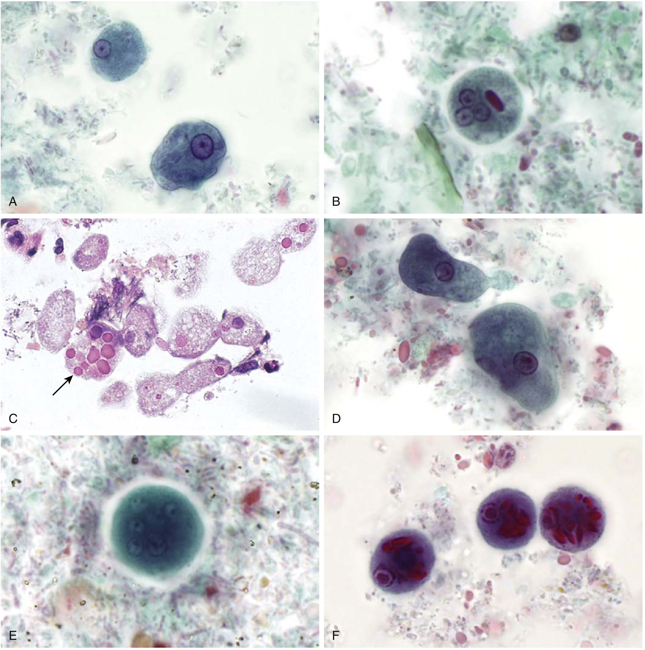
(Henry's Clinical Diagnosis, Fig. 65.10 - A: E. histolytica/dispar trophozoites; B: Cyst with 3 of 4 nuclei and rounded chromatoid bar; C: Trophozoites with ingested RBCs - arrow; D-E: E. coli forms; F: C. polecki cyst)
2. Life Cycle
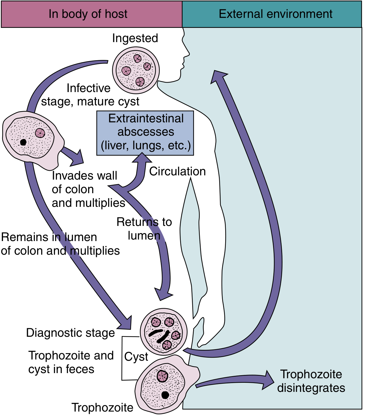
(Medical Microbiology 9e, Fig. 72.1 - Life cycle of E. histolytica)
The life cycle involves two stages: trophozoite and cyst. Humans are the only reservoir of infection.
Step-by-step:
-
Ingestion of mature quadrinucleate cysts - via contaminated food/water (fecal-oral route). Trophozoites cannot survive in the external environment or gastric acid, so cysts are the infective stage.
-
Excystation - Cysts pass through the stomach; exposure to gastric acid stimulates release of the pathogenic trophozoite in the duodenum. Each quadrinucleate cyst produces 8 trophozoites (4 nuclei → 8 cells by binary fission).
-
Colonization of large intestine - Trophozoites descend to the large intestine (primarily caecum and ascending colon, then rectosigmoid). They dwell in the colon, multiply by binary fission.
-
Two possible outcomes:
- Non-invasive: Trophozoites remain in the lumen → encyst → cysts passed in formed stool → environmental contamination → transmission
- Invasive: Trophozoites invade the bowel wall → ulceration → may enter portal circulation → liver (most common extraintestinal site), then lungs, brain, heart, skin
-
Encystment - When conditions become unfavorable (temperature drop, desiccation), trophozoites retract pseudopodia, become spherical, and encyst (uninucleate → quadrinucleate cyst)
-
Excretion - Cysts excreted in stool; carriers can shed up to 1.5 × 10⁷ cysts/day. Cysts survive in feces, water, and soil for several days at low temperature/moisture.
Cyst survival: Survive in moist conditions for days to weeks; resistant to chlorination at standard water treatment doses; killed by drying, heating to ~55°C, or freezing. Sand filtration and boiling are effective.
3. Pathogenesis
Mechanisms of tissue invasion
After excystation, invasive trophozoites use multiple mechanisms to destroy tissue:
-
Galactose-inhibitable adherence protein (lectin) - Trophozoites attach to colonic epithelial cells via a Gal/GalNAc-specific lectin on their surface. This attachment is required for cytolysis to occur.
-
Cytotoxin production - After attachment, trophozoites secrete toxins that cause lytic necrosis (not inflammatory necrosis initially). This causes a lethal alteration in host cell membrane permeability → irreversible rise in intracellular calcium → cell death.
-
Lysing of immune cells - Trophozoites lyse colonic epithelial cells, neutrophils, lymphocytes, and monocytes. Release of toxic neutrophil constituents after neutrophil lysis amplifies tissue destruction.
-
Anaerobic preference - Amebae are killed by ambient oxygen concentrations; they thrive in low-oxygen environments (colonic lumen, abscess cavities).
Pathological lesions
Intestinal (colonic) lesions:
- Initial lesion: Pinhead-sized ulcer with raised edges in the mucosa; mucus, necrotic cells, and amebae exude
- Trophozoites multiply above the muscularis mucosae, spreading laterally → undermine mucosa
- Classic "flask-shaped" (collar-button) ulcer: Narrow neck through mucosa leading to an expanded necrotic area in the submucosa - small point of entry, large necrotic base. This is the hallmark lesion of amoebiasis.
- Bacterial superinfection occurs in larger, established ulcers
- Coalescence of ulcers → large denuded mucosal areas
- Ameboma (amoebic granuloma): Granulomatous tumorlike mass on intestinal wall, can obstruct the lumen and mimic carcinoma - produces "napkin ring lesion"
- Perforation into peritoneal cavity is possible
Extraintestinal spread:
- Trophozoites penetrate the muscularis, enter mesenteric venules → portal blood → liver
- Amebic liver abscess (ALA): Most common extraintestinal form (~5% of intestinal cases). Predominantly right lobe, usually single. Aspirate is classically "anchovy sauce" / chocolate-colored pus (liquefied hepatic parenchyma). Trophozoites found at the margin of abscess, not in the center.
- Hematogenous/contiguous spread → lungs (rupture across diaphragm), brain, pericardium, skin (perianal ulcers)
Factors determining invasion:
- Number of amebae ingested
- Pathogenic capacity of the strain (zymodeme)
- Host immunity and gut motility
- Enteric bacteria (enhance amebic growth)
4. Laboratory Diagnosis
A. Microscopy (Stool Examination)
- Gold standard for intestinal amoebiasis
- Examine at least 3 stool specimens on separate days (parasites not homogeneously distributed)
- Fresh liquid/semi-liquid stools → look for motile trophozoites (survive only a few hours outside body; must be examined immediately)
- Formed stools → cysts (more stable)
Wet mount: Direct or concentration methods
- Trophozoites show rapid, progressive, unidirectional motility via hyaline pseudopodia; sharp ectoplasm-endoplasm demarcation
Permanent stained smear (Trichrome or iron-hematoxylin stain):
- Best method for detailed morphologic identification
- Shows nuclear detail: uniform peripheral chromatin + central karyosome
- Ingested RBCs in trophozoites = pathognomonic for invasive E. histolytica
Concentration techniques (formalin-ethyl acetate): For cyst detection
PAS stain on tissue sections: Trophozoites stain prominently
Note: E. histolytica is morphologically indistinguishable from E. dispar, E. moshkovskii, and E. bangladeshi - RBC ingestion is the only microscopic pathognomonic feature.
B. Culture
- Robinson's medium / Jones' medium / Boeck-Drbohlav medium
- Not widely used for diagnosis; mainly for research
- Useful for zymodeme analysis
C. Antigen Detection (EIA/ELISA)
- Enzyme immunoassay (EIA) on stool specimens
- Highly specific; can differentiate E. histolytica from E. dispar
- Commercially available (e.g., E. histolytica II ELISA, TechLab)
- Sensitivity ~85-90%, specificity ~99%
- Preferred over microscopy for species-level diagnosis
D. Serology (Antibody Detection)
- Detects host anti-amebic IgG antibodies
- Most useful for extraintestinal amoebiasis (ALA)
- ~95% of ALA patients are seropositive
- ~70% seropositive in active intestinal infection
- ~10% seropositive in asymptomatic carriers
- Tests: Indirect hemagglutination (IHA) - most sensitive; ELISA; counterimmunoelectrophoresis (CIE); indirect fluorescent antibody (IFA); latex agglutination
- Limitation: Titers persist months-to-years after treatment, so cannot distinguish current from past infection; less useful in endemic areas
E. Molecular Methods (PCR / DNA probes)
- Most sensitive and specific method
- Can definitively distinguish E. histolytica from E. dispar and other species
- FDA-approved multiplex panels: BioFire FilmArray GI Panel, BD MAX Enteric Parasite Panel, xTag GI Pathogen Panel
- Useful when serology/antigen tests are inconclusive
F. Aspiration of Liver Abscess
- Aspirate is chocolate-brown ("anchovy sauce") pus
- Trophozoites more likely in last aspirated material (at abscess wall margin)
- Direct microscopy or permanently stained slides
- PCR amplification of amebic 16S rRNA from aspirate is highly reliable
G. Imaging
- Ultrasound, CT scan, MRI of liver: Show abscess (usually single, right lobe)
- Cannot distinguish amebic from pyogenic abscess on imaging alone - requires serology
Summary Table: Lab Diagnosis
| Test | Best for | Notes |
|---|---|---|
| Stool microscopy (wet mount + trichrome) | Intestinal amoebiasis | 3 specimens; RBC phagocytosis = pathognomonic |
| Antigen EIA on stool | Intestinal amoebiasis | Differentiates E. histolytica vs E. dispar |
| Serology (IHA, ELISA) | ALA; extraintestinal disease | 95% positive in ALA; persists after cure |
| PCR / molecular | Species differentiation | Most specific; FDA-approved panels |
| Liver aspirate microscopy | ALA | Last aspirated material; margins of abscess |
| Culture | Research only | Zymodeme analysis |
Epidemiology (Quick Review)
- Worldwide distribution; highest in tropical/subtropical regions with poor sanitation
- Global: ~50 million infected; ~100,000 deaths/year
- India: ~15% prevalence (range 3.6-47.4%)
- Incubation period: 2-4 weeks (can be longer)
- Transmission: fecal-oral (contaminated water/food), oral-anal sexual contact, mechanical vectors (flies, cockroaches)
- Cysts resist standard chlorination; sand filtration and boiling are effective
Treatment (Brief)
- Invasive/systemic disease: Metronidazole or tinidazole (systemic penetration) → followed by luminal agent
- Luminal agents (to eradicate cysts/asymptomatic carriage): Diloxanide furoate, paromomycin, iodoquinol
- Treatment of asymptomatic carriers prevents environmental shedding
Sources: Medical Microbiology 9e (Murray); Henry's Clinical Diagnosis 23e; Park's Textbook of Preventive and Social Medicine; Jawetz Medical Microbiology 28e; Sleisenger & Fordtran's GI and Liver Disease; Robbins Pathologic Basis of Disease
This is a shared conversation. Sign in to Orris to start your own chat.