
Dermatitis vs fungal infection how to differentiate
Reading File
Now let me search the medical library and web in parallel for comprehensive information on differentiating dermatitis from fungal infections.
Finding Sources
Finding Sources
Searching the Web
dermatitis vs fungal infection clinical differentiation diagnosis features
Finding Sources
Finding Sources
Good - found relevant sections. Let me read the key textbook content now.
Reading File
Reading File
Reading File
Reading File
Finding Sources
I now have enough comprehensive information. Let me also fetch the clinical image of atopic dermatitis from the textbook.
Loading Image
I now have comprehensive information to write a detailed, well-structured answer. Here it is:
Dermatitis vs Fungal Infection: How to Differentiate
These two conditions overlap significantly in appearance - both produce red, scaly, itchy skin - and can even coexist. The table and framework below covers clinical, investigative, and contextual features that help distinguish them.
1. Quick Clinical Summary
| Feature | Dermatitis (Eczema) | Fungal Infection (Tinea/Candida) |
|---|---|---|
| Lesion border | Ill-defined, blends with normal skin | Sharply defined, raised, active border |
| Shape | Irregular, diffuse | Annular/circular with central clearing (tinea corporis) |
| Scaling | Fine, diffuse | Peripheral/leading edge scales |
| Vesicles | Common (dyshidrosis: deep-seated) | Can occur (tinea pedis moccasin/vesicular type) |
| Lichenification | Common in chronic cases | Absent |
| Symmetry | Often bilateral (both hands, both feet) | Often unilateral or asymmetric |
| Satellite lesions | Absent | Present in candidal infections |
| Contagious | No | Yes (person-to-person, animals, fomites) |
| Nail involvement | Rare (pitting in psoriatic overlap) | Onychomycosis common |
| Atopy history | Frequent (asthma, rhinitis, family hx) | Not associated |
2. Clinical Features in Detail
Dermatitis (Eczema variants)
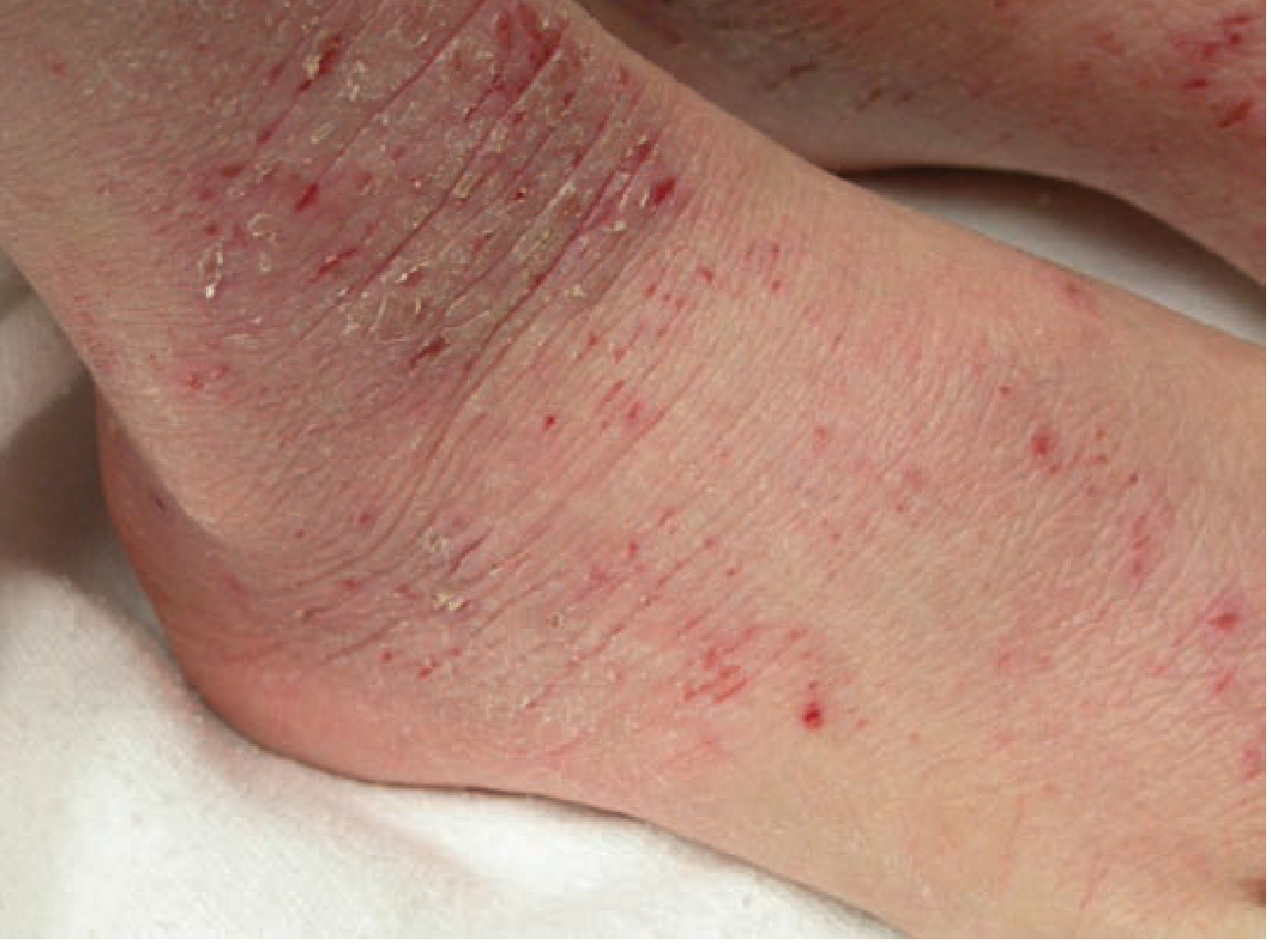
Atopic dermatitis presents as erythematous, pruritic, scaly patches with prominent dorsal surface involvement of hands and feet. Chronic cases develop hyperpigmentation, lichenification, and fissuring. Classic body distribution includes antecubital/popliteal fossae, posterior neck, wrists, and ankles (see image below).
Dyshidrosis (pompholyx) begins as deep-seated pruritic vesicles on the lateral aspects of palms and soles - no erythema initially. Chronic cases develop erythema and scales that can be very hard to distinguish from tinea pedis.
Contact dermatitis (allergic/irritant) follows the pattern of allergen exposure. Chemical irritant dermatitis often involves the dorsa of the feet (from shoes), whereas tinea pedis is uncommon in children and rarely involves the dorsum.
Fungal Infections
Tinea corporis: Annular lesion with central clearing, raised scaly border. The advancing border is the key - scraping from this edge gives the best KOH yield.
Tinea pedis: Three patterns:
- Moccasin type - diffuse scaling of the entire sole, easy to confuse with dermatitis
- Interdigital type - maceration and fissuring between toes
- Vesicular type - vesicles on the instep/sole
Tinea cruris: Sharply marginated plaques in the groin, with scalloped border; spares the scrotum (unlike candidal intertrigo which involves the scrotum).
Candidal intertrigo: Beefy red erythema in skin folds + satellite pustules beyond the main plaque - satellite lesions are a hallmark feature.
3. The Diagnostic Key: KOH Preparation
The single most important differentiating test is the potassium hydroxide (KOH) preparation. This is the definitive bedside test that can confirm or exclude a fungal infection when clinical features are ambiguous.
"Always consider a fungal infection and a potassium hydroxide preparation can exclude this possibility. A biopsy cannot differentiate between the different types of dermatitis."
- Tintinalli's Emergency Medicine, p. 1702
How to perform:
- Scrape the leading (active) edge of the lesion with a #15 scalpel or slide edge
- Apply 2 drops of KOH (with or without fungal stain) under a coverslip
- Examine at 10x first, then 40x to confirm true hyphae/arthroconidia
- True fungal hyphae cross cell borders; "mosaic false hyphae" (cell borders) are the most common artifact
KOH sensitivity: 77-88%; specificity: 62-95% (varies by technique and stain use)
If KOH is negative but suspicion remains, send for fungal culture - visible growth within 1-2 weeks.
4. Critical Pitfall: Tinea Incognita
When tinea is treated with topical steroids (misdiagnosed as eczema), it transforms into tinea incognita - an atypical presentation that looks like eczema:
- Widespread lesions
- Loss of the raised, scaling advancing border
- Central area becomes eczematous rather than clearing
- Multiple irregular edges may appear
- KOH examination or biopsy confirms fungus
This is a common and clinically dangerous scenario. Combination steroid-antifungal creams (e.g., clotrimazole/betamethasone) frequently lead to widespread tinea and fungal folliculitis and should be avoided.
5. Site-Specific Differential Points
| Body Site | Dermatitis Clue | Fungal Clue |
|---|---|---|
| Scalp | Seborrheic: greasy yellow scales, diffuse | Tinea capitis: broken hairs, KOH+ on moist swab; T. tonsurans mimics seborrheic scale |
| Feet/Hands | Bilateral symmetry; dyshidrotic vesicles deep-seated | Unilateral; moccasin type; toe web maceration |
| Groin | Contact/irritant; involves scrotum | Tinea cruris: spares scrotum; sharp border |
| Groin (yeast) | - | Candida: involves scrotum + satellite pustules |
| Body | Nummular dermatitis: coin-shaped but no advancing edge | Tinea corporis: annular, central clearing, KOH+ |
| Nails | Pitting (psoriasis) | Onychomycosis: subungual debris, thickening |
| Skin folds | Seborrhea, intertrigo | Candida: satellite lesions, beefy red |
6. Investigations Summary
| Test | Use |
|---|---|
| KOH prep | First-line; confirms hyphae/spores in fungal infection |
| Fungal culture | If KOH negative but suspicion persists; species ID |
| Wood's lamp | Microsporum fluoresces green; tinea versicolor fluoresces yellow-orange; most Trichophyton do NOT fluoresce |
| Patch testing | For suspected allergic contact dermatitis |
| Skin biopsy | Helpful for tinea incognita; cannot differentiate types of dermatitis from each other |
| Total IgE / eosinophils | Support atopic dermatitis but not diagnostic |
7. Treatment Response as a Diagnostic Tool
- If a lesion worsens or spreads with topical steroids alone - suspect tinea (tinea incognita pattern)
- If a lesion improves with antifungals - confirms fungal etiology
- If improving with steroids but stalling - consider mixed/concurrent condition (coexistence is common)
Sources: Andrews' Diseases of the Skin (Clinical Dermatology); Tintinalli's Emergency Medicine; Textbook of Family Medicine 9e; Goodman & Gilman's Pharmacological Basis of Therapeutics
This is a shared conversation. Sign in to Orris to start your own chat.