
Cell adhesive molecule
Reading File
Cell adhesion molecules (CAMs) are proteins on cell surfaces that mediate binding between cells and between cells and the extracellular matrix. Major families include integrins, selectins, immunoglobulin superfamily members (ICAM, VCAM, NCAM), and cadherins. They play roles in development, immunity, inflammation, wound healing, and cancer metastasis.
Searching sources...
Finding Sources
Finding Sources
Good results. Now let me read the most relevant sections - from Rheumatology (IgSF receptors), Histology (multiadhesive glycoproteins), Dermatology (leukocyte-endothelial interactions), and Neuroscience (CAMs in neural connectivity), plus Harper's Biochemistry (cancer/metastasis).
Reading File
Reading File
Reading File
Reading File
Now let me get the dermatology section on leukocyte-endothelial interactions and the selectins section, plus the Rheumatology integrin section for a fuller picture.
Reading File
Reading File
Finding Sources
Excellent. I now have rich content from multiple textbooks. Let me also fetch the key diagram of leukocyte extravasation steps and the multiadhesive glycoprotein figure.
Loading Image
Loading Image
I now have comprehensive textbook content and two excellent diagrams. Here is a thorough answer:
Cell Adhesion Molecules (CAMs)
Cell adhesion molecules are cell surface glycoproteins that mediate binding of cells to other cells and to the extracellular matrix (ECM). They are transmembrane proteins that also transduce intracellular signals regulating proliferation, survival, migration, and differentiation.
Major Families
1. Integrins
Integrins are heterodimeric transmembrane receptors composed of non-covalently associated α and β subunits. There are 18 α and 8 β subunits, forming at least 24 different αβ combinations. They are the primary receptors linking the ECM to the intracellular actin cytoskeleton.
Key features:
- Require divalent cations (Mg²+, Ca²+) for ligand binding
- Can exist in low-affinity or high-affinity conformations (inside-out signaling by chemokines converts them)
- Ligand binding triggers outside-in signaling - formation of focal adhesion complexes that activate cascades regulating proliferation, survival, and cytokine secretion
Major integrin ligands and functions (from Firestein & Kelley's Textbook of Rheumatology):
| Integrin | Alternative Name | Ligands |
|---|---|---|
| α₁β₁ | VLA-1 | Laminin, collagen |
| α₂β₁ | VLA-2 | Laminin, collagen |
| α₅β₁ | VLA-5 | Fibronectin |
| α₆β₁ | VLA-6 | Laminin |
| αLβ₂ | LFA-1, CD11a/CD18 | ICAM-1, ICAM-2, ICAM-3, JAM-A |
| αMβ₂ | Mac-1, CR3, CD11b/CD18 | ICAM-2, iC3b, fibrinogen |
| αvβ₃ | Vitronectin receptor | Vitronectin, fibronectin, osteopontin |
| α₄β₇ | CD49d | Fibronectin, VCAM-1, MAdCAM-1 |
2. Selectins
Selectins mediate the initial tethering and rolling of leukocytes on the endothelium - the first step of extravasation. They are C-type lectins that bind carbohydrate ligands on opposing cells.
Three members:
| Selectin | Expression | Key Ligands |
|---|---|---|
| P-selectin | Endothelium (Weibel-Palade bodies), platelets | PSGL-1 (P-selectin glycoprotein ligand-1) |
| E-selectin | Activated endothelium | ESL-1, CLA (cutaneous lymphocyte antigen) |
| L-selectin | Leukocytes | GlyCAM-1, CD34, MAdCAM-1 |
Selectins bind sialylated, fucosylated carbohydrates (e.g., sialyl Lewis x, sLex). Their interactions are of relatively low affinity, which is precisely why they allow rolling rather than firm adhesion.
3. Immunoglobulin Superfamily (IgSF) CAMs
Defined by one or more Ig-like repeats of 60-100 amino acids stabilized by a disulfide bond. They mediate the firm adhesion step of leukocyte extravasation by binding integrins on the leukocyte surface.
Key members:
| Molecule | Full Name | Integrin Ligand | Function |
|---|---|---|---|
| ICAM-1 (CD54) | Intercellular adhesion molecule-1 | LFA-1 (αLβ₂), Mac-1 | Firm adhesion, expressed on endothelium after activation |
| ICAM-2 (CD102) | Intercellular adhesion molecule-2 | LFA-1, Mac-1 | Constitutively expressed |
| ICAM-3 (CD50) | Intercellular adhesion molecule-3 | LFA-1 | Leukocyte-leukocyte adhesion |
| VCAM-1 (CD106) | Vascular cell adhesion molecule-1 | VLA-4 (α₄β₁), α₄β₇ | Inflammation, atherogenesis |
| PECAM-1 (CD31) | Platelet-endothelial CAM-1 | αvβ₃ | Transmigration through endothelial junctions |
| MAdCAM-1 | Mucosal addressin CAM-1 | α₄β₇ | Lymphocyte homing to gut |
| NCAM | Neural cell adhesion molecule | - | Axon guidance, neural development |
4. Cadherins
Cadherins mediate homotypic, calcium-dependent adhesion - they bind the same cadherin species on adjacent cells. Classic cadherins have:
- 5 extracellular domains
- Single transmembrane domain
- Cytoplasmic tail that binds β-catenin → α-catenin → actin cytoskeleton
Key members and roles:
| Cadherin | Location | Role |
|---|---|---|
| E-cadherin | Epithelial cells | Maintains epithelial integrity; loss triggers EMT in cancer |
| N-cadherin | Neurons, muscle | Neural development and synaptogenesis |
| VE-cadherin | Vascular endothelium | Maintains tight endothelial junctions; opens transiently during leukocyte transmigration |
| P-cadherin | Placenta, epithelium | Epithelial morphogenesis |
Leukocyte Extravasation: The Multi-Step Cascade
This is the best-studied physiological role of CAMs. The process proceeds in sequential steps, each dependent on distinct CAM families:
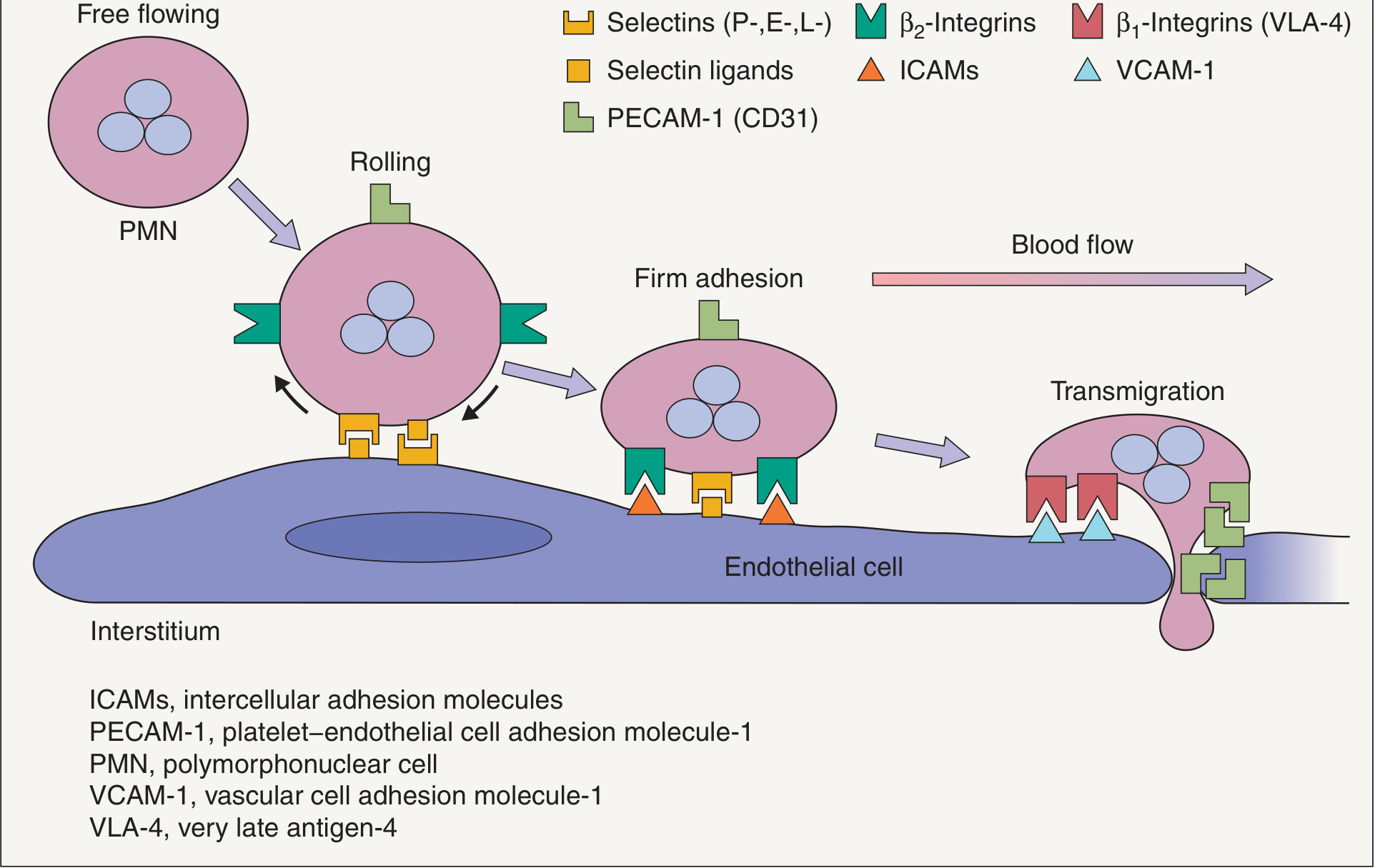
Step 1 - Tethering and Rolling: Selectins (P-, E-, L-) interact with their carbohydrate ligands (PSGL-1, ESL-1, CLA). This produces slow rolling of the PMN along the endothelium.
Step 2 - Activation: Chemokines presented on the endothelial surface activate G-protein-coupled receptors on the leukocyte, triggering inside-out signaling that converts integrins from low-affinity to high-affinity conformation.
Step 3 - Firm Adhesion: High-affinity β₂-integrins (LFA-1, Mac-1) bind ICAM-1 and ICAM-2 on the endothelium, stopping the leukocyte completely.
Step 4 - Transmigration (Diapedesis): The leukocyte squeezes between endothelial cells. VE-cadherin junctions and junctional adhesion molecules (JAMs) must open transiently, a process regulated by vascular endothelial protein tyrosine phosphatase (VE-PTP). PECAM-1 (CD31) on both the leukocyte and the endothelial junction facilitates passage.
- Dermatology 2-Volume Set 5e, p. 2133
- Firestein & Kelley's Textbook of Rheumatology, p. 304
Multiadhesive Glycoproteins (ECM-Associated CAMs)
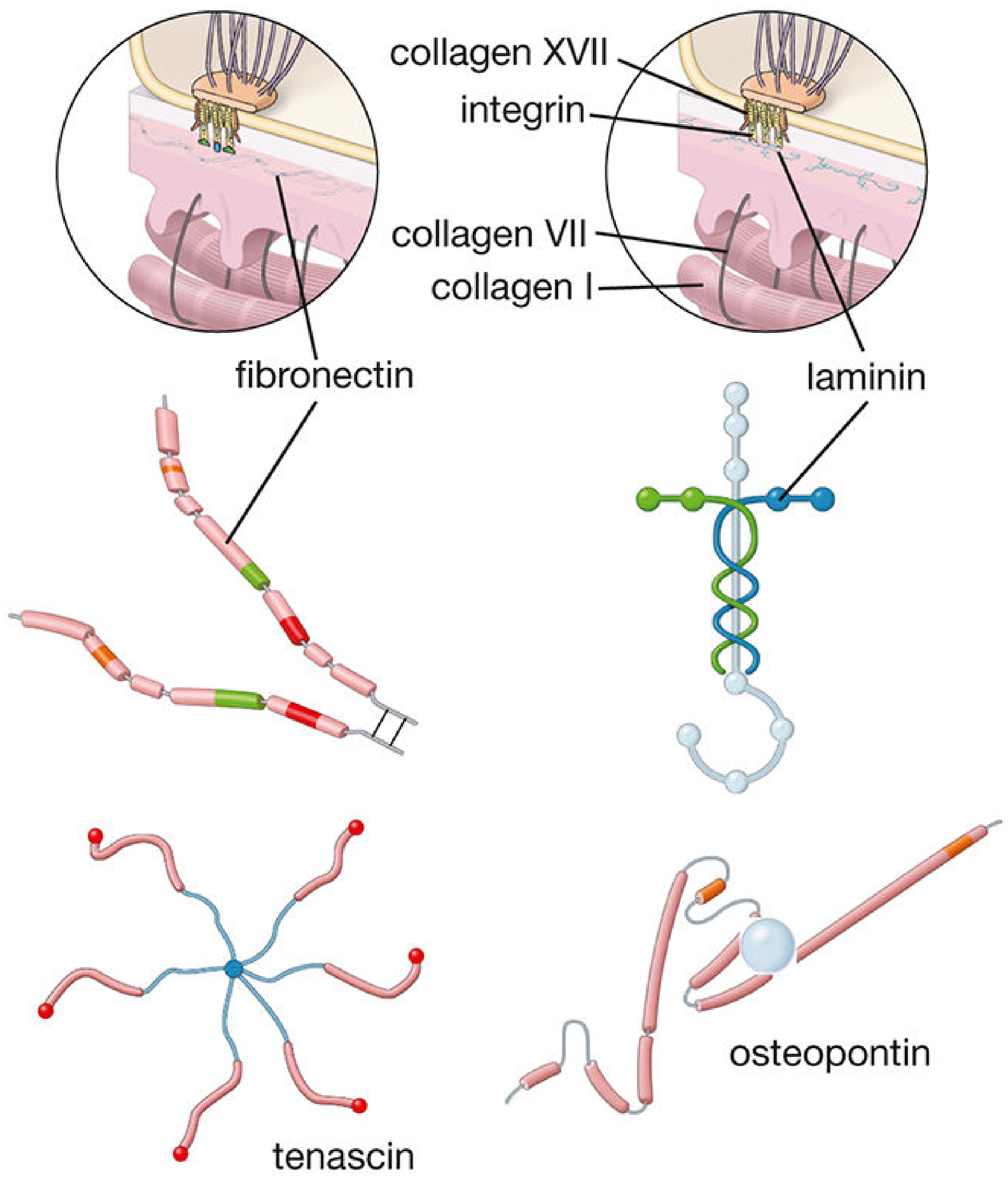
These are ECM-resident proteins with multiple binding domains that link the ECM to cell-surface receptors:
-
Fibronectin (250-280 kDa): Most abundant glycoprotein in connective tissue. Dimer linked by disulfide bonds. Has binding domains for heparan sulfate, collagen I/II/III, fibrin, hyaluronan, and integrin receptors. Activates cell attachment to ECM.
-
Laminin (140-400 kDa): Present in basal laminae. Binds collagen IV, heparan sulfate, entactin, and laminin receptors on cell surfaces.
-
Tenascin (280 kDa/monomer): Expressed during embryogenesis and wound healing; re-expressed in malignant tumors. Hexameric structure; binds fibrinogen, heparin, and EGF-like growth factors.
-
Osteopontin (44 kDa): Found in bone ECM. Binds osteoclasts via integrin αvβ₃ to anchor them to bone surface.
-
Histology: A Text and Atlas with Correlated Cell and Molecular Biology, p. 496-497
Syndecans
Syndecans are four single-pass transmembrane proteoglycans carrying heparan sulfate and chondroitin sulfate chains. They serve as co-receptors integrating adhesive contacts with growth factor signaling (binding FGF, VEGF, TGF-β, fibronectin). Syndecan-4 knockout mice show impaired wound healing and altered fibronectin responses.
Homophilic vs. Heterophilic Binding
| Type | Description | Example |
|---|---|---|
| Homophilic | CAM binds identical molecule on neighbor | Cadherins |
| Heterophilic | CAM binds a different molecule | Selectins (bind carbohydrates), Integrins (bind ECM proteins) |
| Both | Context-dependent | IgSF members (ICAM can bind both) |
- Harper's Illustrated Biochemistry, 32nd Ed, p. (cancer chapter)
CAMs in Disease
Inflammation
ICAM-1 and VCAM-1 are upregulated on endothelium by TNF-α, IL-1, and LPS - they serve as the molecular "landing pad" for leukocytes in inflammatory diseases including rheumatoid arthritis, asthma, and IBD.
Cancer and Metastasis
CAM changes are central to metastasis:
- Loss of E-cadherin allows cells to detach from the primary tumor (epithelial-mesenchymal transition, EMT)
- Upregulation of N-cadherin increases motility ("cadherin switching")
- Changes in integrin expression alter attachment to distant organ ECM, contributing to organ-specific metastatic tropism (e.g., prostate cancer to bone via αvβ₃/osteopontin)
- MMPs degrade ECM to create paths for migrating tumor cells
- Harper's Illustrated Biochemistry, 32nd Ed
Neural Development
NCAM (neural CAM) and other IgSF members guide axon fasciculation and growth cone navigation. Ephrins (chemorepellents) and netrins (chemoattractants) collaborate with CAMs in establishing neural circuits.
- Neuroscience: Exploring the Brain, 5th Ed
Congenital Tufting Enteropathy
Loss-of-function mutations in EpCAM (epithelial cell adhesion molecule) cause autosomal recessive tufting enteropathy - a lethal diarrhea in newborns due to disrupted intestinal epithelial integrity.
- Yamada's Textbook of Gastroenterology, 7th Ed
Summary Table
| Family | Binding Type | Ligand Type | Ca²+ Required? | Key Role |
|---|---|---|---|---|
| Integrins | Heterophilic | ECM proteins, IgSF | Yes (Mg²+/Ca²+) | Firm adhesion, signaling, ECM attachment |
| Selectins | Heterophilic | Carbohydrates (sLex) | Yes | Leukocyte rolling |
| IgSF (ICAM, VCAM) | Homo/Heterophilic | Integrins, IgSF | No | Firm adhesion, immune recognition |
| Cadherins | Homophilic | Same cadherin | Yes | Cell-cell adhesion, tissue integrity, EMT |
| Syndecans | Heterophilic | ECM proteins, growth factors | No | Co-receptor, wound healing |
This is a shared conversation. Sign in to Orris to start your own chat.