
Abnormal uterine bleeding and mass per abdomen theory and clinical
Reading File
Now let me search the medical library for both topics in parallel.
Finding Sources
Finding Sources
Finding Sources
Finding Sources
Finding Sources
Reading File
Reading File
Finding Sources
Reading File
Finding Sources
Finding Sources
Reading File
Reading File
Reading File
Reading File
Reading File
Finding Sources
Reading File
Reading File
Reading File
Loading Image
I now have comprehensive information from multiple authoritative sources. Let me compile the full answer.
Abnormal Uterine Bleeding (AUB) and Mass Per Abdomen (Uterine Fibroids)
PART 1: ABNORMAL UTERINE BLEEDING (AUB)
Definition and Normal Parameters
AUB is defined as any change in the frequency, regularity, duration, or volume of uterine bleeding outside normal parameters. A normal menstrual cycle:
- Frequency: every 24-38 days
- Duration of flow: 4-8 days
- Cycle shorter than 21 days or longer than 35 days is abnormal
- Flow <2 days or >7 days is abnormal
The older terms "menorrhagia," "metrorrhagia," "menometrorrhagia," and "dysfunctional uterine bleeding (DUB)" are obsolete and replaced by the PALM-COEIN system. The term DUB should no longer be used.
The PALM-COEIN Classification (FIGO 2011)
Established by the FIGO Working Group on Menstrual Disorders. Each letter represents a cause:
PALM - Structural Causes (diagnosed by imaging or histology)
| Letter | Cause | Notes |
|---|---|---|
| P | Polyp (AUB-P) | Endometrial or endocervical polyp |
| A | Adenomyosis (AUB-A) | Ectopic endometrial glands in myometrium |
| L | Leiomyoma (AUB-L) | Submucosal (AUB-LSM) vs. other (AUB-LO) |
| M | Malignancy & Hyperplasia (AUB-M) | Endometrial carcinoma, hyperplasia |
COEIN - Non-structural Causes (medical/functional)
| Letter | Cause | Notes |
|---|---|---|
| C | Coagulopathy (AUB-C) | Von Willebrand disease, thrombocytopenia |
| O | Ovulatory Dysfunction (AUB-O) | PCOS, perimenopause, anovulation |
| E | Endometrial (AUB-E) | Endometritis, endometrial defects |
| I | Iatrogenic (AUB-I) | IUCD, exogenous hormones, tamoxifen |
| N | Not yet classified (AUB-N) | Arteriovenous malformations, etc. |
(Source: ROSEN's Emergency Medicine; Sabiston Textbook of Surgery)
AUB by Age Group (Robbins Basic Pathology)
| Age Group | Causes |
|---|---|
| Prepuberty | Precocious puberty (hypothalamic, pituitary, or ovarian origin) |
| Adolescence | Anovulatory cycle, coagulation disorders |
| Reproductive age | Pregnancy complications (abortion, ectopic, trophoblastic disease), anatomic lesions (leiomyoma, adenomyosis, polyps, hyperplasia, carcinoma), dysfunctional uterine bleeding |
| Perimenopause | Anovulatory cycles, ovulatory dysfunction, inadequate luteal phase |
| Postmenopause | Anatomic lesions (carcinoma, hyperplasia, polyps), endometrial atrophy |
Normal Menstrual Cycle - Hormonal Basis
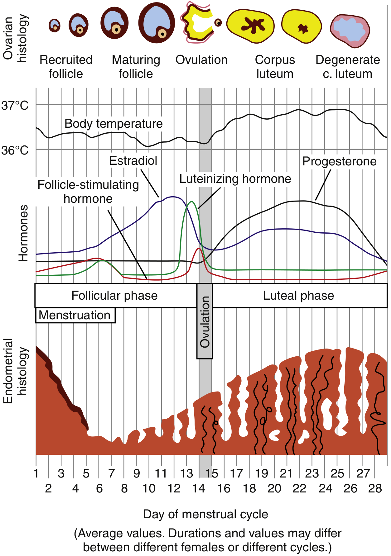
The menstrual cycle begins on Day 1 of menses. Estrogen dominates the follicular phase, causing endometrial proliferation. After ovulation (Day ~14), the corpus luteum produces progesterone, which matures the endometrium. If implantation does not occur, the corpus luteum degenerates, progesterone and estrogen fall sharply, and menstruation occurs. This normally occurs ~14 days after ovulation.
Pathophysiology of Dysfunctional/Ovulatory AUB
Approximately 50% of excessive menstruation falls under ovulatory dysfunction (AUB-O):
- If a woman does not ovulate, there is no corpus luteum and therefore no progesterone
- Unopposed estrogen causes the endometrium to proliferate until it becomes unstable and breaks down
- Results in irregular, unpredictable, and often heavy bleeding
- Anovulatory cycles are most common at menarche and perimenopause due to HPO axis fluctuations
Other causes of anovulation include:
- PCOS (polycystic ovary syndrome)
- Pituitary tumors secreting prolactin (disrupts GnRH)
- Hyperprolactinemia, hypothalamic dysfunction (anorexia nervosa)
- Obesity, malnutrition, chronic systemic disorders (liver, thyroid disease)
(Source: Robbins Basic Pathology; ROSEN's Emergency Medicine)
Types of Bleeding Pattern by Cause
| Pattern | Causes |
|---|---|
| Menstrual (heavy) | Endometrial polyp/malignancy, fibroids |
| Intermenstrual | Vaginal/cervical trauma or malignancy, cervical polyp, endometrial polyp/malignancy |
| Postcoital | Cervical ectropion, cervical polyp/malignancy, vaginal malignancy |
| Postmenopausal | Endometrial atrophy (most common), endometrial carcinoma, polyps, hyperplasia |
Special Cause: Tamoxifen
Women on tamoxifen (used for breast cancer) are a special group:
- Tamoxifen can induce endometrial abnormalities in 10-40% of women
- Causes: endometrial polyps, hyperplasia, cancer, and rarely uterine sarcomas
- Risk doubles in the first year and quadruples after 5 years
- Risk is time-dependent but dose-independent
- Aromatase inhibitors (anastrozole, letrozole, exemestane) have less endometrial effect and may reverse tamoxifen-induced abnormalities
(Source: Bailey and Love's Short Practice of Surgery)
Clinical Evaluation
History:
- Volume, frequency, duration, and relationship to menstrual cycle
- Age of menarche or menopause
- Sexual history, risk factors for STIs
- History of easy bruising/bleeding (family history of coagulopathy)
- Medications (hormonal contraceptives, IUCD, tamoxifen)
- Systemic disease: thyroid, liver, metabolic syndrome, obesity
- Prior cesarean section (cesarean scar defect)
Physical Examination:
- Signs of hypovolemia and anemia in acute bleeding
- PCOS signs: obesity, acne, hirsutism, acanthosis nigricans
- Thyroid examination (nodules)
- Skin: petechiae, ecchymoses (coagulopathy)
- Speculum: vaginal/cervical lesions
- Bimanual exam: uterine enlargement, tenderness, palpable fibroid
Investigations:
- Pregnancy test (urine or serum β-hCG) - first and always
- Full blood count - hemoglobin, platelets
- Pelvic ultrasound - first-line imaging (2D/3D, saline sonogram)
- Thyroid function tests, prolactin level
- Coagulation studies - if heavy bleeding since menarche, family history, or signs of systemic bleeding
- Cervical cancer screening if not up to date
- STI screening
- Endometrial biopsy (Pipelle or hysteroscopic)
Indications for endometrial biopsy:
- All women >45 years with AUB (including intermenstrual bleeding)
- Women <45 years with: obesity/unopposed estrogen, persistent AUB, AUB refractory to medical treatment, elevated familial cancer risk
- Any postmenopausal bleeding
- Suspected endometrial pathology
- Failed medical treatment
Postmenopausal bleeding is its own diagnosis - the most common cause is endometrial atrophy, but endometrial carcinoma must be excluded.
(Source: Sabiston Textbook of Surgery; Bailey and Love's Short Practice of Surgery)
Endometrial Hyperplasia (precursor to carcinoma)
Mechanism: Prolonged unopposed estrogen causes exaggerated endometrial proliferation.
Risk factors for estrogen excess: obesity (adipose converts steroids to estrogens), exogenous estrogens without progesterone, estrogen-producing ovarian lesions (PCOS, granulosa-theca cell tumors).
Types:
- Hyperplasia without atypia: increased gland-to-stroma ratio; low risk of progression to carcinoma (1-3%)
- Hyperplasia with atypia (Endometrial Intraepithelial Neoplasia - EIN): complex gland proliferation with nuclear atypia + PTEN mutations; considered true precancer. Treatment: hysterectomy (fertility desired: high-dose progestins)
Endometrial Carcinoma
- Most common gynecologic cancer in high-income countries
- Type 1 (Endometrioid): 80% of cases; estrogen-related; arises from hyperplasia; PTEN and mismatch repair gene mutations; associated with Lynch syndrome, Cowden syndrome
- Type 2 (Serous): 15% of cases; arises in atrophic endometrium of older postmenopausal women; TP53 mutations; far more aggressive
PART 2: MASS PER ABDOMEN - UTERINE FIBROIDS (LEIOMYOMA)
Definition and Epidemiology
Uterine fibroids (leiomyomata) are benign, well-circumscribed smooth muscle tumors of the uterus. They are the most common pelvic tumors in women.
- Present in up to 40% of women during reproductive years
- Present in 70-80% of women by age 50
- Black/African-Caribbean women are disproportionately affected (2-3x higher rates)
- Incidence highest in women aged 45-49 years
- Require estrogen and progesterone to grow; grow rapidly in early pregnancy; shrink after menopause
- Malignant transformation to leiomyosarcoma in <1% (approximately 1 in 1100-1500 women under 45)
Genetics:
- Germline mutations in FH (fumarate hydratase) gene - associated with early onset uterine + cutaneous leiomyomas + renal cell cancer
- Somatic mutations in MED12 gene (X chromosome)
(Source: Goldman-Cecil Medicine; Bailey and Love's Short Practice of Surgery)
Classification by Location
| Type | Location | Clinical Significance |
|---|---|---|
| Submucosal | Projects into uterine cavity | Heavy/irregular periods, infertility, recurrent pregnancy loss; if pedunculated, may extrude through cervical os |
| Intramural | Within myometrial wall | Pressure symptoms; heavy periods if distorts endometrium; infertility |
| Subserosal | On outer surface of uterus | Pressure symptoms; if pedunculated, may mimic ovarian tumour |
| Broad ligament / Cervical | Rare locations | Unusual presentations |
The submucosal fibroid is most responsible for AUB because it increases endometrial surface area and prevents adequate uterine contractions from compressing endometrial vessels.
Clinical Presentation
Symptoms (many fibroids are asymptomatic):
- Menstrual disturbance: heavy and/or irregular menstrual bleeding (most common symptom)
- Anaemia (from chronic blood loss)
- Pelvic/abdominal mass - may present as mass per abdomen
- Pressure symptoms:
- Pelvic discomfort and fullness
- Urinary frequency, urgency, retention (anterior fibroids compressing bladder)
- Constipation, backache (posterior fibroids)
- Varicosities (due to back-pressure from large fibroids)
- Infertility (especially with submucosal or intramural fibroids distorting the cavity)
- Recurrent pregnancy loss
- Acute pain (rare): torsion of pedunculated fibroid, red degeneration (especially in pregnancy due to rapid growth outstripping blood supply)
Examination Findings - Mass Per Abdomen
On abdominal examination:
- Enlarged uterus felt arising from the pelvis
- Smooth, firm, irregular ("knobby") surface
- Non-tender (unless red degeneration)
- Cannot get below the mass (it's arising from the pelvis)
On bimanual/vaginal examination:
- Enlarged uterus with attached firm swellings
- The uterus and mass move together
- Cervix moves with the mass (differentiates from ovarian tumour)
Differential diagnosis of mass per abdomen (uterine vs. ovarian):
- In an ovarian tumour: uterus is felt separately from the mass on vaginal examination (unless adherent)
- In a fibroid uterus: the uterus and swelling move as one unit
- Pedunculated subserosal fibroid may closely mimic an ovarian tumour
Investigations
- Pelvic ultrasound (first-line) - high sensitivity and specificity for fibroids
- MRI - if ultrasound is inconclusive or declined; better delineation of fibroid size, number, and location
- Saline infusion sonogram (SIS/SHG) - for submucosal fibroids
- Hysteroscopy - for submucosal fibroids; combines diagnosis + treatment
- FBC - assess anaemia
- Tumour markers (CA-125) - if ovarian pathology needs to be excluded
- Endometrial biopsy - if AUB present and malignancy suspected
Treatment
Choice depends on: age, fertility intentions, size/number/location of fibroids, and severity of symptoms.
Conservative (Asymptomatic)
- Watchful waiting - fibroids shrink after menopause
Medical (Reduce Bleeding, Symptom Control)
| Drug | Mechanism/Use |
|---|---|
| NSAIDs (mefenamic acid, naproxen) | Reduce dysmenorrhoea and heavy bleeding |
| Combined OCP / Progestins | Suppress endometrial proliferation |
| Levonorgestrel-IUS (Mirena) | Reduces menstrual blood loss |
| Mifepristone (anti-progesterone) | 5 mg/day x 6 months or 10 mg/day x 3 months - reduces fibroid size and symptoms (transient effect) |
| Ulipristal acetate (SPRM) | 5-10 mg/day x 13 weeks - reduces fibroid size |
| GnRH agonists (leuprorelin) | Downregulate HPO axis - shrink fibroids pre-operatively (short-term, bone loss risk) |
| GnRH antagonists (elagolix 300 mg BD, relugolix 40 mg OD) | Combined with estradiol 1 mg + norethindrone 0.5 mg to reduce HMB and preserve bone density |
Surgical - Uterus-Preserving
- Hysteroscopic myomectomy - submucosal fibroids; preserves fertility; better outcomes vs. uterine artery embolization
- Open/laparoscopic myomectomy - intramural/subserosal; preserves uterus; vasopressin injection aids haemostasis
- Uterine artery embolization (UAE) - minimally invasive; less effective than myomectomy
- Focused ultrasound surgery / Radiofrequency ablation - newer ablative options
- Endometrial ablation - if AUB without desire for fertility (not for large fibroids)
Note: Power morcellation has fallen out of favour because of the risk of disseminating occult malignant tissue that can appear like fibroids on imaging.
Surgical - Non-Uterus-Preserving
- Hysterectomy - definitive treatment; options: abdominal, vaginal, laparoscopic
Emergency surgery is only required for uncontrollable menstrual bleeding or severe uncontrollable pain - these are rare.
(Source: Bailey and Love's Short Practice of Surgery; Goldman-Cecil Medicine; Sabiston Textbook of Surgery)
Clinical Summary Table: AUB vs. Fibroid
| Feature | AUB (functional/medical) | Fibroid (structural/mass) |
|---|---|---|
| Age | Any (most common perimenopausal) | Reproductive age (peak 40-49) |
| Presentation | Irregular/heavy bleeding | Heavy bleeding + pelvic mass |
| Examination | Normal or enlarged uterus | Enlarged, irregular, firm uterus |
| Key investigation | Pelvic USS + endometrial biopsy | Pelvic USS / MRI |
| Treatment | Medical (hormones, NSAIDs) | Conservative / Medical / Surgical |
| Definitive surgery | Endometrial ablation / Hysterectomy | Myomectomy / Hysterectomy |
Sources: Sabiston Textbook of Surgery; Bailey and Love's Short Practice of Surgery (28th Ed.); ROSEN's Emergency Medicine; Robbins & Kumar Basic Pathology; Goldman-Cecil Medicine; Berek & Novak's Gynecology.
This is a shared conversation. Sign in to Orris to start your own chat.