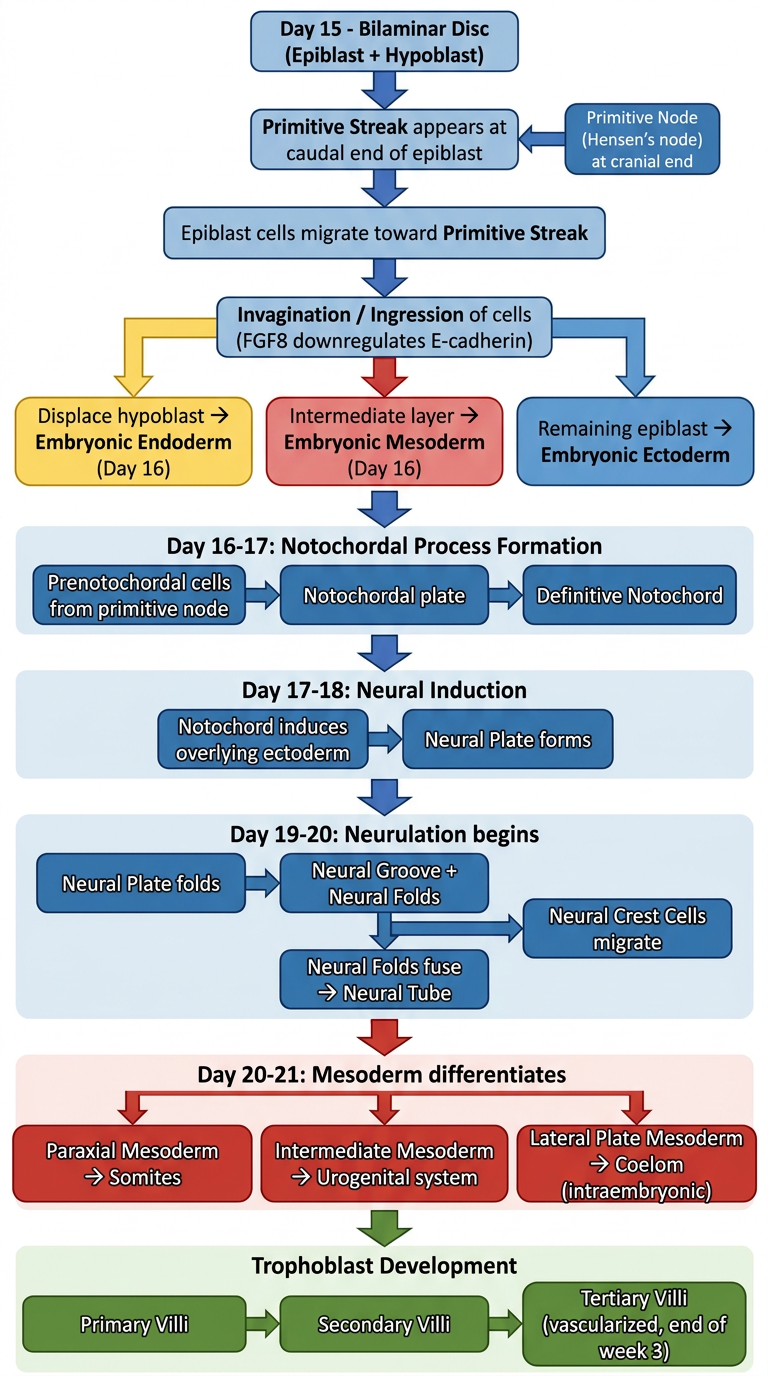
Third Week of Embryonic Development
The third week (days 15–21) is the most eventful period of early embryogenesis. It coincides with the week after the first missed menstrual period (~5 weeks after LMP). The defining event is gastrulation — the transformation of the bilaminar disc into the trilaminar germ disc.
Textbook Timeline & Events
Day 14–15 | Starting Point: Bilaminar Disc
The embryo arrives at week 3 as a bilaminar embryonic disc consisting of:
- Epiblast — upper layer (columnar cells)
- Hypoblast — lower layer (cuboidal cells)
Day 15–16 | Gastrulation Begins: Primitive Streak
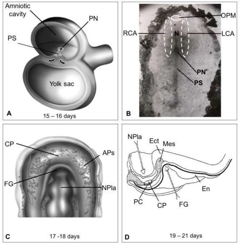
Primitive streak appears at the caudal end of the epiblast dorsal surface — initially a vague thickening, but by day 15–16 it is a clearly defined narrow groove with bulging lateral ridges.
- The cranial end of the streak forms the primitive node (Hensen's node), which surrounds the primitive pit
- Key molecular drivers: FGF8 (synthesized by streak cells) downregulates E-cadherin, releasing cells from epiblast adhesion
- BRACHYURY (T-box gene) controls cell migration and mesoderm specification
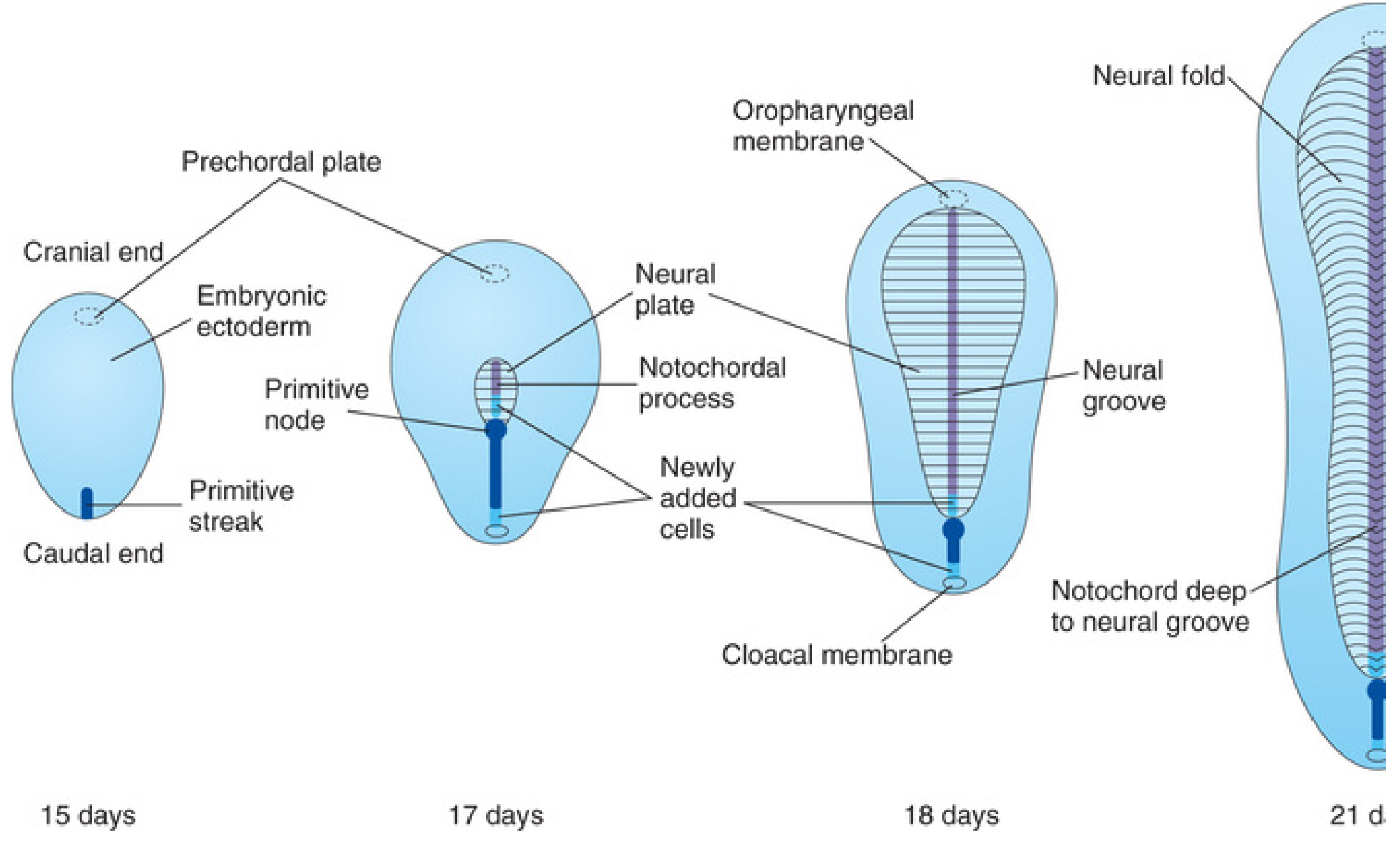
Day-by-day development of the embryonic disc showing appearance of primitive streak, notochordal process, neural plate, neural groove, and notochord — The Developing Human
Day 16 | Three Germ Layers Established (Trilaminar Disc)
Epiblast cells migrate toward the primitive streak, invaginate (become flask-shaped), and slip beneath:
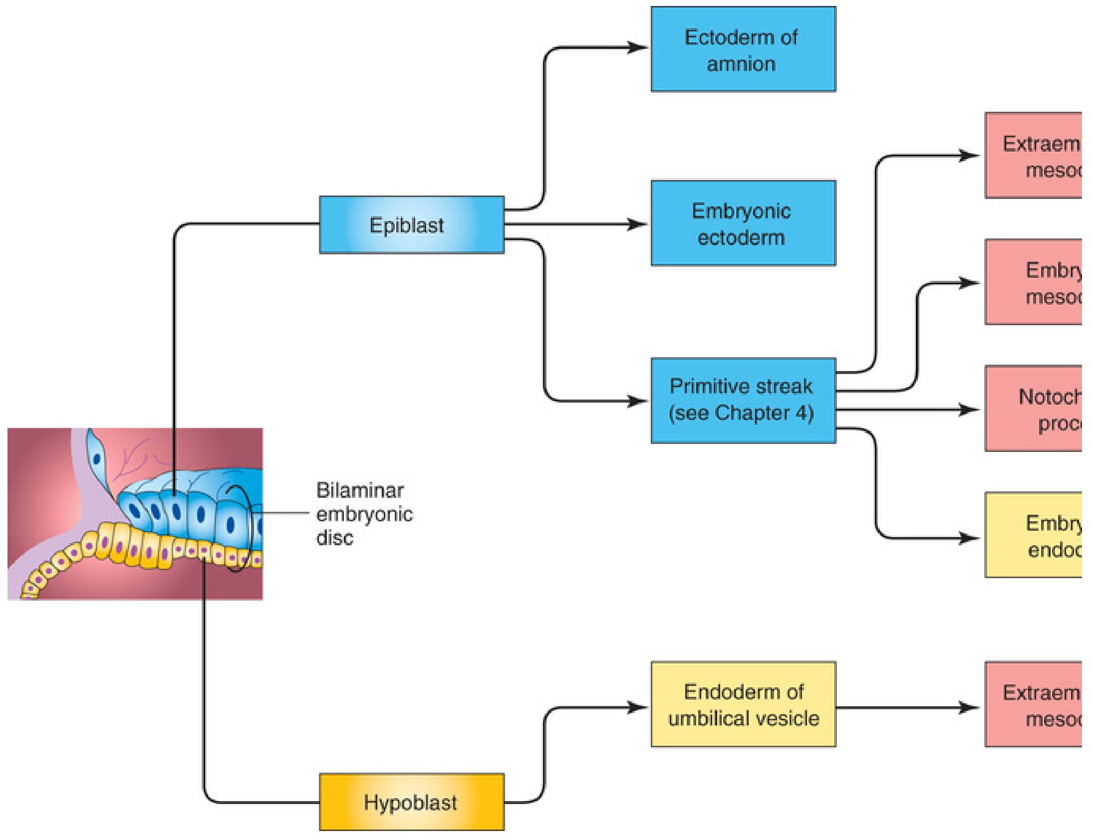
Derivation of all three germ layers from the epiblast through gastrulation — The Developing Human, FIG. 4.2
| Migration Fate | Germ Layer | Key Derivatives |
|---|
| Cells displace hypoblast | Endoderm | GI epithelium, respiratory tract, liver, pancreas |
| Cells between epiblast & endoderm | Mesoderm | Muscle, bone, cardiovascular, kidney, gonads |
| Cells remaining in epiblast | Ectoderm | Skin epidermis, entire nervous system, eyes, ears |
"The epiblast, through the process of gastrulation, is the source of all three germ layers." — Langman's Medical Embryology
Day 16–17 | Notochord Formation
- Prenotochordal cells from the primitive node migrate cranially in the midline toward the prechordal plate
- These cells intercalate into the hypoblast → form the notochordal plate (temporary bilaminar midline)
- As endoderm replaces hypoblast, notochordal plate detaches → forms the definitive notochord (solid cord)
Functions of the notochord:
- Defines the longitudinal axis of the embryo
- Signaling center — induces formation of the axial skeleton (vertebral column)
- Induces overlying ectoderm to form the neural plate (primary neurulation)
Boundaries established:
- Oropharyngeal membrane (cranial) — tightly adherent ecto- and endoderm; future oral opening; no mesoderm here
- Cloacal membrane (caudal) — future anal/urogenital opening
Day 17–18 | Neural Induction & Allantois
Embryonic disc at 15–21 days: PS = primitive streak, PN = primitive node, N = notochord, OPM = oropharyngeal membrane, cardiogenic areas (RCA/LCA), and neural plate (NPla) — Days 15–21
- The notochord induces overlying embryonic ectoderm → thickens into the neural plate
- Allantois appears as a small diverticulum from the caudal wall of the yolk sac into the connecting stalk
- Involved in early blood formation and development of the urinary bladder
Day 18–21 | Neurulation (Neural Tube Formation)
- Neural plate folds longitudinally → neural groove forms in the midline
- Lateral edges elevate → neural folds rise and converge
- Neural folds fuse (starting at cervical level, proceeding cranially & caudally) → neural tube
- Neural crest cells undergo epithelial-to-mesenchymal transition and migrate away bilaterally
Neural tube defects result from failure of fusion (e.g., spina bifida = caudal failure; anencephaly = cranial failure)
Day 20–21 | Mesoderm Differentiation & Somitogenesis
The intraembryonic mesoderm differentiates into three zones on each side of the notochord:
| Zone | Name | Derivatives |
|---|
| Adjacent to notochord | Paraxial mesoderm | Somites → dermis (dermatome), skeletal muscle (myotome), vertebrae & ribs (sclerotome) |
| Intermediate | Intermediate mesoderm | Kidneys, ureters, gonads, adrenal cortex |
| Lateral | Lateral plate mesoderm | Splits into somatic (body wall) + splanchnic (visceral) layers; intraembryonic coelom forms between them |
First pair of somites appears around day 20. ~3 pairs form per day thereafter. Somite count is used to date embryos.
Throughout Week 3 | Trophoblast (Chorionic Villi) Development
| Stage | Structure | Timing |
|---|
| Primary villus | Cytotrophoblast core + syncytial covering | Start of week 3 |
| Secondary villus | Mesoderm invades core | Mid-week 3 |
| Tertiary villus | Mesoderm differentiates into blood vessels (villous capillary system) | End of week 3 |
Summary Flowchart (Text)
BILAMINAR DISC (Day 14)
       │
       ▼
PRIMITIVE STREAK forms at caudal epiblast (Day 15)
  └─ Primitive node (cranial) + Primitive pit
       │  (FGF8, BRACHYURY, NODAL, BMP4)
       ▼
EPIBLAST CELLS INVAGINATE through streak (Day 15–16)
  ├──→ Displace hypoblast ────────→ ENDODERM
  ├──→ Middle layer ───────────────→ MESODERM
  └──→ Remaining epiblast ─────────→ ECTODERM
                            ↓
                  TRILAMINAR DISC (Day 16)
       │
       ▼
NOTOCHORD FORMATION (Day 16–17)
  Primitive node cells → Notochordal process → Notochordal plate → Definitive Notochord
       │
       ▼
NEURAL INDUCTION (Day 17–18)
  Notochord induces ectoderm → Neural Plate
       │
       ▼
NEURULATION (Day 18–21)
  Neural plate → Neural groove → Neural folds → NEURAL TUBE
                                              └── Neural Crest cells migrate
       │
       ▼
MESODERM DIFFERENTIATION (Day 20–21)
  ├── Paraxial → SOMITES (sclerotome, myotome, dermatome)
  ├── Intermediate → Urogenital primordia
  └── Lateral plate → Intraembryonic COELOM
       │
       ▼
TROPHOBLAST: Primary → Secondary → TERTIARY VILLI (vascularized)
Clinical Correlates
| Event | Clinical Significance |
|---|
| Primitive streak persists | Sacrococcygeal teratoma (remnant of primitive streak) |
| Failed gastrulation | Sirenomelia (caudal regression), structural malformations |
| Neural tube closure defect | Anencephaly (cranial), Spina bifida (caudal) |
| Disrupted laterality (5-HT signaling) | Situs inversus, dextrocardia, heart defects |
| SSRIs in pregnancy (disrupts serotonin) | Increased risk of cardiac malformations |
| Absent oropharyngeal membrane breakdown | Failure of oral cavity formation |
Sources:
- The Developing Human: Clinically Oriented Embryology (Moore & Persaud), Ch. 4 — Third Week
- Langman's Medical Embryology (Sadler), Ch. 5 — Third Week: Trilaminar Germ Disc