Show me the 10 most common heart disease with ecg pattern
ECG patterns common heart diseases electrocardiogram
ST elevation myocardial infarction STEMI ECG leads
atrial fibrillation ECG irregular rhythm no P waves
heart block complete AV block third degree ECG bradycardia
left ventricular hypertrophy ECG voltage criteria Sokolow
ventricular tachycardia wide QRS tachycardia ECG
pericarditis saddle-shaped ST elevation diffuse ECG
hypertrophic cardiomyopathy ECG deep Q waves LVH
pulmonary embolism ECG S1Q3T3 right heart strain sinus tachycardia
Wolff-Parkinson-White WPW delta wave short PR ECG pre-excitation
10 Common Heart Diseases & Their ECG Patterns
1. 🫀 ST-Elevation Myocardial Infarction (STEMI)
- ST elevation ≥1 mm in ≥2 contiguous limb leads, or ≥2 mm in ≥2 contiguous precordial leads
- Hyperacute T waves (tall, peaked) in early phase
- Pathological Q waves (>40 ms wide, >25% of R-wave height) in evolved MI
- Reciprocal ST depression in opposing leads
- "Tombstone" morphology in severe proximal LAD occlusion
| Territory | Leads with ST Elevation | Artery |
|---|---|---|
| Anterior | V1–V4 | LAD |
| Inferior | II, III, aVF | RCA |
| Lateral | I, aVL, V5–V6 | LCx |
| Posterior | ST depression V1–V2 + tall R | RCA/LCx |
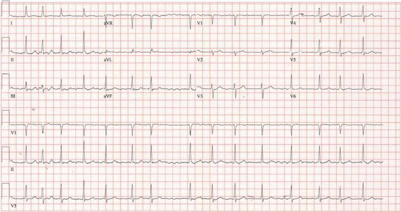
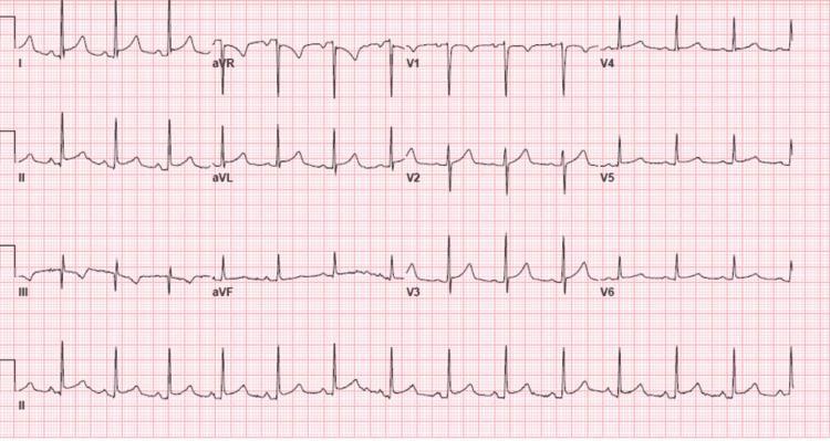
2. 🫀 Atrial Fibrillation (AF)
- Irregularly irregular R-R intervals — the hallmark
- Absent P waves replaced by fine/coarse fibrillatory (f) waves (best seen in V1)
- Narrow QRS complexes (unless aberrant conduction or bundle branch block)
- Rapid ventricular response if uncontrolled (>100 bpm)
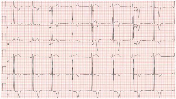
3. 🫀 Complete (3rd-Degree) AV Heart Block
- AV dissociation — P waves and QRS complexes fire completely independently
- Atrial rate faster than ventricular rate
- Regular, slow ventricular escape rhythm (~20–40 bpm)
- Narrow QRS = nodal escape; Wide QRS = ventricular/infranodal escape
- No fixed PR interval
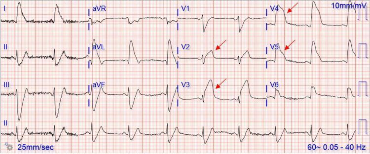
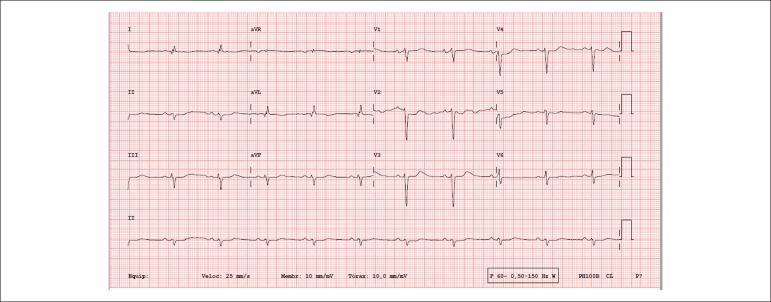
4. 🫀 Left Ventricular Hypertrophy (LVH) — Hypertensive Heart Disease
- Sokolow-Lyon criteria: S in V1 + R in V5 or V6 ≥35 mm
- Cornell criteria: R in aVL + S in V3 >28 mm (men), >20 mm (women)
- LV strain pattern: ST depression + T-wave inversion in I, aVL, V5–V6
- Left axis deviation
- Broadened QRS (not always frank LBBB)
5. 🫀 Ventricular Tachycardia (VT)
- Wide QRS tachycardia (QRS >120 ms) at rate ≥100 bpm
- Regular or slightly irregular rhythm
- AV dissociation (P waves independent of QRS)
- Fusion beats and capture beats (pathognomonic)
- Concordance in precordial leads (all positive or all negative)
- Northwest/superior axis deviation
| Feature | Points toward VT |
|---|---|
| QRS >160 ms | ✓ |
| Positive concordance V1–V6 | ✓ |
| AV dissociation | ✓ (pathognomonic) |
| Brugada/Wellens criteria | ✓ |

6. 🫀 Acute Pericarditis
- Stage 1: Diffuse saddle-shaped (concave) ST elevation in almost all leads; PR depression; ST elevation in aVR absent or with PR elevation
- Stage 2: ST normalizes, T waves flatten
- Stage 3: Diffuse T-wave inversions
- Stage 4: Normalization
- Key differentiator from STEMI: Diffuse (not localised), no reciprocal changes except aVR, PR segment depression
7. 🫀 Pulmonary Embolism (PE)
- Most common: Sinus tachycardia (non-specific)
- Classic S₁Q₃T₃ pattern: Deep S in I, Q wave in III, inverted T in III
- Right bundle branch block (complete or incomplete)
- T-wave inversions in V1–V4 (right ventricular strain)
- Right axis deviation
- P pulmonale (peaked P waves in II ≥2.5 mm)
- New AF in massive PE

8. 🫀 Hypertrophic Cardiomyopathy (HCM)
- LVH voltage criteria (tall R in lateral leads, deep S in right leads)
- Deep narrow Q waves in lateral (I, aVL, V5–V6) and inferior leads — due to septal hypertrophy
- Apical HCM (Yamaguchi): Giant deep symmetric T-wave inversions in V3–V5 (≥10 mm depth)
- ST depression in lateral leads
- Left axis deviation
- AF occurs in 20–25% of patients
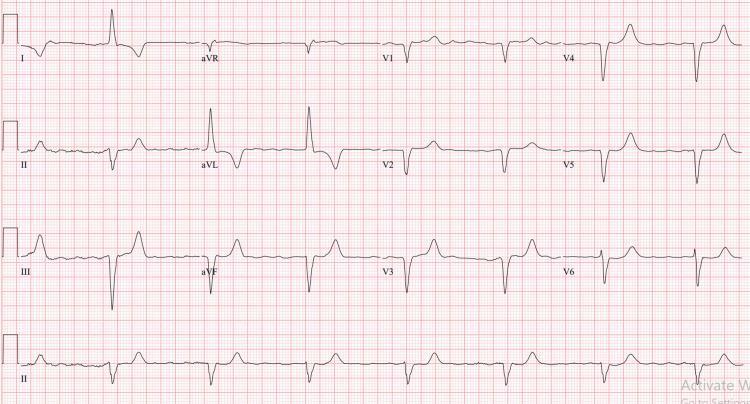
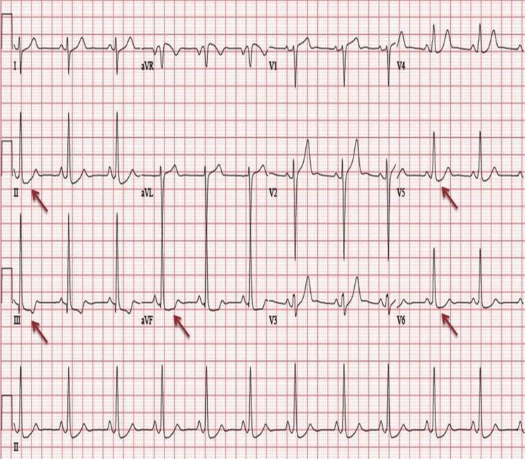
9. 🫀 Wolff-Parkinson-White (WPW) Syndrome
- Short PR interval (<120 ms)
- Delta wave — slurred initial upstroke of QRS
- Widened QRS (>120 ms) due to pre-excitation
- Secondary ST-T changes discordant with delta wave direction
- Risk: can precipitate AF → rapid conduction via accessory pathway → VF
| Pathway Location | Delta Polarity |
|---|---|
| Left lateral | Negative in V1, positive in I |
| Posteroseptal | Negative in II, III, aVF |
| Anteroseptal | Positive in V1 |

10. 🫀 Dilated Cardiomyopathy (DCM)
- Left bundle branch block (LBBB) — most characteristic
- QRS ≥120 ms, broad notched R in I/aVL/V5–V6, deep S/QS in V1–V3
- Diffuse ST-T changes (secondary to LBBB or ischaemia)
- Left axis deviation
- AF is common (30–40%)
- Low voltage QRS (if severe fibrosis or pericardial effusion co-exists)
- P mitrale (broad bifid P waves in II) — left atrial enlargement
- Pseudo-infarction Q waves mimicking prior MI
Quick Reference Summary
| # | Condition | Key ECG Finding |
|---|---|---|
| 1 | STEMI | ST elevation, Q waves, reciprocal depression |
| 2 | Atrial Fibrillation | Irregularly irregular, no P waves |
| 3 | Complete Heart Block | AV dissociation, slow escape rhythm |
| 4 | LVH / HTN Heart Disease | High voltage + strain pattern |
| 5 | Ventricular Tachycardia | Wide QRS ≥120 ms, AV dissociation |
| 6 | Acute Pericarditis | Diffuse saddle ST elevation, PR depression |
| 7 | Pulmonary Embolism | Sinus tachycardia, S₁Q₃T₃, RV strain |
| 8 | Hypertrophic Cardiomyopathy | Giant T inversions, deep Q waves |
| 9 | WPW Syndrome | Short PR, delta wave, widened QRS |
| 10 | Dilated Cardiomyopathy | LBBB, AF, low voltage |
Sources: Braunwald's Heart Disease (Elsevier), ROSEN's Emergency Medicine; ECG images from peer-reviewed clinical literature via PMC.
Convert this information into pdf
ls -la /home/daytona/workspace/ 2>/dev/null || echo "workspace empty or not foun…
mkdir -p /home/daytona/workspace/ecg-guide && python3 -c "from reportlab.lib.pag…
~/ecg-guide/generate_pdf.py
import requests
import os
from io import BytesIO
from reportlab.lib.pagesizes import A4
from reportlab.lib import colors
from reportlab.lib.units import mm, cm
from reportlab.lib.styles import getSampleStyleSheet, ParagraphStyle
from reportlab.lib.enums import TA_LEFT, TA_CENTER, TA_JUSTIFY
from reportlab.platypus import (
    SimpleDocTemplate, Paragraph, Spacer, Image, Table, TableStyle,
    HRFlowable, PageBreak, KeepTogether
)
from reportlab.platypus.flowables import BalancedColumns
from reportlab.graphics.shapes import Drawing, Rect
from reportlab.pdfgen import canvas

OUTPUT_PATH = "/home/daytona/workspace/ecg-guide/Heart_Diseases_ECG_Guide.pdf"

# ─── Colour palette ────────────────────────────────────────────────────────────
DARK_RED   = colors.HexColor("#8B0000")
MED_RED    = colors.HexColor("#C0392B")
LIGHT_RED  = colors.HexColor("#FADBD8")
DARK_GREY  = colors.HexColor("#2C3E50")
MID_GREY   = colors.HexColor("#5D6D7E")
LIGHT_GREY = colors.HexColor("#EAF0FB")
TABLE_HDR  = colors.HexColor("#1A3A5C")
TABLE_ALT  = colors.HexColor("#EBF5FB")
WHITE      = colors.white
BLACK      = colors.black

# ─── ECG Data ──────────────────────────────────────────────────────────────────
DISEASES = [
    {
        "num": 1,
        "name": "ST-Elevation Myocardial Infarction (STEMI)",
        "icon": "❤",
        "key_findings": [
            "ST elevation ≥1 mm in ≥2 contiguous limb leads; ≥2 mm in precordial leads",
            "Hyperacute T waves (tall, peaked) in early phase",
            "Pathological Q waves (>40 ms, >25% R height) in evolved MI",
            "Reciprocal ST depression in opposing leads",
            '"Tombstone" morphology in proximal LAD occlusion',
        ],
        "table": {
            "headers": ["Territory", "Leads", "Artery"],
            "rows": [
                ["Anterior", "V1–V4", "LAD"],
                ["Inferior", "II, III, aVF", "RCA"],
                ["Lateral", "I, aVL, V5–V6", "LCx"],
                ["Posterior", "ST↓ V1–V2 + tall R", "RCA/LCx"],
            ]
        },
        "img_url": "https://cdn.orris.care/cdss_images/pmc_clinical_VQA_0f5964997b5f333f6a7b13ecdc70a73ef6e81127eaee03ef38d9c724b26bbf31.jpg",
        "img_caption": "Anterior STEMI — tombstone ST elevation V2–V5 with reciprocal inferior depression",
    },
    {
        "num": 2,
        "name": "Atrial Fibrillation (AF)",
        "icon": "❤",
        "key_findings": [
            "Irregularly irregular R-R intervals — the hallmark",
            "Absent P waves replaced by fine/coarse fibrillatory (f) waves (best in V1)",
            "Narrow QRS complexes unless aberrant conduction or BBB",
            "Rapid ventricular response if uncontrolled (>100 bpm)",
        ],
        "table": None,
        "img_url": "https://cdn.orris.care/cdss_images/pmc_clinical_VQA_a8e7a35a18d282cc6de8c169a0213a6712f9fe9e4a7253f7828e22c867033097.jpg",
        "img_caption": "AF — irregularly irregular rhythm, absent P waves, fine fibrillatory baseline",
    },
    {
        "num": 3,
        "name": "Complete (3rd-Degree) AV Heart Block",
        "icon": "❤",
        "key_findings": [
            "AV dissociation — P waves and QRS complexes fire independently",
            "Atrial rate faster than ventricular rate",
            "Regular, slow ventricular escape rhythm (~20–40 bpm)",
            "Narrow QRS = nodal escape; Wide QRS = ventricular/infranodal escape",
            "No fixed PR interval",
        ],
        "table": None,
        "img_url": "https://cdn.orris.care/cdss_images/pmc_clinical_VQA_c76f7d8cf3869000720e9ce457857cf031f9b360704aa4236ad85275ba26169e.jpg",
        "img_caption": "3rd-degree AV block — P waves march through independently of slow wide QRS escape",
    },
    {
        "num": 4,
        "name": "Left Ventricular Hypertrophy (LVH) — Hypertensive Heart Disease",
        "icon": "❤",
        "key_findings": [
            "Sokolow-Lyon: S in V1 + R in V5/V6 ≥35 mm",
            "Cornell: R in aVL + S in V3 >28 mm (men), >20 mm (women)",
            "LV strain pattern: ST depression + T-wave inversion in I, aVL, V5–V6",
            "Left axis deviation",
            "Broadened QRS (not always frank LBBB)",
        ],
        "table": None,
        "img_url": "https://cdn.orris.care/cdss_images/pmc_clinical_VQA_03152b722b1cb86ef1d57f4d755aa687989488df7da9defa36d7d35f9ab80e28.jpg",
        "img_caption": "LVH with strain pattern — high voltage laterally with ST depression and T-wave inversions",
    },
    {
        "num": 5,
        "name": "Ventricular Tachycardia (VT)",
        "icon": "❤",
        "key_findings": [
            "Wide QRS tachycardia (QRS >120 ms) at rate ≥100 bpm",
            "Regular or slightly irregular rhythm",
            "AV dissociation (P waves independent of QRS)",
            "Fusion beats and capture beats (pathognomonic)",
            "Concordance in precordial leads (all positive or all negative)",
            "Northwest/superior axis deviation",
        ],
        "table": {
            "headers": ["Feature", "Points toward VT"],
            "rows": [
                ["QRS >160 ms", "✓"],
                ["Positive concordance V1–V6", "✓"],
                ["AV dissociation", "✓ (pathognomonic)"],
                ["Brugada/Wellens criteria", "✓"],
            ]
        },
        "img_url": "https://cdn.orris.care/cdss_images/pmc_clinical_VQA_d15a92de4e89b46f273b22d254d1e30fcd6a190c6d581b193e6dd338338c6bb6.jpg",
        "img_caption": "Monomorphic VT — wide complex tachycardia with positive concordance, no P waves",
    },
    {
        "num": 6,
        "name": "Acute Pericarditis",
        "icon": "❤",
        "key_findings": [
            "Stage 1: Diffuse saddle-shaped (concave) ST elevation + PR depression",
            "Stage 2: ST normalises, T waves flatten",
            "Stage 3: Diffuse T-wave inversions",
            "Stage 4: Full normalisation",
            "Key differentiator from STEMI: diffuse, no territory, PR depression present",
        ],
        "table": None,
        "img_url": "https://cdn.orris.care/cdss_images/pmc_clinical_VQA_0be5b02bdb0710f3bd3fcdbdc88b2944c0c2c3f7e84ae335cad7b36d24ffb97c.jpg",
        "img_caption": "Acute pericarditis — diffuse saddle-shaped ST elevation + PR depression across multiple leads",
    },
    {
        "num": 7,
        "name": "Pulmonary Embolism (PE)",
        "icon": "❤",
        "key_findings": [
            "Most common: Sinus tachycardia (non-specific but sensitive)",
            "Classic S₁Q₃T₃ pattern: deep S in I, Q wave in III, inverted T in III",
            "Right bundle branch block (complete or incomplete)",
            "T-wave inversions in V1–V4 (right ventricular strain)",
            "Right axis deviation; P pulmonale (peaked P ≥2.5 mm in II)",
            "New AF in massive PE",
        ],
        "table": None,
        "img_url": "https://cdn.orris.care/cdss_images/pmc_clinical_VQA_7cfb4819f88c84d02f863ca2f948165b6f4f6fafd405ca0b0a7df41e2d8d8e2a.jpg",
        "img_caption": "Pulmonary embolism — sinus tachycardia, S1Q3T3 pattern, RV strain T inversions V1–V3",
    },
    {
        "num": 8,
        "name": "Hypertrophic Cardiomyopathy (HCM)",
        "icon": "❤",
        "key_findings": [
            "LVH voltage criteria (tall R laterally, deep S right leads)",
            "Deep narrow Q waves in lateral (I, aVL, V5–V6) and inferior leads",
            "Apical HCM (Yamaguchi): giant deep symmetric T inversions in V3–V5 (≥10 mm)",
            "ST depression in lateral leads",
            "Left axis deviation; AF in 20–25% of patients",
        ],
        "table": None,
        "img_url": "https://cdn.orris.care/cdss_images/pmc_clinical_VQA_3d2687643839a530919d460d4461031928da6585c23315b742b24d71296327a0.jpg",
        "img_caption": "Apical HCM (Yamaguchi) — giant symmetric T-wave inversions reaching 15 mm depth in V2–V4",
    },
    {
        "num": 9,
        "name": "Wolff-Parkinson-White (WPW) Syndrome",
        "icon": "❤",
        "key_findings": [
            "Short PR interval (<120 ms)",
            "Delta wave — slurred initial upstroke of the QRS",
            "Widened QRS (>120 ms) due to ventricular pre-excitation",
            "Secondary ST-T changes discordant with delta wave direction",
            "Risk: AF → rapid accessory pathway conduction → VF",
        ],
        "table": {
            "headers": ["Pathway Location", "Delta Polarity"],
            "rows": [
                ["Left lateral", "Negative in V1, positive in I"],
                ["Posteroseptal", "Negative in II, III, aVF"],
                ["Anteroseptal", "Positive in V1"],
            ]
        },
        "img_url": "https://cdn.orris.care/cdss_images/pmc_clinical_VQA_fd0d58cc1913acd56cbc62cbf42657e4a465f052a9bb2ea0d3893584ef6f70e6.jpg",
        "img_caption": "WPW syndrome — short PR interval, prominent delta waves, widened QRS complexes",
    },
    {
        "num": 10,
        "name": "Dilated Cardiomyopathy (DCM)",
        "icon": "❤",
        "key_findings": [
            "Left bundle branch block (LBBB) — most characteristic (QRS ≥120 ms)",
            "Broad notched R in I/aVL/V5–V6; deep QS in V1–V3",
            "Diffuse ST-T changes secondary to LBBB",
            "Left axis deviation; AF common (30–40%)",
            "Low voltage QRS (if fibrosis or pericardial effusion)",
            "Pseudo-infarction Q waves mimicking prior MI",
        ],
        "table": None,
        "img_url": "https://cdn.orris.care/cdss_images/pmc_clinical_VQA_3adea767053955d22dff275d5b4935c292277e5153fb88fae914ccff7abedb82.jpg",
        "img_caption": "DCM/LBBB pattern — low voltage QRS, broad notched morphology, secondary ST-T changes",
    },
]

SUMMARY_ROWS = [
    ["1", "STEMI", "ST elevation, Q waves, reciprocal depression"],
    ["2", "Atrial Fibrillation", "Irregularly irregular, no P waves"],
    ["3", "Complete Heart Block", "AV dissociation, slow escape rhythm"],
    ["4", "LVH / HTN Heart Disease", "High voltage + lateral strain pattern"],
    ["5", "Ventricular Tachycardia", "Wide QRS ≥120 ms, AV dissociation"],
    ["6", "Acute Pericarditis", "Diffuse saddle ST elevation, PR depression"],
    ["7", "Pulmonary Embolism", "Sinus tachycardia, S₁Q₃T₃, RV strain"],
    ["8", "Hypertrophic Cardiomyopathy", "Giant T inversions, deep Q waves"],
    ["9", "WPW Syndrome", "Short PR, delta wave, widened QRS"],
    ["10", "Dilated Cardiomyopathy", "LBBB, AF, low voltage"],
]


# ─── Helper: download image ─────────────────────────────────────────────────
def fetch_image_bytes(url, timeout=15):
    try:
        r = requests.get(url, timeout=timeout)
        r.raise_for_status()
        return BytesIO(r.content)
    except Exception as e:
        print(f"  [WARN] Could not fetch {url}: {e}")
        return None


# ─── Page template with header/footer ──────────────────────────────────────
def make_canvas_callback(title_text):
    def on_page(canv, doc):
        canv.saveState()
        w, h = A4
        # Header bar
        canv.setFillColor(DARK_RED)
        canv.rect(0, h - 22*mm, w, 22*mm, fill=1, stroke=0)
        canv.setFont("Helvetica-Bold", 11)
        canv.setFillColor(WHITE)
        canv.drawString(15*mm, h - 14*mm, title_text)
        canv.setFont("Helvetica", 9)
        canv.drawRightString(w - 15*mm, h - 14*mm, "Clinical ECG Reference Guide")
        # Footer bar
        canv.setFillColor(DARK_GREY)
        canv.rect(0, 0, w, 12*mm, fill=1, stroke=0)
        canv.setFont("Helvetica", 8)
        canv.setFillColor(WHITE)
        canv.drawString(15*mm, 4*mm, "Source: Braunwald's Heart Disease | ROSEN's Emergency Medicine")
        canv.drawRightString(w - 15*mm, 4*mm, f"Page {doc.page}")
        canv.restoreState()
    return on_page


# ─── Styles ─────────────────────────────────────────────────────────────────
styles = getSampleStyleSheet()

title_style = ParagraphStyle(
    "MainTitle", fontSize=28, fontName="Helvetica-Bold",
    textColor=WHITE, alignment=TA_CENTER, spaceAfter=6
)
subtitle_style = ParagraphStyle(
    "Subtitle", fontSize=14, fontName="Helvetica",
    textColor=colors.HexColor("#FADBD8"), alignment=TA_CENTER, spaceAfter=4
)
disease_title_style = ParagraphStyle(
    "DiseaseTitle", fontSize=15, fontName="Helvetica-Bold",
    textColor=WHITE, alignment=TA_LEFT, spaceAfter=0, leading=18
)
section_num_style = ParagraphStyle(
    "SectionNum", fontSize=30, fontName="Helvetica-Bold",
    textColor=colors.HexColor("#FADBD8"), alignment=TA_LEFT
)
bullet_style = ParagraphStyle(
    "Bullet", fontSize=9.5, fontName="Helvetica",
    textColor=DARK_GREY, leftIndent=12, bulletIndent=0,
    spaceAfter=3, leading=13
)
caption_style = ParagraphStyle(
    "Caption", fontSize=8, fontName="Helvetica-Oblique",
    textColor=MID_GREY, alignment=TA_CENTER, spaceAfter=4
)
body_style = ParagraphStyle(
    "Body", fontSize=9.5, fontName="Helvetica",
    textColor=DARK_GREY, spaceAfter=4, leading=13
)
table_hdr_style = ParagraphStyle(
    "TblHdr", fontSize=9, fontName="Helvetica-Bold",
    textColor=WHITE, alignment=TA_CENTER
)
table_cell_style = ParagraphStyle(
    "TblCell", fontSize=9, fontName="Helvetica",
    textColor=DARK_GREY, alignment=TA_LEFT
)
summary_hdr_style = ParagraphStyle(
    "SumHdr", fontSize=10, fontName="Helvetica-Bold",
    textColor=WHITE, alignment=TA_CENTER
)
summary_cell_style = ParagraphStyle(
    "SumCell", fontSize=9, fontName="Helvetica",
    textColor=DARK_GREY
)


# ─── Build ───────────────────────────────────────────────────────────────────
doc = SimpleDocTemplate(
    OUTPUT_PATH,
    pagesize=A4,
    leftMargin=18*mm, rightMargin=18*mm,
    topMargin=30*mm, bottomMargin=20*mm,
    title="10 Common Heart Diseases & ECG Patterns",
    author="Orris Medical Reference",
)

story = []
PAGE_W = A4[0] - 36*mm   # usable width


# ════════════════════ COVER PAGE ════════════════════════════════════════════
# Big coloured cover block drawn via a custom flowable trick —
# we use a table with coloured background instead.

cover_title_style = ParagraphStyle(
    "CoverTitle", fontSize=32, fontName="Helvetica-Bold",
    textColor=WHITE, alignment=TA_CENTER, leading=38, spaceAfter=8
)
cover_sub_style = ParagraphStyle(
    "CoverSub", fontSize=14, fontName="Helvetica",
    textColor=colors.HexColor("#FADBD8"), alignment=TA_CENTER, spaceAfter=6
)
cover_body_style = ParagraphStyle(
    "CoverBody", fontSize=11, fontName="Helvetica",
    textColor=colors.HexColor("#D5E8F5"), alignment=TA_CENTER, spaceAfter=4
)

cover_content = [
    [Paragraph("10 Common Heart Diseases", cover_title_style)],
    [Paragraph("& Their ECG Patterns", cover_title_style)],
    [Spacer(1, 8*mm)],
    [Paragraph("A Clinical Reference Guide", cover_sub_style)],
    [Spacer(1, 6*mm)],
    [Paragraph(
        "STEMI  •  Atrial Fibrillation  •  Heart Block  •  LVH  •  VT<br/>"
        "Pericarditis  •  Pulmonary Embolism  •  HCM  •  WPW  •  DCM",
        cover_body_style)],
    [Spacer(1, 10*mm)],
    [Paragraph("Source: Braunwald's Heart Disease | ROSEN's Emergency Medicine", ParagraphStyle(
        "src", fontSize=9, textColor=colors.HexColor("#A9CCE3"), alignment=TA_CENTER
    ))],
]

cover_table = Table(cover_content, colWidths=[PAGE_W])
cover_table.setStyle(TableStyle([
    ("BACKGROUND", (0, 0), (-1, -1), DARK_RED),
    ("TOPPADDING",    (0, 0), (-1, -1), 8),
    ("BOTTOMPADDING", (0, 0), (-1, -1), 8),
    ("LEFTPADDING",   (0, 0), (-1, -1), 20),
    ("RIGHTPADDING",  (0, 0), (-1, -1), 20),
    ("VALIGN",        (0, 0), (-1, -1), "MIDDLE"),
    ("ROWBACKGROUNDS",(0, 0), (-1, -1), [DARK_RED]),
]))

# Outer wrapper to add a red box that fills most of the page
story.append(Spacer(1, 25*mm))
story.append(cover_table)
story.append(Spacer(1, 12*mm))

intro_style = ParagraphStyle(
    "Intro", fontSize=10, fontName="Helvetica",
    textColor=DARK_GREY, alignment=TA_JUSTIFY, leading=15
)
story.append(Paragraph(
    "Electrocardiography remains the fastest, most accessible, and most widely used tool in cardiovascular "
    "diagnosis. This guide presents the 10 most clinically encountered heart diseases alongside their "
    "characteristic ECG patterns, diagnostic criteria, and representative tracings sourced from peer-reviewed "
    "clinical literature. Each section includes key findings, localisation tables where applicable, and an "
    "annotated 12-lead ECG image.",
    intro_style
))
story.append(PageBreak())


# ════════════════════ DISEASE SECTIONS ══════════════════════════════════════
for d in DISEASES:
    print(f"Building section {d['num']}: {d['name']} ...")

    # ── Section header bar ──────────────────────────────────────────────────
    num_para  = Paragraph(f"{d['num']:02d}", ParagraphStyle(
        "Num", fontSize=36, fontName="Helvetica-Bold",
        textColor=colors.HexColor("#FADBD8"), alignment=TA_LEFT
    ))
    name_para = Paragraph(d["name"], disease_title_style)

    header_data = [[num_para, name_para]]
    header_table = Table(header_data, colWidths=[22*mm, PAGE_W - 22*mm])
    header_table.setStyle(TableStyle([
        ("BACKGROUND",    (0, 0), (-1, -1), DARK_RED),
        ("VALIGN",        (0, 0), (-1, -1), "MIDDLE"),
        ("LEFTPADDING",   (0, 0), (0, 0),   6),
        ("LEFTPADDING",   (1, 0), (1, 0),   4),
        ("RIGHTPADDING",  (0, 0), (-1, -1), 8),
        ("TOPPADDING",    (0, 0), (-1, -1), 6),
        ("BOTTOMPADDING", (0, 0), (-1, -1), 6),
        ("ROUNDEDCORNERS",(0, 0), (-1, -1), [4, 4, 4, 4]),
    ]))

    # ── Key findings ────────────────────────────────────────────────────────
    findings_paras = [Paragraph("<b>Key ECG Findings</b>", ParagraphStyle(
        "FH", fontSize=10, fontName="Helvetica-Bold",
        textColor=DARK_RED, spaceAfter=4
    ))]
    for f in d["key_findings"]:
        findings_paras.append(Paragraph(f"• {f}", bullet_style))

    # ── Optional table ──────────────────────────────────────────────────────
    tbl_element = None
    if d["table"]:
        td = d["table"]
        t_data = [[Paragraph(h, table_hdr_style) for h in td["headers"]]]
        for i, row in enumerate(td["rows"]):
            bg = TABLE_ALT if i % 2 == 0 else WHITE
            t_data.append([Paragraph(cell, table_cell_style) for cell in row])

        col_count = len(td["headers"])
        col_w = PAGE_W / col_count
        tbl = Table(t_data, colWidths=[col_w] * col_count)
        tbl.setStyle(TableStyle([
            ("BACKGROUND",    (0, 0), (-1, 0),  TABLE_HDR),
            ("ROWBACKGROUNDS",(0, 1), (-1, -1), [TABLE_ALT, WHITE]),
            ("GRID",          (0, 0), (-1, -1), 0.5, colors.HexColor("#BFC9CA")),
            ("TOPPADDING",    (0, 0), (-1, -1), 4),
            ("BOTTOMPADDING", (0, 0), (-1, -1), 4),
            ("LEFTPADDING",   (0, 0), (-1, -1), 6),
            ("RIGHTPADDING",  (0, 0), (-1, -1), 6),
            ("VALIGN",        (0, 0), (-1, -1), "MIDDLE"),
        ]))
        tbl_element = tbl

    # ── ECG Image ────────────────────────────────────────────────────────────
    img_bytes = fetch_image_bytes(d["img_url"])
    img_element = None
    if img_bytes:
        img_max_w = PAGE_W
        img_max_h = 55*mm
        img_element = Image(img_bytes, width=img_max_w, height=img_max_h)
        img_element.hAlign = "CENTER"

    # ── Assemble section ────────────────────────────────────────────────────
    section_parts = [
        header_table,
        Spacer(1, 4*mm),
    ]
    section_parts.extend(findings_paras)
    if tbl_element:
        section_parts.append(Spacer(1, 3*mm))
        section_parts.append(tbl_element)
    if img_element:
        section_parts.append(Spacer(1, 5*mm))
        section_parts.append(img_element)
        section_parts.append(Paragraph(d["img_caption"], caption_style))
    section_parts.append(HRFlowable(width=PAGE_W, thickness=0.5, color=colors.HexColor("#BFC9CA")))
    section_parts.append(Spacer(1, 6*mm))

    story.extend(section_parts)

    # Page break after every 2 diseases to keep layout clean
    if d["num"] % 2 == 0 and d["num"] < 10:
        story.append(PageBreak())


# ════════════════════ SUMMARY TABLE PAGE ════════════════════════════════════
story.append(PageBreak())

story.append(Paragraph("Quick Reference Summary", ParagraphStyle(
    "SumTitle", fontSize=18, fontName="Helvetica-Bold",
    textColor=DARK_RED, spaceAfter=8, alignment=TA_CENTER
)))
story.append(HRFlowable(width=PAGE_W, thickness=2, color=DARK_RED))
story.append(Spacer(1, 4*mm))

sum_data = [[
    Paragraph("#", summary_hdr_style),
    Paragraph("Condition", summary_hdr_style),
    Paragraph("Hallmark ECG Finding", summary_hdr_style),
]]
for row in SUMMARY_ROWS:
    sum_data.append([
        Paragraph(row[0], ParagraphStyle("SC", fontSize=10, fontName="Helvetica-Bold",
                                         textColor=DARK_RED, alignment=TA_CENTER)),
        Paragraph(row[1], summary_cell_style),
        Paragraph(row[2], summary_cell_style),
    ])

sum_table = Table(sum_data, colWidths=[12*mm, 65*mm, PAGE_W - 77*mm])
sum_table.setStyle(TableStyle([
    ("BACKGROUND",    (0, 0), (-1, 0),  TABLE_HDR),
    ("ROWBACKGROUNDS",(0, 1), (-1, -1), [TABLE_ALT, WHITE]),
    ("GRID",          (0, 0), (-1, -1), 0.5, colors.HexColor("#BFC9CA")),
    ("TOPPADDING",    (0, 0), (-1, -1), 6),
    ("BOTTOMPADDING", (0, 0), (-1, -1), 6),
    ("LEFTPADDING",   (0, 0), (-1, -1), 8),
    ("RIGHTPADDING",  (0, 0), (-1, -1), 8),
    ("VALIGN",        (0, 0), (-1, -1), "MIDDLE"),
    ("FONTSIZE",      (0, 1), (-1, -1), 9.5),
]))
story.append(sum_table)

story.append(Spacer(1, 10*mm))
story.append(Paragraph(
    "<b>Sources:</b> Braunwald's Heart Disease: A Textbook of Cardiovascular Medicine (Elsevier, 10th ed.) | "
    "ROSEN's Emergency Medicine: Concepts and Clinical Practice (Elsevier) | "
    "ECG images sourced from peer-reviewed PMC clinical literature.",
    ParagraphStyle("Sources", fontSize=8, textColor=MID_GREY, leading=12, alignment=TA_JUSTIFY)
))


# ─── Build PDF ───────────────────────────────────────────────────────────────
doc.build(
    story,
    onFirstPage=make_canvas_callback("10 Common Heart Diseases & ECG Patterns"),
    onLaterPages=make_canvas_callback("10 Common Heart Diseases & ECG Patterns"),
)
print(f"\n✓ PDF saved to: {OUTPUT_PATH}")
cd /home/daytona/workspace/ecg-guide && python3 generate_pdf.py 2>&1
ls -lh /home/daytona/workspace/ecg-guide/Heart_Diseases_ECG_Guide.pdf
Heart Diseases ECG Guide
PDF Document · PDF
- Cover page with title and condition list on a deep red background
- 10 individual sections, each with:
- Numbered header bar (red)
- Bullet-point key ECG findings
- Localisation/criteria table (where applicable — STEMI, VT, WPW)
- Full-width real 12-lead ECG image from peer-reviewed clinical literature
- Annotated caption describing the ECG
- Quick Reference Summary table at the end listing all 10 conditions and their hallmark ECG finding
- Running header and page footer with source attribution on every page