
Hey orris
Hey! How can I help you today?
Share me some general anatomy notes on topic joints for 1st year BPT student
Joints (articulations) are connections between two or more bones. For a 1st year BPT student, understanding joint classification, structure, and movement is foundational for all physiotherapy work.
Searching sources...
Reading File
Finding Sources
Finding Sources
Reading File
Finding Sources
Reading File
Finding Sources
Loading Image
Finding Sources
Reading File
Finding Sources
Reading File
Loading Image
Reading File
Excellent - I have rich textbook content. Let me now compile the complete, well-structured notes.
Anatomy Notes: Joints (Articulations)
For 1st Year BPT Students
1. Definition
A joint (articulation) is a point of contact between two or more bones of the skeleton. Joints are essential for skeletal motion, function, and quality of life. Each joint is uniquely shaped to suit its anatomical location and required range of motion.
- Firestein & Kelley's Textbook of Rheumatology
2. Classification of Joints
Joints are classified in two main ways: by structure (histological composition) and by function (degree of movement). These two systems correlate with each other.
A. Structural Classification
| Type | Also Known As | Connecting Tissue | Movement | Examples |
|---|---|---|---|---|
| Fibrous | Synarthroses | Dense connective tissue | Minimal/none | Skull sutures, gomphoses, syndesmoses |
| Cartilaginous | Amphiarthroses | Hyaline cartilage or fibrocartilage | Slight | Pubic symphysis, intervertebral discs, 1st rib-sternum joint |
| Synovial | Diarthroses | Synovial cavity + fluid | Freely moveable | Knee, shoulder, hip, elbow, wrist |
B. Functional Classification
| Functional Name | Movement | Structural Equivalent |
|---|---|---|
| Synarthrosis | Immobile | Fibrous joints |
| Amphiarthrosis | Slightly mobile | Cartilaginous joints |
| Diarthrosis | Freely mobile | Synovial joints |
- Imaging Anatomy Text and Atlas Vol. 3 - Bones, Joints, Vessels and Nerves
3. Types of Joints in Detail
A. Fibrous Joints
No joint cavity. Bones are united directly by fibrous connective tissue. Three subtypes:
- Sutures - Found between skull bones. Minimally flexible at birth (allow brain growth), eventually ossify (= synostosis). E.g., coronal suture, sagittal suture.
- Syndesmoses - Bones joined by thick ligaments or an interosseous membrane. Classified as amphiarthrosis (slightly moveable). E.g., inferior tibiofibular joint, interosseous membrane between radius and ulna.
- Gomphoses - Peg-in-socket joints. The periodontal ligament connects the tooth root to its bony socket. E.g., teeth in jaw sockets.
B. Cartilaginous Joints
No joint cavity. Bones are united by cartilage. Two subtypes:
- Primary cartilaginous joints (Synchondroses) - United by hyaline cartilage. E.g., epiphyseal growth plates (temporary), 1st rib-sternum joint (permanent).
- Secondary cartilaginous joints (Symphyses) - United by fibrocartilage. E.g., pubic symphysis, intervertebral discs (nucleus pulposus + annulus fibrosus).
C. Synovial Joints (Most Important for BPT)
The most common type of joint in the body. Characterized by a joint cavity filled with synovial fluid.
4. Structure of a Synovial Joint
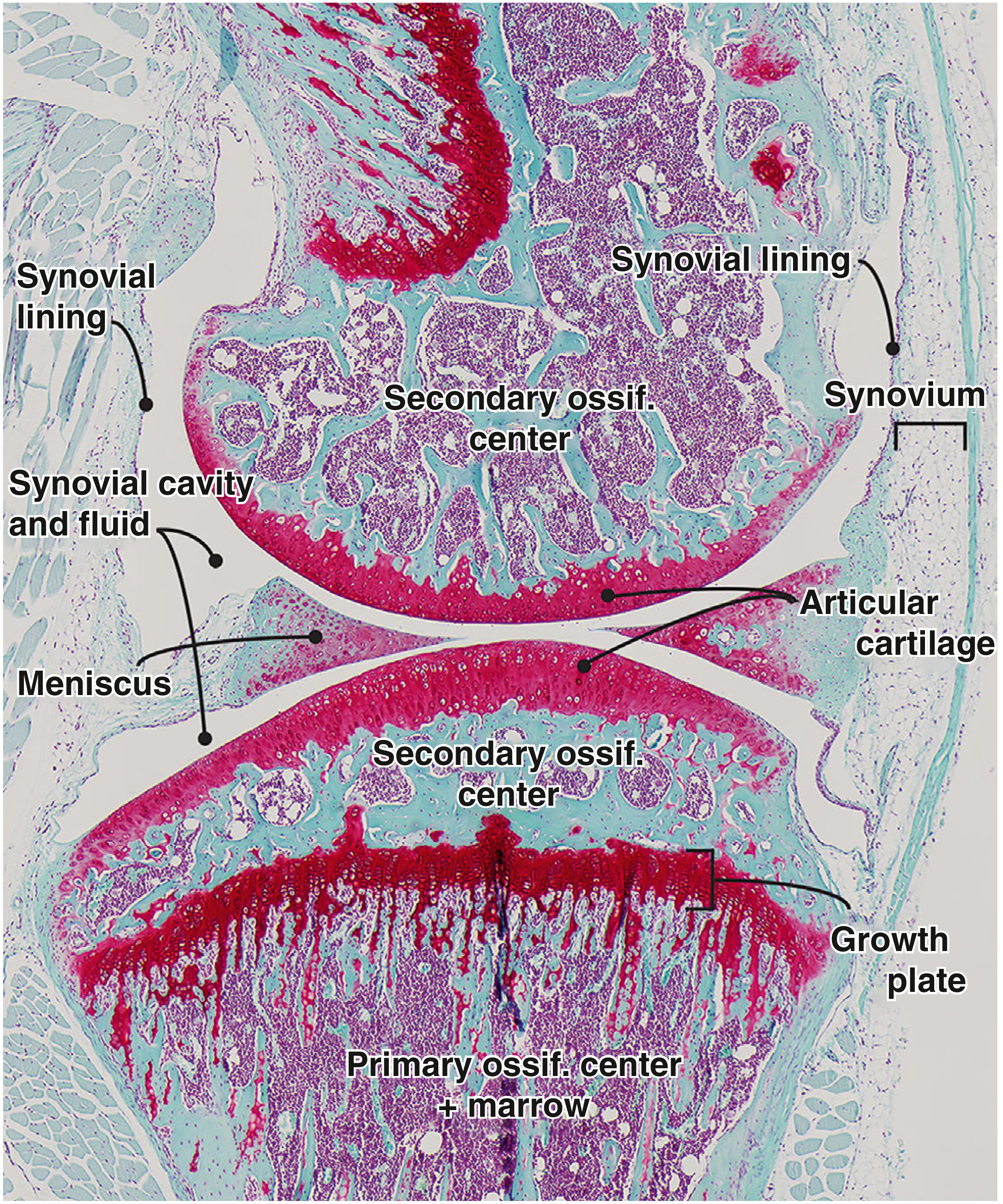
Key components:
| Component | Description |
|---|---|
| Articular cartilage | Hyaline cartilage covering bone ends; 1-2 mm (phalanges) to 5-7 mm (femoropatellar); avascular, nourished by synovial fluid |
| Joint capsule | Encloses the joint; has two layers: outer fibrous membrane + inner synovial membrane |
| Synovial membrane | Inner lining; secretes synovial fluid; contains Type A (macrophage-like) and Type B (fibroblast-like) synoviocytes |
| Synovial fluid | Lubricating fluid containing water, hyaluronic acid, lubricin, and phospholipids |
| Ligaments | Intracapsular (inside capsule, e.g., ACL) or extracapsular (outside, e.g., MCL) |
| Bursae | Fluid-filled connective tissue sacs that reduce friction near tendons and muscles |
| Menisci/Discs | Fibrocartilage pads that improve joint congruence (e.g., knee menisci, TMJ disc) |
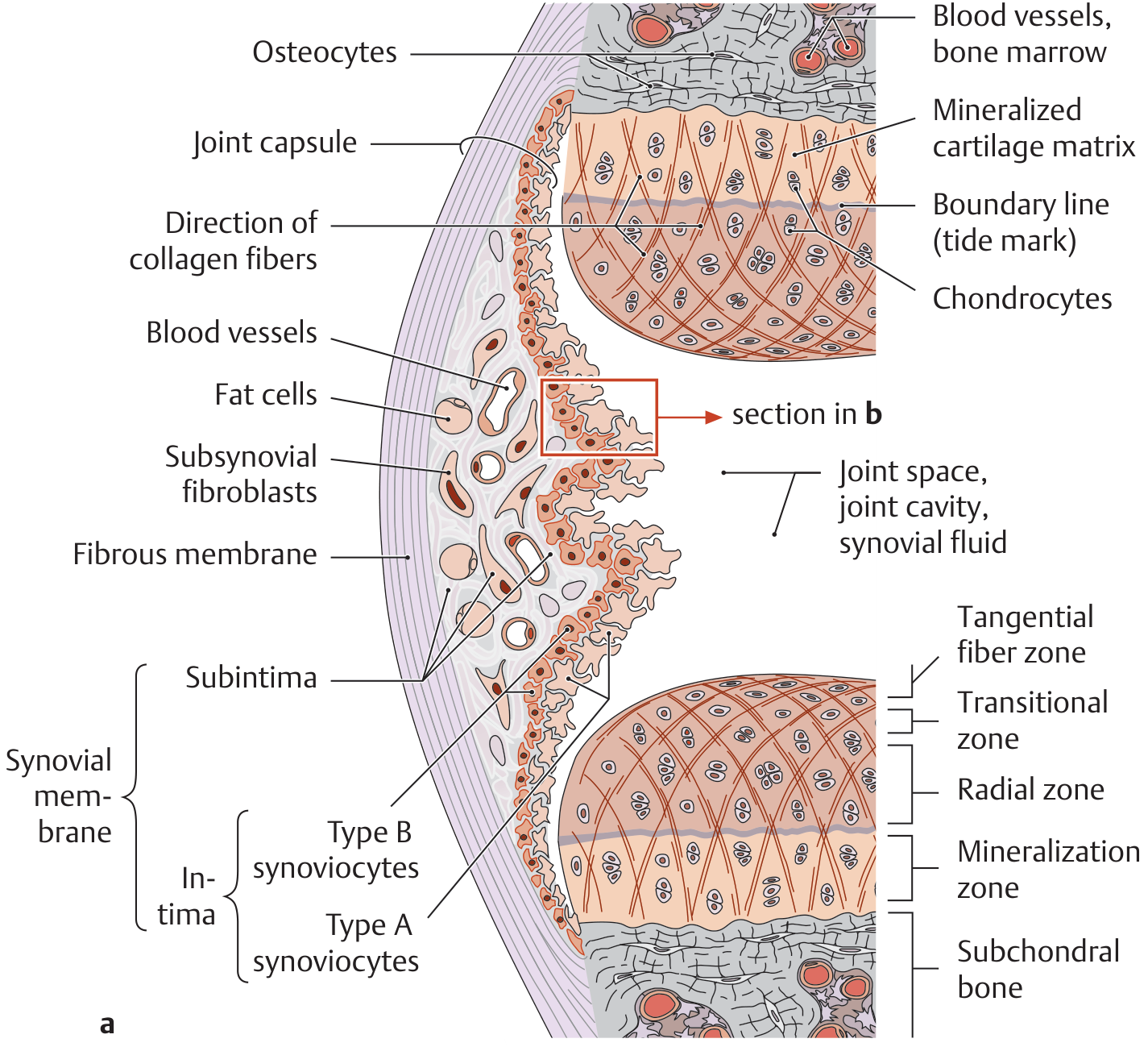
Key note for BPT: The synovial membrane can regenerate even after surgical removal (synovectomy). Hyaline cartilage, however, has very limited regenerative capacity since it is avascular and lacks a perichondrium.
5. Types of Synovial Joints
Classified by number of axes of motion and shape of articular surfaces:
By Axes of Motion:
- Uniaxial - Movement in 1 plane
- Biaxial - Movement in 2 planes
- Multiaxial - Movement in multiple directions
By Shape (6 Types):
| Type | Axes | Movement | Examples |
|---|---|---|---|
| Hinge | Uniaxial | Flexion/Extension only | Elbow (humeroulnar), knee, ankle, interphalangeal joints |
| Pivot | Uniaxial | Rotation only | Atlantoaxial (C1-C2), proximal radioulnar joint |
| Plane (Gliding) | Multiaxial | Gliding/sliding | Intercarpal, intertarsal, acromioclavicular, facet joints of vertebrae |
| Saddle | Biaxial | Flexion/Extension + Abduction/Adduction | 1st carpometacarpal (thumb), sternoclavicular joint |
| Condyloid (Ellipsoid) | Biaxial | Flexion/Extension + side-to-side | Metacarpophalangeal, radiocarpal (wrist), metatarsophalangeal joints |
| Ball-and-Socket | Multiaxial | All directions including rotation | Hip (acetabulofemoral), shoulder (glenohumeral) |
- Imaging Anatomy Text and Atlas Vol. 3
6. Movements at Synovial Joints
| Movement | Description |
|---|---|
| Flexion | Decreasing angle between bones |
| Extension | Increasing angle between bones |
| Abduction | Moving away from the midline |
| Adduction | Moving toward the midline |
| Rotation | Spinning around a longitudinal axis (medial/lateral) |
| Circumduction | Combination of flexion, extension, abduction, adduction (circular movement) |
| Pronation/Supination | Forearm rotation |
| Inversion/Eversion | Foot movements |
| Protraction/Retraction | Forward/backward movement (jaw, shoulder) |
| Elevation/Depression | Upward/downward movement (jaw, shoulder blade) |
7. Factors Affecting Joint Stability
- Shape of articular surfaces - Deeper sockets = more stable (e.g., hip > shoulder)
- Ligaments - Primary stabilizers; intracapsular and extracapsular
- Muscle tone - Muscles crossing the joint provide dynamic stability
- Atmospheric pressure - Especially important in the shoulder joint
- Fibrocartilaginous structures - Labrum (hip/shoulder), menisci (knee)
8. Clinical Relevance for BPT
| Condition | Joints Involved | Relevance |
|---|---|---|
| Osteoarthritis | Knee, hip, finger joints | Degeneration of articular cartilage |
| Rheumatoid Arthritis | Synovial joints (symmetrical) | Synovial membrane inflammation |
| Sprains | Ankle, knee | Ligament injury |
| Dislocations | Shoulder (most common) | Loss of joint congruence |
| Effusion (Haemarthrosis) | Knee | Bloody fluid from cruciate ligament injury |
| Synovectomy | Any synovial joint | Surgical removal of synovial membrane in chronic arthritis |
9. Quick Memory Tips
- "FBI" for fibrous = Fibrous joints, But Immobile (sutures, syndesmoses, gomphoses)
- "CAS" for cartilaginous = Cartilage, A bit mobile, Synchondroses/Symphyses
- "SHIP" for synovial types = Saddle, Hinge, Pivot; add Plane, Condyloid, Ball-and-socket
- Diarthrosis = Di (two/freely) = Diarthrotic joints are freely moveable
- The shoulder has the greatest ROM but is the most commonly dislocated joint - relevant for your clinical work!
Sources: Firestein & Kelley's Textbook of Rheumatology; THIEME Atlas of General Anatomy and Musculoskeletal System; Imaging Anatomy Text and Atlas Vol. 3 - Bones, Joints, Vessels and Nerves
Share me some general anatomy easy to understand notes on topic joints for 1st year BPT student by context of Bd chaurasis book
BD Chaurasia's Human Anatomy is the gold standard reference for Indian medical and BPT students. It covers joints under the structural classification - fibrous, cartilaginous, and synovial - with clear clinical notes.
Searching sources...
Finding Sources
Searching the Web
BD Chaurasia Human Anatomy joints classification fibrous cartilaginous synovial notes BPT
BD Chaurasia's book is not in the library database, but the library has the THIEME Atlas of General Anatomy - which follows the same classification system used by BD Chaurasia. I'll now compile notes in BD Chaurasia's style and terminology, which is very familiar to Indian BPT students.
Finding Sources
Finding Sources
Finding Sources
Finding Sources
Reading File
Loading Image
Reading File
I now have all the content needed. Let me compile clean, easy-to-understand notes in BD Chaurasia's style and terminology.
🦴 JOINTS (ARTICULATIONS)
General Anatomy Notes - BD Chaurasia Style
For 1st Year BPT Students
DEFINITION
"A joint is a junction between two or more bones (or cartilages) of the skeleton."
- BD Chaurasia, Human Anatomy
Joints are also called articulations. They allow the bones to move while keeping the skeleton together. Without joints, our body would be one solid rigid mass!
CLASSIFICATION OF JOINTS
BD Chaurasia classifies joints in TWO WAYS:
Way 1 - By STRUCTURE (What they are made of)
Way 2 - By FUNCTION (How much they can move)
These two systems always go hand in hand.
CLASSIFICATION TABLE (As per BD Chaurasia)
| Structural Name | Functional Name | Movement | Made of |
|---|---|---|---|
| Fibrous Joints | Synarthrosis | No movement / minimal | Fibrous connective tissue |
| Cartilaginous Joints | Amphiarthrosis | Slight movement | Hyaline cartilage / fibrocartilage |
| Synovial Joints | Diarthrosis | Free movement | Joint cavity + synovial fluid |
PART 1 - FIBROUS JOINTS (Synarthroses)
Simple meaning: Bones are glued together by tough fibrous tissue. Barely any movement.
BD Chaurasia divides fibrous joints into 3 types:
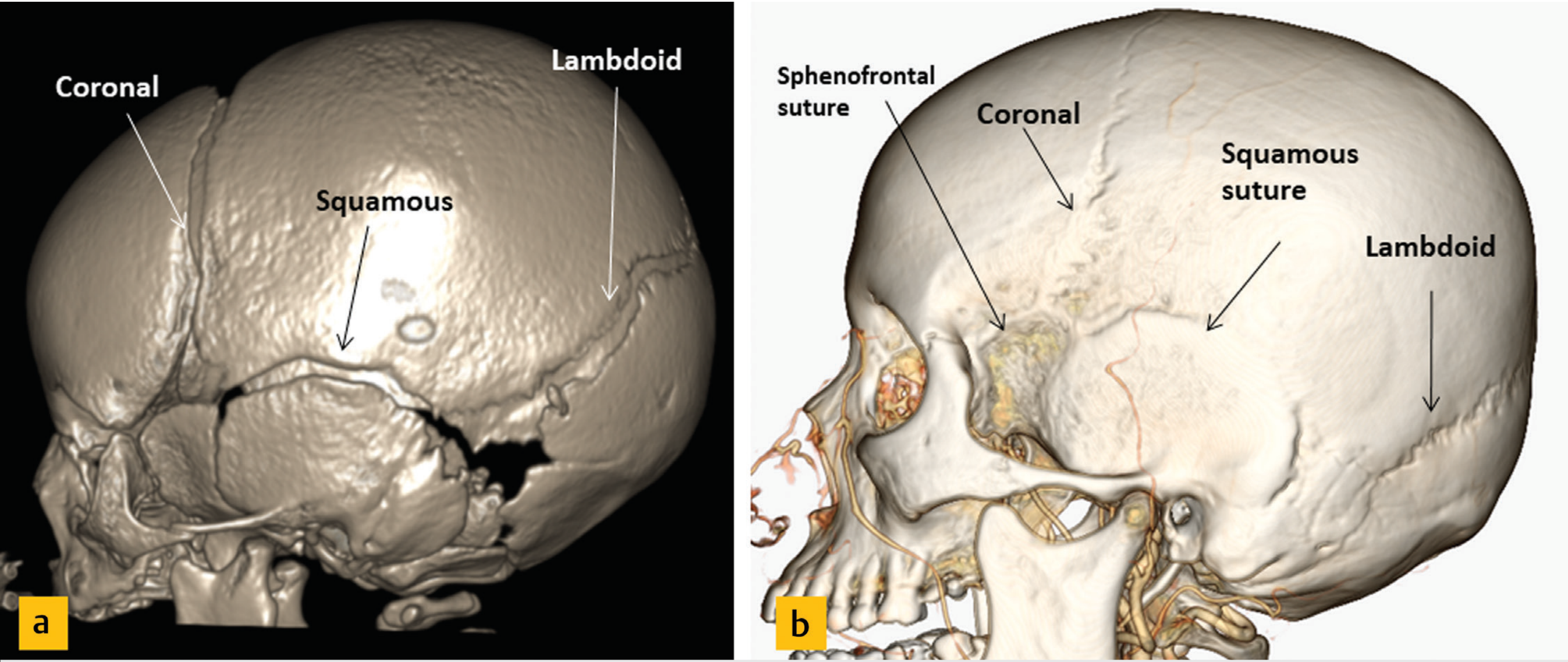
1. SUTURES
- Found only in the skull
- Bones are held together by a thin layer of fibrous tissue
- In babies - sutures are slightly flexible to allow passage through the birth canal
- In adults - sutures become rigid (synarthrosis)
- In old age - sutures may completely fuse into bone = called SYNOSTOSIS
Types of Sutures (BD Chaurasia lists these):
| Type | Description | Example |
|---|---|---|
| Plane/Flat | Edges are straight | Internasal suture |
| Squamous | Edges overlap like fish scales | Temporoparietal suture |
| Serrate | Edges are saw-toothed | Sagittal suture |
| Dentate | Deep interlocking teeth | Coronal suture |
2. SYNDESMOSIS
- Bones joined by a sheet of fibrous tissue called an interosseous membrane OR thick ligaments
- Allows a little movement (amphiarthrosis)
- Easy example: The forearm membrane between radius and ulna; the leg membrane between tibia and fibula
Trick to remember: Syndesmosis = "Syndesmos" (Greek) = Ligament
3. GOMPHOSIS
- A "peg in socket" type joint - unique joint!
- The tooth root (peg) fits into the jaw socket
- Held by periodontal ligament
- Example: Teeth fixed in the maxilla and mandible
- No movement = Synarthrosis
Trick to remember: Gomphosis = "Gomphos" (Greek) = Bolt/Nail (like nailing a peg)
PART 2 - CARTILAGINOUS JOINTS (Amphiarthroses)
Simple meaning: Bones are joined by cartilage. Allow a little bit of movement.
BD Chaurasia divides cartilaginous joints into 2 types:
A. PRIMARY CARTILAGINOUS JOINT = SYNCHONDROSIS
- Bones connected by hyaline cartilage
- No movement (Synarthrosis)
- Can be temporary or permanent
| Type | Example | Fate |
|---|---|---|
| Temporary | Epiphyseal growth plate (between shaft and end of long bone) | Ossifies after growth stops (around 18-25 yrs) |
| Temporary | Y-shaped cartilage of hip bone (joins ilium, ischium, pubis) | Ossifies by age 25 |
| Permanent | First sternocostal joint (1st rib + sternum) | Remains as cartilage throughout life |
| Permanent | Costochondral junctions | Remains as cartilage |
BPT Clinical note: Injury to the epiphyseal growth plate in a growing child can cause shortening or deformity of that limb - always remember this in your paediatric physiotherapy postings!
B. SECONDARY CARTILAGINOUS JOINT = SYMPHYSIS
- Bones connected by a fibrocartilage disc with surrounding ligaments
- Slight movement allowed (Amphiarthrosis)
- Always found in the midline of the body
Examples:
- Pubic symphysis (between two pubic bones)
- Intervertebral discs (between vertebral bodies)
- Manubriosternal joint (between manubrium and body of sternum)
- Symphysis menti (between two halves of the mandible - fuses by 1-2 yrs)
BPT Clinical note: The pubic symphysis separates slightly during pregnancy (due to relaxin hormone) to allow delivery - it can cause pelvic girdle pain in pregnant women, an important physiotherapy condition!
PART 3 - SYNOVIAL JOINTS (Diarthroses)
Simple meaning: The most important joints! Freely moveable. Have a special fluid-filled space between bones.
These are the joints you will treat most as a physiotherapist.
STRUCTURE OF A SYNOVIAL JOINT
BD Chaurasia describes these essential features:
1. ARTICULAR SURFACES
- Bone ends are covered by hyaline cartilage (articular cartilage)
- Smooth, slippery, whitish in colour
- Avascular (no blood supply) - nourished by synovial fluid only
- Thickness: 1-2 mm (small joints) to 5-7 mm (knee/femoropatellar joint)
- Cannot regenerate if damaged - that's why cartilage injuries are serious!
- Exception: Jaw (TMJ) and sternoclavicular joint are covered by fibrocartilage, not hyaline
2. JOINT CAPSULE (Articular Capsule)
Two layers:
- Outer layer = Fibrous membrane - Thick, tough, made of white fibrous tissue; gives strength to the joint
- Inner layer = Synovial membrane - Thin, pink, vascular lining; secretes synovial fluid
3. SYNOVIAL MEMBRANE
- Lines the inside of the capsule (but NOT the articular cartilage)
- Has two cell types:
- Type A synoviocytes - act like macrophages; clean up debris
- Type B synoviocytes - fibroblast-like; produce synovial fluid components
- Can regenerate even in old age (unlike cartilage)
- In rheumatoid arthritis, the synovial membrane becomes inflamed and destroys the joint = Synovitis
4. SYNOVIAL FLUID
- A clear, viscous, straw-coloured fluid (like egg white - "synovia" = egg)
- Contains: Water + Hyaluronic acid + Lubricin + Phospholipids
- Functions:
- Lubrication (reduces friction to nearly zero)
- Nutrition to articular cartilage (diffusion)
- Shock absorption
5. LIGAMENTS
- Strengthen the joint capsule
- Two types:
- Intracapsular ligaments - Inside the capsule (e.g., ACL and PCL of knee)
- Extracapsular ligaments - Outside the capsule (e.g., medial collateral ligament of knee)
6. BURSAE
- Small fluid-filled connective tissue sacs
- Found near joints to reduce friction between tendons/muscles and bone
- Can get inflamed = Bursitis (common in shoulder, knee, hip)
7. ACCESSORY STRUCTURES (in some joints)
- Articular discs / Menisci - Fibrocartilage pads to improve joint fit (e.g., knee menisci, TMJ disc)
- Labrum - Fibrocartilage rim to deepen shallow sockets (e.g., glenoid labrum in shoulder, acetabular labrum in hip)
- Fat pads - Cushion inside the joint (e.g., infrapatellar fat pad in the knee)
TYPES OF SYNOVIAL JOINTS (BD Chaurasia's Classification)
BD Chaurasia describes 7 types of synovial joints (note: he includes "Bicondylar" which other books may not separately list):
| # | Type | Shape | Axes | Movements | Examples |
|---|---|---|---|---|---|
| 1 | Plane (Gliding) | Flat/slightly curved surfaces | Multiaxial (glide) | Gliding only | Intercarpal, intertarsal, acromioclavicular, facet joints of spine |
| 2 | Hinge | Convex cylinder in concave groove | Uniaxial | Flexion + Extension only | Elbow (humeroulnar), ankle, interphalangeal joints |
| 3 | Pivot | Peg in a ring | Uniaxial | Rotation only | Atlantoaxial (C1-C2), proximal radioulnar joint |
| 4 | Bicondylar | Two condyles fit into two sockets | Biaxial (mainly) | Flexion/Extension + slight rotation | Knee joint, temporomandibular joint |
| 5 | Ellipsoid (Condyloid) | Oval convex surface in oval concave socket | Biaxial | Flexion/Extension + Abduction/Adduction | Wrist (radiocarpal), metacarpophalangeal joints |
| 6 | Saddle | Like a horse saddle - both surfaces concavo-convex | Biaxial | Flexion/Extension + Abduction/Adduction + some rotation | 1st carpometacarpal (thumb base), sternoclavicular |
| 7 | Ball & Socket | Ball fits in a cup | Multiaxial | ALL movements including rotation + circumduction | Hip, Shoulder (glenohumeral) |
MOVEMENTS AT SYNOVIAL JOINTS
BD Chaurasia describes these movements:
| Movement | Simple Meaning | Example |
|---|---|---|
| Flexion | Bending - angle decreases | Bending the elbow |
| Extension | Straightening - angle increases | Straightening the elbow |
| Hyperextension | Extension beyond normal | Bending the wrist backward |
| Abduction | Moving AWAY from midline | Raising arm sideways |
| Adduction | Moving TOWARDS midline | Bringing arm back to side |
| Circumduction | Circular movement (cone shape) | Swinging arm in a circle |
| Medial (Internal) Rotation | Rotating inward | Turning palm to face back |
| Lateral (External) Rotation | Rotating outward | Turning palm to face front |
| Supination | Forearm rotation - palm up | Holding a bowl of soup |
| Pronation | Forearm rotation - palm down | Typing on a keyboard |
| Inversion | Sole faces inward | "Pigeon-toed" foot |
| Eversion | Sole faces outward | Foot rolling outward |
| Protraction | Forward movement | Jutting jaw forward |
| Retraction | Backward movement | Pulling jaw back |
| Elevation | Moving upward | Shrugging shoulders |
| Depression | Moving downward | Dropping shoulders down |
| Opposition | Thumb meets little finger | Pinching grip |
FACTORS MAINTAINING JOINT STABILITY
BD Chaurasia lists these in order of importance:
- Shape of articular surfaces - Deep socket = more stable (hip > shoulder). Shallow socket = more mobile but less stable.
- Ligaments - Primary static stabilizers. Prevent excessive movement.
- Muscles and their tendons - Dynamic stabilizers. Muscles around a joint hold it together (e.g., rotator cuff of shoulder).
- Atmospheric pressure - Important in the shoulder joint; helps "suck" the ball into the socket.
- Gravity - Helps in weight-bearing joints like hip and knee.
CLINICAL POINTS FOR BPT (BD Chaurasia Style)
| Condition | Joint | What happens | Physiotherapy relevance |
|---|---|---|---|
| Osteoarthritis (OA) | Knee, hip, fingers | Articular cartilage wears away; bone rubs on bone | Most common condition you will treat |
| Rheumatoid Arthritis (RA) | All synovial joints | Synovial membrane inflames and destroys joint | Look for symmetrical joint swelling |
| Bursitis | Shoulder, knee (prepatellar), hip | Bursa gets inflamed | "Housemaid's knee" = prepatellar bursitis |
| Haemarthrosis | Knee | Blood in joint cavity (e.g., after ACL rupture) | Swollen, painful, warm knee after injury |
| Ankylosis | Any joint | Joint becomes stiff/fused after injury/inflammation | Prevent by early mobilization in physiotherapy |
| Sprain | Ankle, knee | Ligament injury at a joint | Grade I, II, III sprains |
| Dislocation | Shoulder (most common) | Bone comes out of joint socket | Shoulder dislocates anteroinferiorly most often |
| Synovitis | Any synovial joint | Inflamed synovial membrane = excess fluid | Treated with RICE + physiotherapy |
| Craniosynostosis | Skull sutures | Premature fusion of skull sutures in infants | Results in skull deformity |
QUICK REVISION SUMMARY
JOINTS
│
├── FIBROUS (Synarthrosis - No movement)
│ ├── Sutures (Skull) → Plane, Squamous, Serrate, Dentate
│ ├── Syndesmosis (Radius-Ulna, Tibia-Fibula)
│ └── Gomphosis (Teeth in jaw)
│
├── CARTILAGINOUS (Amphiarthrosis - Slight movement)
│ ├── Primary = Synchondrosis (Hyaline cartilage)
│ │ ├── Temporary: Growth plate, Y-cartilage of hip
│ │ └── Permanent: 1st sternocostal, Costochondral
│ └── Secondary = Symphysis (Fibrocartilage)
│ → Pubic symphysis, Intervertebral discs
│
└── SYNOVIAL (Diarthrosis - FREE movement) ← Most important!
├── Plane → Intercarpal, facet joints
├── Hinge → Elbow, ankle, IP joints
├── Pivot → Atlantoaxial, proximal radioulnar
├── Bicondylar → Knee, TMJ
├── Ellipsoid → Wrist, MCP joints
├── Saddle → 1st CMC (thumb), sternoclavicular
└── Ball & Socket → Hip, Shoulder (most mobile!)
MEMORY TRICKS (BD Chaurasia exam tips)
- "FBI Goes Somewhere Cold" = Fibrous, Cartilaginous, Synovial - but with 3 sub-types each
- "Some Happy People Have Beautiful Smiling Eyes" = Suture, Syndesmosis, Gomphosis (Fibrous); Synchondrosis, Symphysis (Cartilaginous); Plane, Hinge, Pivot, Bicondylar, Ellipsoid, Saddle, Ball & Socket (Synovial)
- "Hip and Shoulder are Ball & Socket" = Most mobile, least stable = Most commonly dislocated
- "Knee is Bicondylar" = Not a true hinge - it also allows slight rotation (important for locking mechanism)
- Synovial fluid = EGG WHITE = "Synovia" literally means egg in Greek - clear, viscous
Notes prepared in the context of BD Chaurasia's Human Anatomy classification system, supplemented by Imaging Anatomy Text and Atlas Vol. 3 and THIEME Atlas of General Anatomy
This is a shared conversation. Sign in to Orris to start your own chat.