
Explain me the VITAMIN D , metabolism , formation , functions from HARRISONS internal medicine latest edition . Everything relating to VITAMIN D , the disorders relatinh to it . As i am a PG resident pursuing MD Biochemistry .
Reading File
Now let me search the library database for Vitamin D content in Harrison's.
Finding Sources
Finding Sources
Now let me search specifically in Harrison's for Vitamin D sections:
Finding Sources
Finding Sources
Excellent! Found the key blocks. Let me now read the main Vitamin D sections from Harrison's in parallel:
Reading File
Reading File
The block46 offset 0 started on hypertriglyceridemia - I need to find the exact vitamin D section. Let me search for the correct offset and also read the Chapter 421 content which is the core vitamin D chapter:
Finding Sources
Finding Sources
The main Vitamin D chapter (Ch. 421) is in block46. Let me read the relevant sections starting at the VITAMIN D heading (line 816) and onward:
Reading File
Excellent content! I can see there are images too. Let me continue reading the next sections and fetch the images in parallel:
Reading File
Loading Image
Searching PubMed
"vitamin D" deficiency metabolism
Now let me read the core Vitamin D section more carefully - the synthesis/metabolism/actions/disorders part:
Reading File
I have very rich content now. Let me read the vitamin D disorders section (deficiency, rickets, osteomalacia, hypercalcemia) and fetch the second image:
Reading File
I now have comprehensive content. Let me also quickly read the vitamin D-related hypercalcemia and the rickets/osteomalacia treatment sections:
Reading File
I now have comprehensive content from Harrison's 22E. Let me mark the research tasks as complete and compose the full answer.Here is the comprehensive, Harrison's 22E-based breakdown of Vitamin D for an MD Biochemistry resident:
VITAMIN D - Complete Reference
Harrison's Principles of Internal Medicine, 22nd Edition (2025)
1. NATURE AND CLASSIFICATION
Vitamin D and its metabolites are hormones and hormone precursors rather than true vitamins, because in the proper biologic setting they can be synthesized entirely endogenously. The biologically active form, 1,25-dihydroxyvitamin D [1,25(OH)₂D], is the major steroid hormone involved in regulation of mineral ion homeostasis. Two dietary forms exist:
| Form | Source | Notes |
|---|---|---|
| Vitamin D₂ (ergocalciferol) | Plant sources, some supplements | Shorter t½ due to lower affinity for DBP |
| Vitamin D₃ (cholecalciferol) | Animal sources (fish oil, egg yolk, fortified dairy) | Both forms have equivalent biologic activity |
2. SYNTHESIS AND FORMATION
2a. Cutaneous Synthesis (Endogenous Route)
- UV-B radiation (wavelength 290-320 nm) causes a photochemical cleavage of the B-ring of 7-dehydrocholesterol (provitamin D₃) in the skin, forming pre-vitamin D₃, which undergoes thermal isomerization to vitamin D₃.
- Cutaneous production is decreased by:
- Melanin (competes for UV-B photons)
- High SPF sunblocks
- Aging (efficiency of synthesis declines)
- Northern latitudes / reduced solar exposure
2b. Dietary Absorption
- Vitamin D is absorbed in the small intestine as part of the fat-absorption pathway (chylomicrons).
- Terminal ileal disease impairs enterohepatic circulation of vitamin D metabolites.
3. METABOLISM - THE THREE-STEP ACTIVATION PATHWAY
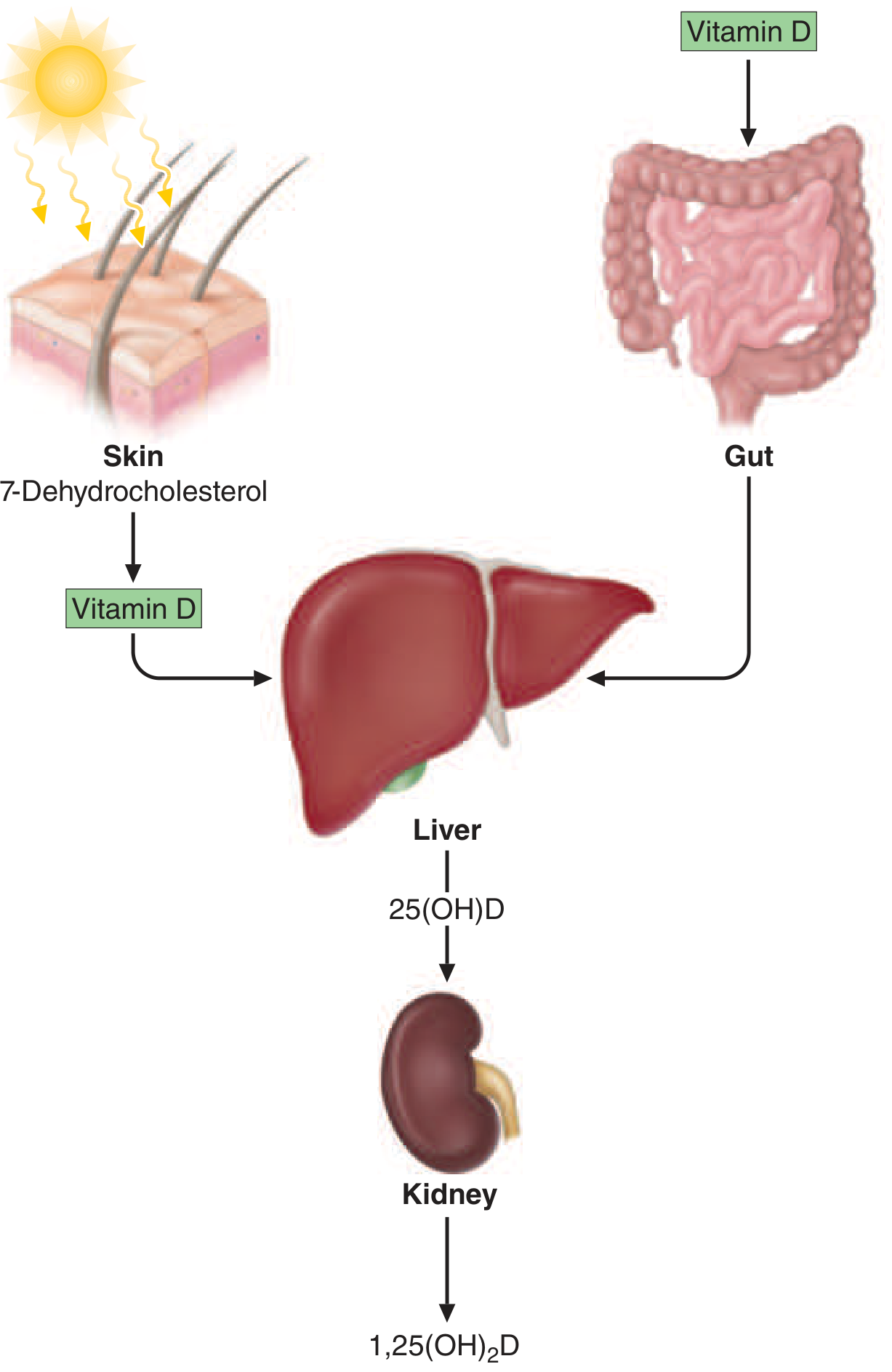
FIGURE 421-4 (Harrison's 22E): Vitamin D is synthesized in the skin in response to ultraviolet radiation and also absorbed from the diet. It is transported to the liver, where it undergoes 25-hydroxylation. This metabolite is the major circulating form of vitamin D. The final step in hormone activation, 1α-hydroxylation, occurs in the kidney.
Step 1 - Transport
After synthesis or intestinal absorption, vitamin D enters the circulation bound to Vitamin D-Binding Protein (DBP), an α-globulin synthesized in the liver.
Step 2 - 25-Hydroxylation in the Liver
| Parameter | Detail |
|---|---|
| Enzyme | Cytochrome P450 oxidase (CYP2R1 is principal enzyme) |
| Location | Liver mitochondria and microsomes |
| Regulation | NOT tightly regulated - substrate-driven |
| Product | 25(OH)D [calcidiol] = major circulating and storage form |
| Measurement | 25(OH)D is the standard clinical marker of vitamin D status |
| Half-life | ~2-3 weeks |
| Protein binding | 88% bound to DBP, ~12% to albumin, only 0.03% free |
Nephrotic syndrome reduces DBP (urinary loss) and greatly shortens the half-life of 25(OH)D.
Step 3 - 1α-Hydroxylation in the Kidney (Rate-Limiting, Regulated Step)
| Parameter | Detail |
|---|---|
| Enzyme | 25-OH-vitamin D-1α-hydroxylase (encoded by CYP27B1 gene) |
| Location | Proximal convoluted tubule cells (primary); also macrophages, keratinocytes, placental trophoblasts |
| Product | 1,25(OH)₂D [calcitriol] = biologically active hormone |
| Inducers | PTH (major), hypophosphatemia, low calcium |
| Repressors | FGF23 (major), calcium, the product itself [1,25(OH)₂D] (negative feedback) |
Key point for Biochemistry: The renal 1α-hydroxylase is subject to tight hormonal regulation (classic endocrine loop), but the extra-renal (macrophage/granuloma) 1α-hydroxylase is induced by interferon-γ and TNF-α and is NOT subject to calcium or phosphorus regulation - this explains hypercalcemia in sarcoidosis.
Step 4 - Inactivation (24-Hydroxylation)
- CYP24A1 (24-hydroxylase) converts both 25(OH)D and 1,25(OH)₂D to 24,25(OH)₂D and 1,24,25(OH)₃D respectively - biologically inactive, water-soluble metabolites excreted in bile.
- This is the primary catabolism pathway and is upregulated by 1,25(OH)₂D itself.
- Loss-of-function mutations in CYP24A1 → impaired catabolism → elevated 1,25(OH)₂D → hypercalciuria and nephrocalcinosis (idiopathic infantile hypercalcemia).
4. MECHANISM OF ACTION - THE VITAMIN D RECEPTOR (VDR)
- 1,25(OH)₂D acts via the nuclear Vitamin D Receptor (VDR), a member of the steroid/thyroid hormone receptor superfamily.
- The VDR forms a heterodimer with the Retinoid X Receptor (RXR).
- This VDR-RXR complex binds to Vitamin D Response Elements (VDREs) in the promoter regions of target genes.
- VDR is expressed in most tissues - intestine, kidney, bone, parathyroid, muscle, immune cells, brain, breast, colon, prostate, keratinocytes.
- Notable exception: Alopecia is seen with mutant VDRs but NOT with vitamin D deficiency - VDR effects on hair follicle are ligand-independent.
5. PHYSIOLOGIC FUNCTIONS
5a. Classical Functions - Mineral Ion Homeostasis
Intestine (primary action):
- Increases transcriptional expression of TRPV6 (apical calcium entry channel), calbindin D-9K (cytosolic calcium ferry), and PMCA1b (basolateral Ca²⁺-ATPase).
- Net effect: markedly increases active calcium absorption.
- Also increases phosphate absorption via NaPi-IIb co-transporter.
Kidney:
- Increases calcium and phosphate reabsorption.
- Suppresses PTH synthesis in parathyroid glands (negative endocrine feedback).
Bone:
- Stimulates osteoblast differentiation and mineralization (provides adequate Ca²⁺ and PO₄³⁻ substrate).
- At pharmacologic doses: stimulates osteoclast activity (bone resorption).
- Regulates normal maturation of the enthesis (bone-tendon attachment site).
Parathyroid:
- 1,25(OH)₂D directly suppresses PTH gene transcription.
- This is the rationale for using calcitriol in secondary hyperparathyroidism of CKD.
5b. Non-Classical (Pleiotropic) Functions
| System | Effect |
|---|---|
| Immune | Modulates innate and adaptive immunity; induces cathelicidins (antimicrobial peptides) |
| Muscle | Required for normal skeletal and cardiac muscle function |
| Cell proliferation | Antiproliferative effect on keratinocytes, breast cancer cells, prostate cancer cells |
| Brain | Possible role in mood regulation; deficiency linked to depression |
| Cardiovascular | Linked to blood pressure regulation (suppresses renin synthesis) |
| Skin | Regulates keratinocyte differentiation |
Harrison's note (22E): Despite compelling observational data, recent placebo-controlled trials did NOT show therapeutic benefit of vitamin D supplementation for cancer prevention, cardiovascular disease, type 2 diabetes, depression, or tuberculosis infection.
6. ASSESSMENT OF VITAMIN D STATUS
| Measure | Details |
|---|---|
| Best marker | Serum 25(OH)D (reflects total body stores) |
| Sufficient | >20 ng/mL (≥50 nmol/L) - adequate for bone health; covers 97.5% of population |
| Some experts recommend | >30 ng/mL for optimal non-skeletal effects |
| Insufficiency | 12-20 ng/mL |
| Deficiency | <12 ng/mL |
| Toxicity marker | 25(OH)D >100 ng/mL |
Note: 1,25(OH)₂D is not the best clinical marker - it may be normal or elevated even in deficiency (secondary hyperparathyroidism drives up renal 1α-hydroxylase).
7. DISORDERS OF VITAMIN D
7a. VITAMIN D DEFICIENCY
Causes (Harrison's Table 421-6):
| Category | Specific Cause |
|---|---|
| Impaired cutaneous production | Lack of sun exposure, dark skin, aging, sunblock use |
| Dietary absence | Exclusively breast-fed infants without supplementation |
| Malabsorption | Short gut syndrome, gastric bypass, fat malabsorption, terminal ileal disease |
| Accelerated inactivation | Barbiturates, phenytoin, rifampin (induce hepatic CYP P450 - increase catabolism) |
| Impaired enterohepatic circulation | Biliary disease |
| Impaired 25-hydroxylation | Severe liver disease, isoniazid, 25-hydroxylase gene mutation |
| Impaired 1α-hydroxylation | Hypoparathyroidism, CKD, CYP27B1 mutation (VDDR Type I), FGF23 excess |
| FGF23 excess | Oncogenic osteomalacia, X-linked/autosomal hypophosphatemic rickets, fibrous dysplasia |
| Target organ resistance | VDR mutation (VDDR Type II), phenytoin |
| Other | Obesity (sequestration in fat), nephrotic syndrome |
Clinical Manifestations:
- Muscle soreness, weakness, bone pain
- Hypocalcemia, hypophosphatemia, secondary hyperparathyroidism
- Rickets in children; Osteomalacia in adults
- Increased risk of fractures
High-risk populations: Elderly, nursing home residents, dark-skinned individuals at northern latitudes, obese individuals, post-gastric bypass patients, neonates of deficient mothers
Treatment:
- Nutritional deficiency: Vitamin D₃ 600-800 IU/day (National Academy of Medicine recommendation); higher doses for treatment of established deficiency
- Glucocorticoid-induced osteoporosis prevention: Calcium 1000-1200 mg/day + Vitamin D 600-800 IU/day + weight-bearing exercise
7b. RICKETS (Pediatric)
Pathophysiology: Failure of normal mineralization of the growth plate cartilage and osteoid.
Causes by mechanism:
- Calcium-deficiency rickets - nutritional vitamin D deficiency (most common globally)
- Phosphopenic rickets - FGF23-mediated phosphate wasting (see hereditary forms below)
- Vitamin D-Dependent Rickets (VDDR):
| Type | Defect | Gene | Key Features |
|---|---|---|---|
| VDDR Type I (Pseudovitamin D-deficiency rickets) | Defective 1α-hydroxylase | CYP27B1 mutation | Low 1,25(OH)₂D, responds to calcitriol |
| VDDR Type II (Hereditary vitamin D-resistant rickets) | VDR mutation | VDR gene | Elevated 1,25(OH)₂D, alopecia, requires massive doses |
Clinical features of rickets:
- Craniotabes (softening of skull bones)
- Rachitic rosary (costochondral junction enlargement)
- Harrison's sulcus (indentation along diaphragm insertion)
- Bowing of weight-bearing long bones
- Widened, frayed, cupped metaphyses on X-ray
- Reduced growth, delayed tooth eruption
- Some cases: symptomatic hypocalcemia and markedly elevated PTH
7c. OSTEOMALACIA (Adult)
Definition: Defective mineralization of existing (mature) bone matrix (osteoid).
Key difference from osteoporosis: Osteoporosis = reduced bone mass but normal mineralization; Osteomalacia = normal or increased bone mass but defective mineralization.
Biochemical profile:
- Low/normal serum calcium
- Low/normal serum phosphate
- Elevated ALP
- Elevated PTH (secondary)
- Low 25(OH)D (in nutritional form)
Radiological features:
- Looser's zones (pseudofractures) - pathognomonic - horizontal lucent bands perpendicular to bone cortex
- Coarsened trabecular pattern
Differential diagnosis includes:
- Nutritional vitamin D deficiency
- Malabsorption
- Defective vitamin D metabolism (drug-induced, renal osteodystrophy)
- Phosphate-wasting syndromes (oncogenic osteomalacia, X-linked hypophosphatemia)
- VDDR types I and II
7d. HEREDITARY HYPOPHOSPHATEMIC RICKETS (FGF23-Mediated)
| Disorder | Gene/Mechanism | Biochemical Features |
|---|---|---|
| X-linked hypophosphatemia (XLH) | PHEX mutation → excess FGF23 | Low PO₄, low/inappropriately normal 1,25(OH)₂D, normal Ca²⁺ |
| Autosomal dominant hypophosphatemic rickets (ADHR) | FGF23 mutation (resistant to cleavage) | Same pattern |
| ARHR | DMP1 mutation | Same pattern |
| Oncogenic osteomalacia (TIO) | Tumor secretes FGF23 | Same - resolves with tumor removal |
FGF23 works by:
- Inhibiting renal tubular phosphate reabsorption (downregulates NaPi-IIa/IIc)
- Suppressing renal 1α-hydroxylase (CYP27B1) → reduces 1,25(OH)₂D
7e. VITAMIN D-RELATED HYPERCALCEMIA
Mechanisms:
1. Vitamin D Intoxication:
- Requires chronic ingestion of >10,000 IU/day in otherwise healthy adults
- Mechanism: elevated 25(OH)D (>100 ng/mL) itself has biologic activity at high levels, causing increased intestinal Ca²⁺ absorption and bone resorption
- Note: 1,25(OH)₂D may NOT be elevated (unlike in granulomatous disease)
- Treatment: stop vitamin D, restrict calcium intake, hydration; glucocorticoids (40-100 mg/day prednisone) are effective; fat stores may sustain toxicity for weeks after cessation
2. Granulomatous Disease (Sarcoidosis, TB, Fungal infections, Lymphoma):
- Macrophages in granulomas express 1α-hydroxylase that is induced by interferon-γ and TNF-α
- This extra-renal hydroxylase lacks normal negative feedback from calcium and 1,25(OH)₂D
- Results in unregulated overproduction of 1,25(OH)₂D → hypercalcemia
- Treatment: glucocorticoids (suppress macrophage 1α-hydroxylase activity); also hydroxychloroquine
7f. DEFECTIVE VITAMIN D METABOLISM - DRUG-INDUCED
| Drug | Mechanism |
|---|---|
| Phenytoin, barbiturates, rifampin | Induce hepatic CYP P450 → accelerate catabolism of vitamin D metabolites → functional deficiency |
| Isoniazid | Impairs 25-hydroxylation |
| Ketoconazole | Inhibits 1α-hydroxylase (CYP27B1) |
| Glucocorticoids | Reduce intestinal calcium absorption; also reduce 25-hydroxylase activity |
CYP3A4 gain-of-function mutations: Autosomal recessive form of rickets due to accelerated oxidation/inactivation of vitamin D metabolites - treated with high-dose calcitriol.
7g. VITAMIN D IN CHRONIC KIDNEY DISEASE (CKD)
- Reduced functioning renal mass → reduced 1α-hydroxylase activity → low 1,25(OH)₂D
- Results in: hypocalcemia → secondary hyperparathyroidism → renal osteodystrophy
- Subtypes of renal osteodystrophy: osteitis fibrosa cystica (high-turnover), adynamic bone disease (low-turnover), mixed
- Treatment: calcitriol or active vitamin D analogues (paricalcitol, doxercalciferol); phosphate binders; dialysis
8. RECOMMENDED DAILY INTAKE
| Age Group | RDA |
|---|---|
| 1-70 years | 600 IU/day |
| >70 years | 800 IU/day |
| Upper tolerable limit (adults) | 4000 IU/day (some conservative guidelines say 2000 IU) |
| Therapeutic replacement | 1000-5000 IU/day or more |
9. VITAMIN D PREPARATIONS USED CLINICALLY
| Preparation | Active Form | Key Use |
|---|---|---|
| Cholecalciferol (D₃) | Requires 2 hydroxylations | Nutritional supplementation |
| Ergocalciferol (D₂) | Requires 2 hydroxylations | Nutritional supplementation |
| Calcidiol [25(OH)D₃] | Requires 1α-hydroxylation only | Liver disease, malabsorption |
| Calcitriol [1,25(OH)₂D₃] | Already active | CKD, VDDR Type I, hypoparathyroidism |
| Paricalcitol, Doxercalciferol | VDR agonists with reduced hypercalcemic risk | Secondary hyperparathyroidism in CKD |
10. SUMMARY FLOWCHART OF VITAMIN D AXIS
7-Dehydrocholesterol (skin)
        ↓ UV-B (290-320 nm)
   Vitamin D₃/D₂
        ↓ Liver CYP2R1 (not tightly regulated)
   25(OH)D [Calcidiol] ← main serum marker, half-life 2-3 wk
        ↓ Kidney CYP27B1 (tightly regulated)
               ↑ PTH, ↑ low PO₄
               ↓ FGF23, ↑ Ca²⁺, ↓ 1,25(OH)₂D
   1,25(OH)₂D [Calcitriol] ← active hormone
        ↓ VDR-RXR heterodimer → VDRE → gene transcription
   ↑ Intestinal Ca²⁺ & PO₄ absorption
   ↑ Renal Ca²⁺ & PO₄ reabsorption
   ↑ Bone mineralization
   ↓ PTH synthesis
        ↓ CYP24A1 (catabolism)
   24,25(OH)₂D / 1,24,25(OH)₃D → excretion (bile)
Sources:
- Harrison's Principles of Internal Medicine, 22nd Edition (2025), Chapter 421: "Vitamin D Deficiency and Disorders of Vitamin D Metabolism" (Block 46, pp. 3313-3320)
- Harrison's 22E, Chapter 37: "Vitamin and Trace Mineral Deficiency and Excess" (Block 37)
Recent Literature Note: Recent placebo-controlled trials (including VITAL, D-HEALTH, USPSTF 2021) have not confirmed benefits of vitamin D supplementation for cancer, cardiovascular disease, type 2 diabetes, or respiratory infections in individuals who are not severely deficient - consistent with Harrison's 22E assessment. The evidence remains strongest for bone health and fall prevention in elderly, vitamin D-deficient populations.This is a complete, research-grade breakdown sourced directly from Harrison's Principles of Internal Medicine, 22nd Edition (2025). Here's a quick orientation to the material above:
What's covered and where to focus for MD Biochemistry:
-
Synthesis & Metabolism (Section 2-3) - The three-step activation (skin → liver → kidney), the enzymes (CYP2R1, CYP27B1, CYP24A1), their genes, regulation by PTH/FGF23/calcium, and protein binding via DBP. This is pure biochemistry and is exam-critical.
-
Mechanism of Action (Section 4) - VDR-RXR heterodimer, VDREs, ligand-independent VDR action in hair follicles - these are molecular biochemistry concepts.
-
Functions (Section 5) - Classical (mineral ion homeostasis) and pleiotropic effects, with the important caveat that RCTs have not confirmed non-skeletal benefits of supplementation.
-
Disorders (Section 7) - All disorders organized by pathomechanism:
- VDDR Type I (CYP27B1 mutation) vs. Type II (VDR mutation)
- FGF23-mediated phosphopenic rickets (XLH, ADHR, oncogenic osteomalacia)
- Drug-induced deficiency (phenytoin, barbiturates, rifampin - via CYP induction)
- CYP24A1 loss-of-function → hypercalcemia
- Sarcoidosis hypercalcemia - the unregulated extra-renal 1α-hydroxylase mechanism
-
Clinical preparations (Section 9) - Helps understand which diseases need active calcitriol vs. standard vitamin D.
The biochemistry exam highlight: CYP27B1 (1α-hydroxylase) is the rate-limiting, tightly regulated step, and CYP2R1 (25-hydroxylase) is substrate-driven and not tightly regulated - this distinction explains why 25(OH)D is the best clinical marker and why 1,25(OH)₂D can be paradoxically elevated even in deficiency.
This is a shared conversation. Sign in to Orris to start your own chat.