
Prostratomegaly
prostatomegaly benign prostatic hyperplasia
benign prostatic hyperplasia enlarged prostate diagram
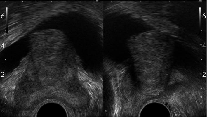
This diagnostic image consists of two grayscale prostate ultrasonography views (likely transverse and sagittal planes) demonstrating Benign Prostatic Hyperplasia (BPH) with significant mid-lobe hypertrophy. The prostate gland appears enlarged with a heterogeneous echotexture. A prominent, solid tissue mass originating from the median lobe is seen protruding superiorly into the anechoic (black) space of the bladder lumen. This intravesical prostatic protrusion (IPP) is a key clinical finding that correlates with bladder outlet obstruction. The image also displays standard sonographic depth markers on the lateral margins and a dark circular acoustic shadow at the bottom center, representing the position of the ultrasound probe. This material is used to teach medical students and urology residents the sonographic evaluation of lower urinary tract symptoms and the anatomical manifestations of prostatic enlargement.
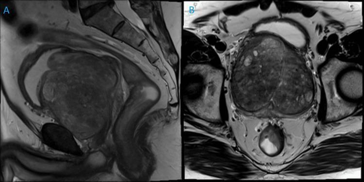
Diagnostic Magnetic Resonance Imaging (MRI) of the male pelvis in sagittal (A) and coronal (B) planes, demonstrating massive benign prostatic hyperplasia (BPH). The prostate gland is severely enlarged, documented at 372g, showing marked expansion of the transition zone. In the sagittal view (A), the prostate is seen inferior to the urinary bladder and anterior to the rectum, occupying a dominant portion of the pelvic cavity and causing superior displacement of the bladder base. The coronal view (B) highlights the heterogeneous signal intensity within the gland, characteristic of stromal and glandular nodules in BPH, without suspicious focal lesions indicative of malignancy. The surrounding pelvic floor musculature and bladder neck are clearly identifiable. This imaging serves as an educational example of extreme prostatic enlargement and its anatomical impact on adjacent pelvic structures, relevant to urological surgical planning such as Holmium Laser Enucleation of the Prostate (HoLEP).
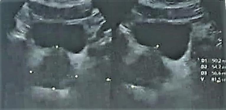
This diagnostic image consists of two side-by-side grayscale ultrasound frames of the male pelvis, specifically demonstrating the prostate gland in a patient with benign prostatic hyperplasia (BPH). The prostate appears enlarged with a heterogeneous echogenicity pattern and a somewhat nodular texture, characteristic of hyperplastic transition zone growth. Caliper markers (small white dots) are positioned across the gland to measure three-dimensional parameters (D1, D2, and D3) to calculate the total prostatic volume (V), shown as numerical values on the right-hand side. The bladder, appearing as a dark, hypoechoic space, is visible superior to the prostate. The image illustrates the typical transrectal ultrasound (TRUS) appearance of BPH, where the transition zone expands and compresses the peripheral zone, often creating a 'surgical capsule' boundary. This visual is used for clinical assessment of prostate volume, which is a key factor in staging the severity of BPH and determining the necessity for medical or surgical intervention.
Prostratomegaly (Prostatomegaly / BPH)
Anatomy - Prostate Zones
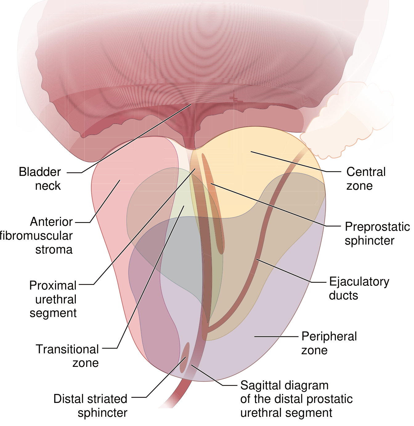
- Peripheral zone - largest, most prostate cancers arise here
- Central zone - surrounds ejaculatory ducts
- Transitional zone - site of BPH origin
- Anterior fibromuscular stroma
Epidemiology
- BPH histology present in ~50% of men >50 years and ~90% of men in their 80s
- Of those with histologic BPH, approximately 50% will develop notable lower urinary tract symptoms (LUTS)
- Prevalence increases linearly between ages 40-80 years
- A major global healthcare challenge with worldwide longevity increasing
Pathogenesis
- Dihydrotestosterone (DHT) is the principal mediator - 10x more potent than testosterone
- DHT is synthesized from circulating testosterone in the prostate by 5α-reductase type 2
- DHT binds nuclear androgen receptors, driving expression of genes that support growth and survival of prostatic epithelial and stromal cells
- DHT-induced growth factors increase stromal cell proliferation and decrease epithelial cell apoptosis
- With aging, testosterone levels decline but estrogen levels remain unchanged or rise (via peripheral conversion); estrogens act synergistically with DHT - both cell types express estrogen receptors
- BPH does not occur in men castrated before puberty or in men with genetic conditions blocking androgen activity
Morphology / Pathology
- Prostate weight increases 3- to 5-fold (60-100 g or greater) in BPH
- Involves the transition zone, compressing the urethra to a slit-like orifice
- Cross-section: hyperplastic nodules of variable color/consistency
- Nodules may be solid or cystic (dilated glands)
- Microscopically: variable proportions of proliferating glandular elements and fibromuscular stroma
- Hyperplastic glands lined by tall columnar epithelial cells + peripheral flattened basal cells (key distinction from carcinoma, which lacks basal cells)
- Glandular lumina may contain corpora amylacea (laminated proteinaceous secretory material)
Clinical Features / LUTS
- Mechanical - physical obstruction by enlarged gland
- Dynamic - smooth muscle-mediated contraction of the stroma
| Symptom Type | Examples |
|---|---|
| Obstructive (voiding) | Weak stream, hesitancy, straining, incomplete emptying, overflow dribbling |
| Irritative (storage) | Frequency, urgency, nocturia, dysuria |
- Residual urine acting as culture medium → recurrent UTIs, cystitis, pyelonephritis
- Bladder hypertrophy and detrusor dysfunction
- Acute urinary retention - painful bladder distension
- Hydronephrosis if obstruction is complete and untreated
Ultrasound Appearance

Management
Medical (first-line)
| Drug Class | Mechanism | Examples |
|---|---|---|
| α1-adrenergic blockers | Relax prostatic smooth muscle by blocking α1 receptors | Tamsulosin, Alfuzosin, Doxazosin |
| 5α-reductase inhibitors | Block DHT synthesis from testosterone; shrink the gland | Finasteride, Dutasteride |
| Combination therapy | Both classes together | More effective for large glands |
| Antimuscarinics / β3 agonists | For storage/overactive bladder component | Solifenacin, Mirabegron |
Surgical (for medically refractory disease)
- TURP (Transurethral Resection of the Prostate) - gold standard
- HoLEP (Holmium Laser Enucleation of the Prostate) - preferred for very large glands (e.g., >372 g)
- High-intensity focused ultrasound (HIFU)
- Laser therapy (PVP)
- Transurethral electrovaporization
- Radiofrequency ablation
- Open prostatectomy (for very large glands with bladder complications)
Key Distinctions: BPH vs Prostate Cancer
| Feature | BPH | Prostate Carcinoma |
|---|---|---|
| Zone affected | Transitional zone | Peripheral zone (mostly) |
| Basal cells | Present | Absent |
| PSA | Mildly elevated | Can be markedly elevated |
| DRE | Smooth, rubbery enlargement | Hard, irregular nodule |
| Risk of malignancy | Does not predispose | Independent entity |
- Robbins & Kumar Basic Pathology, pp. 659-660
- Goldman-Cecil Medicine, pp. 1316-1318
- Campbell-Walsh-Wein Urology (multiple chapters)
- Bailey & Love's Short Practice of Surgery, 28th Ed.
Pituitary apoplexy
pituitary apoplexy MRI hemorrhage tumor
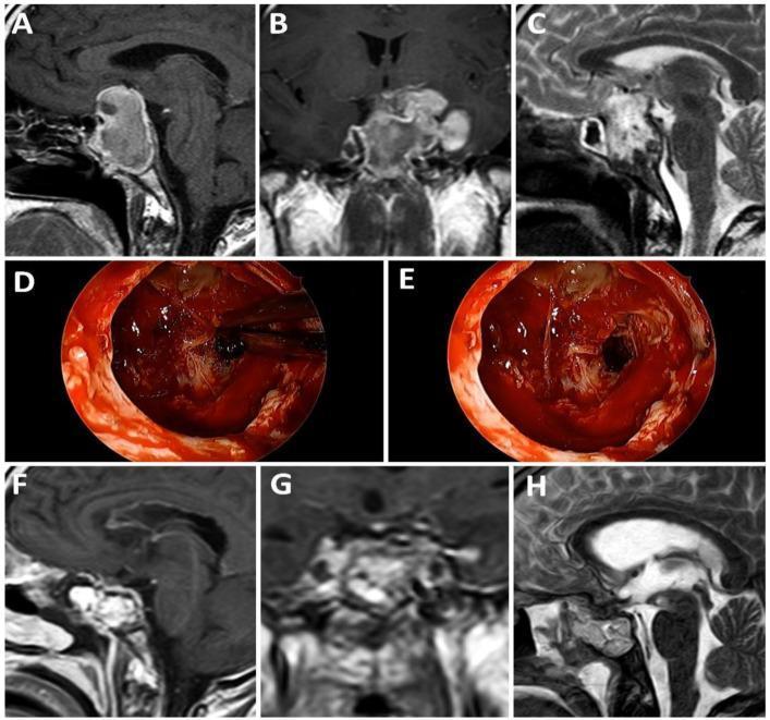
This composite figure illustrates the surgical management of a giant pituitary macroadenoma with apoplexy. Panels A-C display preoperative neuroimaging: (A) T1-weighted sagittal MRI shows a large sellar mass with suprasellar extension and high-signal intensity areas suggesting hemorrhage; (B) T1-weighted post-contrast coronal MRI reveals heterogeneous enhancement and lateral extension toward the cavernous sinuses; (C) T2-weighted sagittal MRI highlights the mass effect on the optic chiasm and third ventricle. Panels D and E are intraoperative endoscopic photographs demonstrating the surgical resection of the hemorrhagic and necrotic tumor tissue via a transsphenoidal approach. Panels F-H show the postoperative MRI results: (F) T1-weighted sagittal, (G) T1-weighted coronal, and (H) T2-weighted sagittal views confirm significant tumor debulking, decompression of the optic chiasm, and restoration of the suprasellar cistern. This case demonstrates the diagnostic findings of pituitary apoplexy and the efficacy of surgical intervention in restoring anatomical relationships in the sellar region.

This composite figure presents a clinical case of pituitary apoplexy through multimodality imaging and histopathology. Panel (a) is an axial non-contrast CT scan showing a hyperdense mass in the sellar/suprasellar region, characteristic of acute intratumoral hemorrhage. Panels (b) and (c) are coronal T1-weighted MRI scans; (b) shows a large, pre-contrast hyperintense pituitary tumor extending into the suprasellar cistern, while (c) demonstrates a lack of internal enhancement following gadolinium administration, indicative of tumor infarction or necrosis. Panel (d) is a coronal T2-weighted MRI revealing a heterogeneous tumor with a prominent low-signal-intensity (hypointense) peripheral rim, a classic sign of hemosiderin deposition or acute-on-chronic bleeding in pituitary apoplexy. Panel (e) provides a histopathological correlation with hematoxylin and eosin (H&E) staining, showing a clear demarcation between viable papillary-patterned pituitary adenoma tissue (left) and areas of extensive hemorrhage and necrosis (right). The sequence illustrates the diagnostic hallmark of sudden hemorrhagic transformation of a macroadenoma.
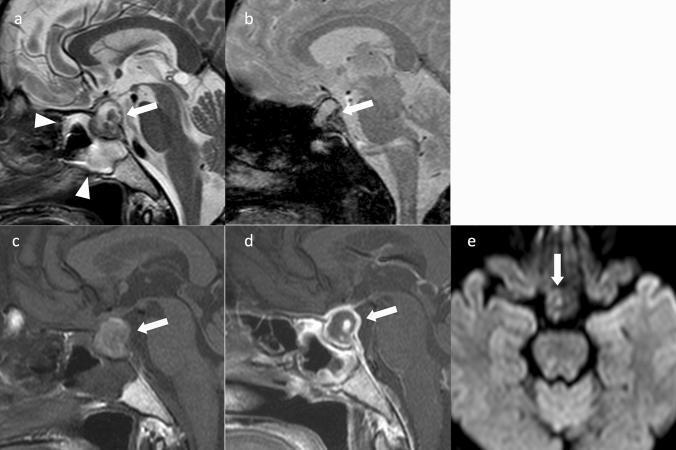
This composite of brain MRI sequences illustrates pituitary apoplexy in a female patient. Panel (a), a sagittal T2-weighted image, shows a heterogeneous mass in the sella turcica (arrow) with mixed hyperintense and hypointense signals. Prominent mucosal thickening is visible in the adjacent sphenoid sinus (arrowheads), a common sign of acute apoplexy. Panel (b), a sagittal T2*-weighted image, reveals a significant hypointense region within the mass (arrow), highly suggestive of acute hemorrhage due to magnetic susceptibility effects. Panel (c) shows a mildly hyperintense signal on sagittal non-contrast T1-weighted imaging, while panel (d) demonstrates peripheral ring enhancement of the sellar mass (arrow) on contrast-enhanced T1-weighted imaging, indicating a central area of necrosis or hemorrhage without vascular supply. Panel (e), an axial diffusion-weighted image (DWI), shows the mass as iso-intense (arrow). These diagnostic findings are classic for pituitary neuroendocrine tumor (PitNET) apoplexy, characterized by sudden intratumoral hemorrhage and infarction.
"pituitary apoplexy" management
PMID: 39033812
Pituitary Apoplexy
Definition
Imaging - Multimodality Appearance
Predisposing Conditions
| Category | Examples |
|---|---|
| Pituitary tumor | Nonfunctioning macroadenoma (most common), Cushing disease, acromegaly |
| Vascular | Hypertension / hypotension (~25% have inadequately treated hypertension) |
| Surgery | Cardiac surgery (cardiopulmonary bypass), major orthopedic procedures |
| Drugs | Cabergoline, bromocriptine, anticoagulants, estrogens |
| Endocrine stimulation tests | TRH stimulation test, insulin tolerance test |
| Head trauma | - |
| Pregnancy | Especially postpartum hemorrhage with hypovolemia (Sheehan syndrome) |
| Infections | Dengue fever, hypophysitis |
| Other | Radiation therapy, poorly controlled diabetes, sickle cell anemia |
Pathophysiology
- Raised intrasellar pressure - compresses pituitary tissue and cavernous sinus structures
- Cranial nerve palsies - compression of CN III, IV, VI (in cavernous sinus) and V1/V2 - causes diplopia (occurs in 40-100%)
- Visual field defects - upward expansion compresses the optic chiasm - bitemporal hemianopsia
- Meningeal irritation - extravasation of blood into subarachnoid space
- Acute hypopituitarism - destruction of glandular tissue - most critically, acute adrenocortical insufficiency
Clinical Features
| Symptom | Approximate Incidence |
|---|---|
| Headache (retro-orbital, unilateral, or bilateral temporal) | 95% |
| Vomiting | 70% |
| Visual field defect (bitemporal hemianopsia) | 65% |
| Decreased visual acuity | 50% |
| Diplopia (CN III, IV, V, VI palsies) | 40-100% |
| Hypotension / cardiovascular collapse | 95% |
| Meningismus | Rare |
| Hemiplegia | Rare |
- Often the first presentation of a previously undiagnosed pituitary tumor
- Can mimic subarachnoid hemorrhage (sudden severe "thunderclap" headache)
- May cause the "triple response" of pituitary AVP disruption: transient diabetes insipidus → SIADH/hyponatraemia → return to DI (as AVP stores are exhausted)
Differential Diagnosis
- Subarachnoid hemorrhage
- Bacterial meningitis
- Cavernous sinus thrombosis
- Migraine
- Rathke cyst hemorrhage
- Hyperemesis gravidarum (if pregnant)
Diagnosis
Imaging (cornerstone)
- MRI (T2-weighted) - preferred - most sensitive; shows hemorrhage, infarction, mass effect on chiasm and cavernous sinuses
- CT scan - useful when MRI is unavailable; hyperdense sellar mass on non-contrast CT = acute hemorrhage
Bloods (urgent)
- Electrolytes, renal function, LFTs, FBC + platelets, prothrombin time
- Random cortisol, free T4, TSH, prolactin (>80% of patients have endocrine dysfunction)
- LH, FSH, testosterone/estradiol, GH, IGF-1 (less urgent)
CSF
- Not usually required if diagnosis is certain
- If performed: red cells, elevated protein, xanthochromia (if blood has entered CSF)
Management
1. Immediate Stabilization
- IV 0.9% NaCl boluses to maintain hemodynamic stability
- High-dose parenteral glucocorticoids: Hydrocortisone 100 mg IV every 8 hours (covers adrenal crisis - the most immediately life-threatening complication)
- Monitor serum and urine electrolytes/osmolalities closely for the triple AVP response
2. Surgical Decompression (Transsphenoidal)
- Progressive or severe visual deterioration
- Worsening neurological impairment (falling consciousness)
- Lack of spontaneous improvement on conservative management
- Larger tumors (more prone to hemorrhage; rapid growth increases apoplexy risk)
3. Conservative Management
- Appropriate for mild/moderate symptoms, stable vision
- High-dose steroids alone with close monitoring
- Evidence: A 2024 meta-analysis of 908 patients found no statistically significant difference between surgical and conservative management for recovery of ophthalmoplegia, visual fields, visual acuity, hypopituitarism, or tumor recurrence - though more severe cases typically undergo surgery
4. Follow-up
- 80% of patients have residual hypopituitarism after apoplexy (with or without surgery)
- Repeat endocrine testing 1 month post-discharge to confirm persistent deficits
- Lifelong hormone replacement if endocrine deficit persists
- MRI pituitary at 3-6 month intervals until anatomy stable, then yearly for 5 years
- Risk of recurrent apoplexy - ongoing surveillance essential
Key Points Summary
- Pituitary apoplexy = hemorrhagic infarction of the pituitary, usually into a macroadenoma
- Classic triad: sudden severe headache + visual disturbance + altered consciousness
- Mimics SAH - always consider in sudden thunderclap headache
- Immediate threat to life is acute adrenal insufficiency - give hydrocortisone empirically
- Cornerstone imaging: urgent MRI T2
- Surgery if visual loss is progressive; conservative management otherwise equally effective
- 80% are left with hypopituitarism - lifelong endocrine follow-up mandatory
- Goldman-Cecil Medicine, pp. 2401-2402 (Table 205-4, Table 205-5)
- Scott-Brown's Otorhinolaryngology Head & Neck Surgery, pp. 960, 975
- Kanski's Clinical Ophthalmology, 10th Ed.
- Marin-Castañeda et al. (2024) - Meta-analysis of 908 patients, World Neurosurgery - surgical vs conservative management: comparable outcomes
How does the triple AVP response present clinically and what's the management of each phase?