
Azelaic acid use
"azelaic acid"[MeSH Terms] AND (acne OR rosacea OR hyperpigmentation)
azelaic acid acne rosacea treatment topical
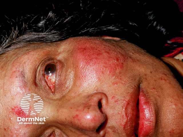
Clinical photography of the face, frontal close-up, high-resolution. Anatomical region: central facial skin including cheeks, nasal dorsum, perioral area, and periocular tissues. Primary subject: papulopustular rosacea with ocular involvement. Appearance: diffuse erythema of the central face with a constellation of inflammatory papules and pustules scattered across the malar region and nose; mild telangiectasia along the cheeks; lips and surrounding skin show no ulceration. Periocular redness and lid-margin irritation are present, compatible with ocular rosacea; eyelids appear mildly edematous without obvious keratitis in this image. Skin texture shows slight roughness and residual flaking. The overall pattern is chronic inflammatory facial dermatitis centered on the midface, rather than acne conglobata or lupus. Diagnostic significance: classic papulopustular rosacea with probable ocular involvement; warrants ophthalmology referral to assess ocular surface, blepharitis, and potential corneal involvement; informs systemic versus topical therapy decisions. Differential considerations: acne vulgaris (less likely distribution), steroid-induced rosacea, seborrheic dermatitis, lupus erythematosus mimicking rosacea, contact dermatitis. Clinical relevance: important for educational purposes, differential diagnosis of facial erythema, guiding treatment planning (topical metronidazole/azelaic acid, doxycycline in adequate dosing, eyelid hygiene) and patient counseling on triggers and chronicity. This image illustrates rosacea heterogeneity and emphasizes the need for multidisciplinary management. Guides comprehensive patient care.
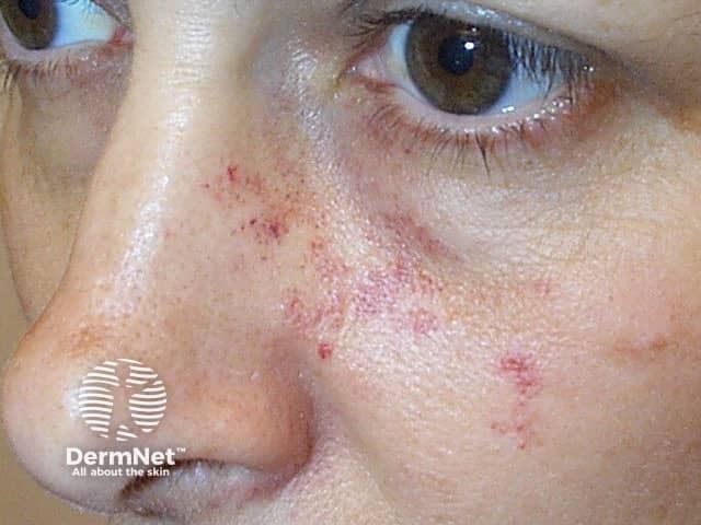
This clinical photograph captures the central facial region, focusing on the nose and malar areas. Modality and technique: noninvasive, standard color dermatologic photography with natural illumination; no dyes or contrast agents used. Anatomical location includes the nasal dorsum, alar rims, and adjacent cheeks in an in vivo adult patient. Visual features: numerous small erythematous papules scattered along the nasal bridge and malar areas, occasional focal crusting, mild surface desquamation, and subtle background erythema; skin texture appears slightly rough with enlarged pores in the nasal region. There are no confluent plaques or obvious pustules of large size evident in this field. Pathology suggested: inflammatory papules with superficial vascular dilation; differential includes papulopustular acne, rosacea, folliculitis, and contact dermatitis. Diagnostic significance: indicates an inflammatory facial dermatosis; may reflect early rosacea or acneiform eruption; the absence of comedones would argue against classic acne. Clinical correlation: patient history (onset, triggers, facial flushing, ocular symptoms) is essential; treatment implications include topical therapies (azelaic acid, metronidazole) or systemic antibiotics for rosacea or acne, with monitoring for response and adverse effects. Potential clinical use: educational reference for dermatology training, differential diagnosis discussion, telemedicine skin assessments, and therapy tracking. Useful for clinical education, case discussions, and image-guided assessment.
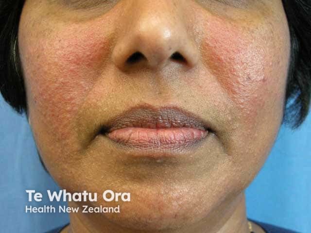
Clinical photography of the face in frontal orientation. The image depicts symmetrical erythematous papules on the malar cheeks with diffuse background erythema, mild textural roughness, and slight scaling. No pustules, comedones, or crusts are conspicuous in this field. The overall appearance is consistent with papulopustular rosacea localized to the central face, particularly the malar regions, with preserved perioral and nasal skin. The lips appear normal aside from mild dryness. Lighting is neutral and color-accurate, allowing reliable assessment of redness intensity and lesion distribution. This photograph documents active inflammatory facial dermatitis and can be used to grade severity, monitor therapeutic response, and educate patients about rosacea triggers. Clinically, the malar predominance helps distinguish rosacea from acne vulgaris, seborrheic dermatitis, or contact dermatitis; absence of comedones further supports rosacea over acne. In practice, management implications include initiation of anti-inflammatory topical therapy (e.g., metronidazole, azelaic acid) or systemic therapy (doxycycline) and strict sun protection. The image supports research in rosacea phenotyping and outcome measures for treatment trials. It is suitable for dermatology training, teledermatology assessments, and patient counseling about disease recognition and prognosis. Additional notes include documenting any ocular symptoms, eyelid involvement, or facial edema to guide comprehensive care for multidisciplinary management planning.
Azelaic Acid - Clinical Overview
What It Is
- 20% cream (Azelex, Skinoren) - Rx
- 15% gel (Finacea) - Rx
- 10-14% formulations - over-the-counter
Indications
| Condition | Notes |
|---|---|
| Acne vulgaris (mild-moderate) | First or second-line; effective for both comedonal and inflammatory acne |
| Rosacea (papulopustular) | SOR: A - strong evidence alongside topical metronidazole |
| Melasma | Inhibits excess pigment production |
| Post-inflammatory hyperpigmentation (PIH) | Targets hyperactive melanocytes selectively |
Mechanisms of Action
-
Antimicrobial - Activity against Cutibacterium (Propionibacterium) acnes and Staphylococcus epidermidis, reducing the bacterial burden that drives acne and rosacea inflammation.
-
Anti-inflammatory - Inhibits free radical oxygen production by neutrophils, reducing oxidative tissue damage at inflammatory sites. Also has a direct anti-inflammatory effect independent of antimicrobial action.
-
Anticomedogenic / normalizes keratinization - Modifies abnormal follicular keratinization, helping prevent comedone formation.
-
Skin-lightening (anti-melanogenic) - Acts as a competitive inhibitor of tyrosinase, the rate-limiting enzyme in melanin synthesis. It also has selective cytotoxic and antiproliferative effects on hyperactive melanocytes with little effect on normal melanocytes - a key advantage over hydroquinone.
-
Enzyme inhibition - Acts as a reversible inhibitor of cytochrome P450 reductase and 5α-reductase in microsomes, and inhibits certain enzymes in the respiratory chain.
Formulation Use by Indication
- Acne: 20% cream or 15% gel. In a randomized controlled study, azelaic acid 15% gel showed non-inferior efficacy and greater tolerability than adapalene 0.1% gel. Often used as a second-line alternative to retinoids or benzoyl peroxide.
- Rosacea: 15% gel (Finacea) is the most studied. The [Textbook of Family Medicine](evidence: van Zuuren et al., 2011) gives this an SOR: A rating alongside metronidazole. Recommended in mild disease; for moderate-severe, combined with low-dose doxycycline.
- Melasma/PIH: 15-20% formulations; a 2024 RCT showed combining with 755-nm picosecond laser improved outcomes vs. azelaic acid alone (PMID: 38656631).
Side Effects
- Mild to moderate irritant contact dermatitis: dryness, peeling, erythema, stinging/burning at application site
- These effects typically subside after 2-4 weeks of continued use
- Rare: depigmentation (vitiligo-like changes) - theoretical, given its melanocyte effects, but uncommon in clinical practice
Pregnancy Safety
Place in Therapy
- Comedonal acne: second-line after topical retinoids
- Papular/pustular acne: second-line alternative to benzoyl peroxide/antimicrobial combinations
- Particularly useful in pregnancy and in patients who cannot tolerate retinoids or benzoyl peroxide
- First-line topical option alongside metronidazole (15% gel preferred)
- Mild disease: topical azelaic acid or metronidazole alone
- Moderate-severe: combine with low-dose doxycycline (Goldman-Cecil Medicine)
- Listed alongside hydroquinone (4% HQ) as first-line topical therapy
- Preferred when hydroquinone is contraindicated (pregnancy, long-term use concerns)
Comparison with Other Agents
| Feature | Azelaic Acid | Hydroquinone | Retinoids | Benzoyl Peroxide |
|---|---|---|---|---|
| Pregnancy safe | Yes (B) | Avoid (C) | No (X) | Category C |
| Antibiotic resistance risk | None | N/A | None | None |
| Anti-inflammatory | Yes | No | Mild | Yes |
| Comedolytic | Mild | No | Strong | Mild |
| Depigmentation | Selective | Broad | Indirect | No |
Recent Evidence (2023-2025)
- A 2023 systematic review (PMID: 37550898) confirmed efficacy of azelaic acid across acne, rosacea, melasma, and skin aging, supporting its broad therapeutic profile.
- A 2025 systematic review and meta-analysis (PMID: 40246142) on topical rosacea therapies includes azelaic acid among effective options alongside metronidazole, ivermectin, and brimonidine.
Sources: Dermatology 2-Volume Set 5e (Bolognia), Fitzpatrick's Dermatology Vol 1-2, Textbook of Family Medicine 9e, Goldman-Cecil Medicine; PMID 37550898, 40246142