
Management of Acute pancreatitis ? With amylase value of 1297 bilirubin of 5
"acute pancreatitis"[MeSH Terms] AND management
acute pancreatitis management guidelines 2024 2025 gallstone pancreatitis elevated bilirubin
acute pancreatitis CT scan necrotizing pancreatitis
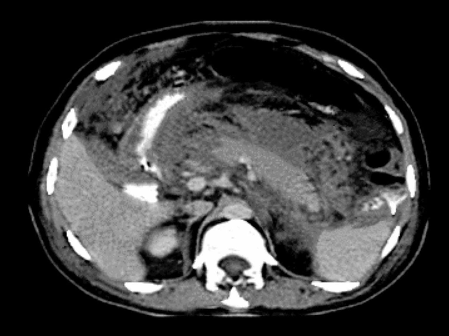
This axial contrast-enhanced computed tomography (CT) scan of the abdomen demonstrates findings consistent with acute necrotizing pancreatitis. The primary diagnostic focus is the peripancreatic region, which shows extensive ill-defined fluid collections and inflammatory changes. These peripancreatic necrotic areas exhibit heterogeneous density, representing a complex mixture of liquid and non-liquid components (solid necrotic fat or debris). In contrast, the pancreatic parenchyma itself maintains relatively uniform enhancement, suggesting preserved perfusion despite the surrounding inflammation. Key anatomical landmarks visible include the vertebral body, portions of the liver, and the stomach displaced by the inflammatory process. This imaging is clinically significant for staging the severity of acute pancreatitis according to the revised Atlanta classification, specifically highlighting the distinction between interstitial edematous pancreatitis and necrotizing pancreatitis based on the presence of extra-pancreatic necrosis.
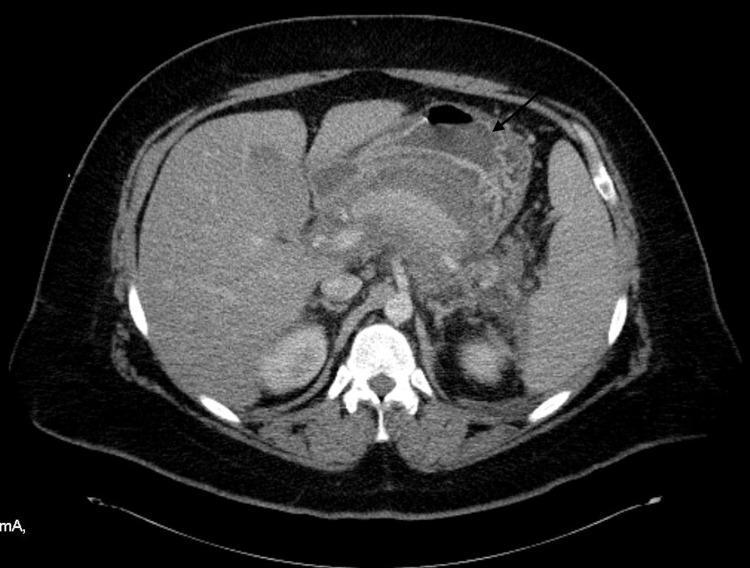
This diagnostic image is an axial contrast-enhanced CT scan of the abdomen demonstrating severe acute necrotizing pancreatitis. The primary finding, indicated by a black arrow, is a distinct area of non-enhancement in the pancreatic tail, which is pathognomonic for pancreatic necrosis (loss of tissue viability/perfusion). The pancreas appears edematous and enlarged. Surrounding the pancreatic parenchyma is an extensive, hyperdense peripancreatic fluid collection; the increased density (high Hounsfield units) of the fluid relative to simple serous fluid is highly suggestive of hemorrhagic pancreatitis. Associated secondary findings include splenomegaly (enlarged spleen) and evidence of splenic vein thrombosis (SVT). This visual case illustrates critical complications of hypertriglyceridemia-induced pancreatitis, specifically the transition from interstitial edematous inflammation to necrotizing and hemorrhagic involvement. The image serves as an educational tool for identifying radiological signs of pancreatic parenchymal death and distinguishing simple peripancreatic fluid from hemorrhagic content in an acute clinical setting.
Management of Acute Pancreatitis
Your Clinical Scenario at a Glance
- Serum Amylase: 1,297 U/L - more than 3x upper limit of normal (ULN ~100-110 U/L), confirming pancreatitis
- Bilirubin: 5 mg/dL - significantly elevated, strongly suggesting gallstone (biliary) pancreatitis with possible common bile duct (CBD) obstruction or concurrent cholangitis
Step 1: Confirm Diagnosis
- Characteristic abdominal pain (epigastric, radiating to back)
- Serum amylase or lipase >3x ULN - your amylase of 1,297 qualifies
- Characteristic findings on CT/MRI
Lipase is more sensitive and specific than amylase; amylase may normalize faster.
Step 2: Assess Severity
Revised Atlanta Classification
| Severity | Features |
|---|---|
| Mild | No organ failure, no local complications. Self-limited (3-7 days) |
| Moderately Severe | Transient organ failure (<48 h) OR local complications |
| Severe | Persistent organ failure (>48 h), one or more organs |
Severity Scoring Tools (at 24h)
- B - BUN >25 mg/dL
- I - Impaired mental status (GCS <15)
- S - SIRS (≥2 of: temp <36° or >38°C; HR >90; RR >20; WBC >12,000 or <4,000)
- A - Age >60 years
- P - Pleural effusion on imaging
- Temp <36°C or >38°C
- HR >90/min
- RR >20/min or PCO2 <32 mmHg
- WBC >12,000 or <4,000/μL or >10% bands
- Hematocrit >44% (hemoconcentration) - risk of necrosis
- Admission BUN >20 mg/dL - rising BUN correlates with mortality
- CRP >100 mg/L (after 48h) - predicts severe disease
- APACHE II score ≥8
Step 3: Initial Management (All Patients)
A. Admission & Monitoring
- Admit to hospital - all patients with acute pancreatitis
- Severe/predicted severe: HDU/ICU with invasive monitoring (vitals, CVP, urine output, blood gases) - Bailey & Love 28e, Table 72.4
- Monitor: FBC, LFTs, urea/creatinine, calcium, glucose, serum lipase, ABG
- A rising BUN during hospitalization = inadequate hydration + higher mortality risk
B. Fluid Resuscitation - MOST IMPORTANT EARLY INTERVENTION
- Balanced crystalloid (Lactated Ringer's preferred over normal saline) - LR reduces the systemic inflammatory response - Schwartz's Surgery 11e, p. 1471
- Rate: 250-500 mL/hour in first 12-24 hours (goal-directed)
- Less aggressive in cardiac, renal, or elderly patients
- Target: normalized BP, HR, urine output >0.5 mL/kg/hr
- Monitor response: A decrease in hematocrit and BUN in first 12-24h confirms adequate resuscitation
Recent ACG 2024 guidelines favor moderate (not over-aggressive) resuscitation in mild cases; vigorous resuscitation (5-10 mL/kg/hr) may be appropriate in severe cases with hemoconcentration.
C. Analgesia
- IV opioids (morphine or hydromorphone) are effective and safe - the old concern about morphine causing sphincter of Oddi spasm is not clinically significant
- NSAIDs (rectal indomethacin) useful adjunct if no contraindications
- Keep patient as comfortable as possible
D. Nil by Mouth vs. Early Oral Feeding
- Mild pancreatitis: Start oral intake (low-fat solid diet) as soon as patient is hungry, pain is improving, and nausea/vomiting resolved - Harrison's 22e
- Do NOT routinely keep NPO - the old "pancreatic rest" concept is abandoned
- Moderate-severe: Enteral nutrition (NG or NJ tube) if patient cannot eat within 72 hours - Schwartz's 11e
- Enteral > Parenteral (TPN): Maintains gut barrier, reduces bacterial translocation, fewer complications, less expensive
E. No Prophylactic Antibiotics (in most cases)
- Prophylactic antibiotics are NOT recommended for predicted severe or necrotizing pancreatitis - Harrison's 22e; Sleisenger & Fordtran's
- When to give antibiotics:
- Confirmed infected pancreatic necrosis (fever + leukocytosis + CT gas in necrotic area)
- Concurrent cholangitis (see ERCP section below)
- Respiratory or urinary infection
- Agents that achieve highest pancreatic tissue concentrations: imipenem, fluoroquinolones (ciprofloxacin + metronidazole) - Bailey & Love 28e
- Duration if used: do not exceed 14 days; guided by cultures
Step 4: ERCP - Critical Decision in Your Case (Bilirubin = 5 mg/dL)
"Urgent ERCP within 24 to 48 hours is indicated if the patient has cholangitis, total serum bilirubin >5 mg/dL, clinical deterioration, or a stone documented in the CBD on imaging." - Sleisenger & Fordtran's GI and Liver Disease
ERCP Indications (Urgent - within 24-72 hours):
- Acute cholangitis (Charcot's triad: fever, jaundice, RUQ pain) - ERCP within 24 hours
- Total bilirubin >5 mg/dL (your patient qualifies)
- CBD stone documented on imaging (ultrasound, MRCP, EUS)
- Clinical deterioration despite supportive care
ERCP Approach:
- Perform biliary sphincterotomy + stone extraction
- Relieving ampullary obstruction reduces pancreatic ductal hypertension
- No benefit of urgent ERCP in mild biliary pancreatitis without cholangitis or biliary obstruction (per AGA meta-analysis of 8 RCTs)
Workup Before ERCP:
- Abdominal ultrasound - assess for gallstones, CBD dilation (>6 mm suspicious), pericholecystic fluid
- MRCP or EUS if US inconclusive for CBD stones (preferred over diagnostic ERCP)
Step 5: Imaging
- Ultrasound (FIRST): Assess for cholelithiasis and CBD dilation - mandatory in all cases
- CT scan (CECT): NOT routine in first 72 hours; indicated if:
- No clinical improvement at 48-72 hours
- Organ failure or clinical deterioration
- Diagnostic uncertainty
- Best evaluated at 3-5 days to assess for necrosis - Harrison's 22e
- CT identifies: interstitial vs. necrotizing pancreatitis, peripancreatic fluid collections, necrosis extent
Step 6: Management of Local Complications
| Complication | Timing | Name | Management |
|---|---|---|---|
| Peripancreatic fluid collection | <4 weeks, no wall | APFC | Often resolves spontaneously |
| Pseudocyst | >4 weeks, defined wall | Pseudocyst | Drain only if symptomatic |
| Necrotic collection | <4 weeks | ANC | Conservative if sterile |
| Walled-off necrosis | >4 weeks | WON | Endoscopic/surgical drainage if infected |
Necrotizing Pancreatitis (5-10% of cases):
- Sterile necrosis: Conservative management unless complications arise
- Infected necrosis: Targeted antibiotics + step-up approach:
- Step 1: Percutaneous or endoscopic drainage
- Step 2 (if needed): Endoscopic or surgical necrosectomy
- Open surgical necrosectomy is the last resort
- Delay intervention by ≥4 weeks to allow collection maturation (for WON)
Step 7: Etiology and Prevention of Recurrence
Confirming Biliary Etiology (Schwartz's 11e):
- More likely in: females, age >50, elevated ALP (>300 IU/L), ALT >100 IU/L, amylase >4,000 IU/L
Cholecystectomy - Mandatory:
- Early laparoscopic cholecystectomy during same admission for mild biliary pancreatitis (reduces recurrence and biliary sepsis) - ACG Guidelines 2024
- Delay until clinical recovery in severe pancreatitis
- Without cholecystectomy, risk of recurrent pancreatitis is 30-50%
If No Gallstones + No Alcohol:
Step 8: Discharge Criteria & Follow-up
- Pain controlled on oral analgesia
- Tolerating oral diet
- No fever, clinically improving
- Amylase/lipase trending down (need not fully normalize before discharge)
- Arrange cholecystectomy if not done during admission
- MRCP or EUS if etiology unclear
Summary Algorithm for Your Patient
Amylase 1,297 + Bilirubin 5 → Likely Acute Biliary Pancreatitis
        ↓
Assess Severity: BISAP, SIRS, BUN, Hematocrit, APACHE II
        ↓
Immediate: IV LR fluids + Analgesia + Monitoring
        ↓
Abdominal Ultrasound → CBD stones? Dilation?
        ↓
Bilirubin ≥5 mg/dL → Urgent ERCP within 24-48h
  (especially if cholangitis signs present)
        ↓
CT scan only if no improvement at 48-72h
        ↓
Same-admission cholecystectomy (if mild)
  OR after recovery (if severe)
Key Sources
- Harrison's Principles of Internal Medicine 22e (2025), Chapter 359
- Schwartz's Principles of Surgery 11e, Chapter 33
- Bailey & Love's Short Practice of Surgery 28e, Chapter 72
- Sleisenger & Fordtran's GI and Liver Disease, Chapter on Acute Pancreatitis
- ACG Guidelines: Management of Acute Pancreatitis (2024) - Tenner et al., Am J Gastroenterol
- IAP/APA Revised Guidelines on Acute Pancreatitis 2025 - Pancreatology, Sep 2025