
Pathogenis of formation of renal stones Surgery
Reading File
Finding Sources
Finding Sources
Searching PubMed
"kidney calculi"[MeSH Terms] AND "pathogenesis"
Reading File
Reading File
Reading File
Reading File
Reading File
Reading File
Pathogenesis of Renal Stone Formation
1. Core principle: supersaturation
Stones form when the urinary concentration of a stone-forming salt exceeds its solubility (supersaturation). Predisposing conditions are high solute concentration, changes in urine pH, and bacterial infection. Risk factors are diet, dehydration, infection and genetic predisposition. Every stone contains an organic matrix of mucoprotein, about 2.5% of its weight (Robbins & Kumar Basic Pathology, Renal Stones).
Normal urine is already supersaturated with calcium oxalate, at about four times its solubility in water. Stones do not form routinely because inhibitors raise the supersaturation needed to start crystallisation. In the presence of these inhibitors, calcium oxalate precipitates only when supersaturation exceeds solubility by 7 to 11 times (Campbell-Walsh Urology, Nucleation and Crystal Growth).
2. Sequence of events
- Supersaturation of urine with stone-forming salts.
- Nucleation:
- Homogeneous nucleation: crystals form in pure solution.
- Heterogeneous nucleation: crystals form on a foreign surface, such as epithelium, debris, another crystal type or a foreign body. For example, monosodium urate can seed calcium oxalate.
- Crystal growth.
- Aggregation of crystals into larger particles.
- Retention in the kidney, through free-particle or fixed-particle mechanisms (section 3).
- Stone growth in layers on the retained nidus.
3. Theories of retention and growth
Free-particle theory
- Crystals nucleate, grow and aggregate in the tubular lumen and are then retained.
- Transit through the nephron takes only 5 to 7 minutes. Early calculations said this was too short for a stone to form.
- Later recalculation showed that crystals can become large enough to be trapped. This is supported by histology showing crystals occluding the inner medullary collecting ducts in some patients.
Fixed-particle theory (Randall's plaque)
- An anchoring site keeps crystals exposed to supersaturated urine for longer.
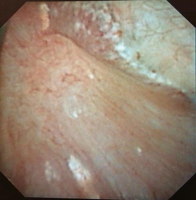
- Randall (1937) found subepithelial plaque on the papillae in 17% of 429 pairs of kidneys at autopsy.
- Papillary plaques were present in 74% of stone formers and 43% of controls (Low and Stoller).
- Evan et al. traced the plaque to the basement membrane of the thin limbs of the loops of Henle. It spreads through the medullary interstitium to a subepithelial position. The plaque is calcium (carbonate) apatite, laid down in layers with organic material, and osteopontin is present on the crystal surface.
- The plaque erodes through the urothelium and exposes a stable surface. Calcium oxalate then nucleates and grows on it as an attached stone.
- This is the main mechanism for idiopathic calcium oxalate stones.
4. Promoters and inhibitors
| Promoters | Inhibitors |
|---|---|
| Low urine volume (dehydration) | Citrate (the most important) |
| High calcium, oxalate, phosphate, uric acid | Magnesium, pyrophosphate |
| Low citrate | Nephrocalcin |
| Urinary pH change | Tamm-Horsfall protein |
| Urinary stasis, foreign bodies | Osteopontin (uropontin) |
| Urea-splitting infection | Bikunin |
| Glycosaminoglycans (chondroitin sulfate, heparan sulfate) |
How citrate acts (Campbell-Walsh):
- It complexes calcium, so less ionic calcium is available to bind oxalate or phosphate.
- It inhibits spontaneous precipitation and agglomeration of calcium oxalate.
- It is strongly effective against calcium phosphate growth.
- It prevents monosodium urate from seeding calcium oxalate.
Magnesium complexes oxalate and so lowers calcium oxalate supersaturation. No specific inhibitor of uric acid crystallisation is known.
5. Pathogenesis by stone type
Calcium stones (about 80%: calcium oxalate, or oxalate mixed with phosphate)
- Hypercalciuria is the commonest metabolic cause. It is seen in absorptive hypercalciuria (increased gut absorption), renal leak, and resorptive causes such as primary hyperparathyroidism and immobilisation.
- Hyperoxaluria can be dietary, enteric (fat malabsorption, ileal disease or bypass) or primary (genetic).
- Hypocitraturia occurs in renal tubular acidosis, chronic diarrhoea and a high-protein diet.
- Hyperuricosuria causes heterogeneous nucleation on urate.
- Urinary calcium and oxalate contribute about equally to calcium oxalate supersaturation (Pak et al.).
Struvite stones (magnesium ammonium phosphate, about 10%)
- Urease-producing bacteria (Proteus, Klebsiella) split urea into ammonia.
- Urine becomes alkaline, and ammonium, magnesium and phosphate precipitate.
- These stones grow quickly and form staghorn calculi.
- Bailey & Love notes that about 25% of paediatric stones are due to UTI with urease-producing organisms.
- The stone harbours bacteria, so infection persists until the stone is completely removed.
Uric acid stones (about 5 to 10%)
- Causes are persistently acidic urine (pH below 5.5), hyperuricosuria and low urine volume. Examples are gout, myeloproliferative disease with high cell turnover, and chemotherapy.
- Humans are prone to these stones because they lack uricase. The end product, uric acid, is 10 to 100 times less soluble than allantoin.
- Uric acid is more soluble in alkaline urine, which is why urinary alkalinisation dissolves these stones.
Cystine stones (1 to 2%)
- Autosomal recessive defect of renal tubular reabsorption of cystine, ornithine, lysine and arginine (COLA).
- Cystine is poorly soluble, especially in acidic urine.
6. Contributing risk factors
- Anatomical stasis: PUJ obstruction, medullary sponge kidney, horseshoe kidney, polycystic kidney, neurogenic bladder.
- Metabolic and systemic: hyperparathyroidism, gout, bowel disease, renal tubular acidosis.
- Lifestyle: low fluid intake, high animal protein and salt intake, hot climate.
- Demographic and genetic: male sex, family history, age and ethnicity.
7. Surgical relevance
- Stone prevention targets the pathogenesis. Measures are high fluid intake, less animal protein and salt, citrate (potassium citrate), thiazides for hypercalciuria, allopurinol for hyperuricosuria, and urinary alkalinisation for uric acid stones (Bailey & Love).
- Infection stones need complete clearance and long-term antibiotics.
- Treatment options are ESWL, ureteroscopy and PCNL, and they are not part of pathogenesis. They are chosen by stone size, location and composition.
Recent evidence check: The PubMed search for recent systematic reviews and meta-analyses returned nothing relevant to renal stone pathogenesis. The results were about vascular calcification in chronic kidney disease. I found no recent evidence that contradicts the textbook account above.
Sources: Campbell-Walsh-Wein Urology (Ch. 91, pp. 2673-2674); Robbins & Kumar Basic Pathology (Renal Stones); Bailey & Love's Short Practice of Surgery, 28th ed. (Urolithiasis). Details on urine pH, struvite and cystine stones, and the stone-type percentages come from my own knowledge, not from the passages I retrieved.
This is a shared conversation. Sign in to Orris to start your own chat.