
Viral Pneumoniae
I'll start by reading the medical library skill, then search for comprehensive information on viral pneumonia.
Reading File
Now I'll run parallel searches across the medical library for viral pneumonia.
Finding Sources
Finding Sources
Searching PubMed
"viral pneumonia" AND management
Excellent sources found. Now reading the key sections in parallel from Murray & Nadel, Rosen's EM, Robbins Pathology, and Goldman-Cecil.
Reading File
Reading File
Reading File
Reading File
Now let me get more depth from Robbins (Influenza pathology), Fishman's, and the Goldman-Cecil inpatient section. Also fetching the key CXR image.
Reading File
Reading File
Reading File
Loading Image
Excellent - I now have comprehensive content from four major textbooks. Let me also get the Fishman's pathogenesis continuation and fetch the viral pneumonia CXR showing hyperinflation.
Reading File
Loading Image
I now have comprehensive material from four major textbooks. Let me compose the full, well-structured answer.
Viral Pneumonia
Definition and Overview
Viral pneumonia is inflammation of the lung parenchyma caused by viral pathogens. It is an extremely important clinical entity that affects a wide variety of hosts - both immunologically intact and immunologically compromised individuals. The burden of disease is highest at the extremes of age (young children and the elderly) and in immunosuppressed patients, but pandemics (e.g., 1918 influenza, H1N1 2009, COVID-19) demonstrate the capacity of viruses to cause devastating disease even in healthy adults.
Common Causative Agents
The spectrum of viruses causing pneumonia depends on age and immune status (Robbins, Cotran & Kumar Pathologic Basis of Disease, p. 663; Fishman's Pulmonary Diseases, p. 2220):
| Host Group | Key Pathogens |
|---|---|
| Children | RSV, Parainfluenza virus, Human Metapneumovirus (hMPV), Adenovirus, Influenza A/B |
| Healthy adults | Influenza A/B (predominant), SARS-CoV-2, Adenovirus |
| Elderly | Influenza A/B, RSV (more severe), SARS-CoV-2 |
| Immunocompromised | CMV, HSV, VZV, RSV, Adenovirus, Influenza, hMPV (broader spectrum) |
| Pandemic/epidemic | Influenza A (H1N1, H3N2), SARS-CoV-2 |
Other agents include rhinovirus, rubeola (measles), and varicella-zoster virus.
Pathogenesis
Three general patterns exist (Fishman's Pulmonary Diseases, p. 2220):
- Primary viral pneumonia - infection initiated in and confined to the respiratory tract (influenza, RSV)
- Respiratory onset with systemic spread - infection begins in the airway but causes systemic disease (measles, varicella)
- Pulmonary involvement secondary to systemic infection - respiratory tract is a secondary target (CMV, disseminated HSV)
Influenza - the Prototype
Using influenza as the model (Fishman's, p. 2220; Robbins, p. 663):
- Virus reaches the lung by contiguous spread from the upper airway or inhalation of small-particle aerosols
- Initial infection of ciliated respiratory epithelium of the trachea, bronchi, and lower respiratory tract causes widespread epithelial cell destruction
- Viral replication inhibits sodium channels → electrolyte/water shifts → fluid accumulation in alveolar lumen
- Infected cells undergo apoptosis (caspase activation) and inhibition of host mRNA translation
- "Danger signals" released from dying cells activate resident macrophages
- Inflammatory mediators (chemokines, cytokines) activate pulmonary endothelium and recruit neutrophils within the first 1-2 days
- Innate immune responses include type I interferon production, which upregulates MX1 gene (encodes GTPase that interferes with viral replication)
- NK cells and cytotoxic T cells kill infected pneumocytes; eventually, antibody responses to hemagglutinin and neuraminidase augment clearance
Histological findings: Hyperemic tracheal/bronchial mucosa with bloody fluid; tracheitis, bronchitis, bronchiolitis; loss of ciliated epithelium; alveolar spaces filled with neutrophils, mononuclear cells, fibrin, and edema fluid; alveolar capillary hyperemia; hyaline membrane formation lining alveolar ducts and alveoli (hallmark of severe primary viral pneumonia).
RSV and Parainfluenza (Children)
Epithelial necrosis, bronchiolar mucus plugging, widespread inflammation and necrosis of lung parenchyma. Fatal RSV pneumonia shows hemorrhagic pneumonia with peribronchial mononuclear infiltration and cytoplasmic inclusion bodies in epithelial cells.
Influenza Hemagglutinin and Neuraminidase
- Hemagglutinin (H1, H2, H3): attaches virus to sialic acid residues on surface polysaccharides; acidic endosome triggers conformational change allowing viral envelope-cell membrane fusion and genome release
- Neuraminidase (N1, N2): cleaves sialic acid to release newly formed virions from infected cells
- Antigenic drift: spontaneous mutations alter H and N epitopes → seasonal epidemics
- Antigenic shift: replacement of H and/or N genes via recombination with animal flu viruses → pandemics (essentially all individuals susceptible)
Clinical Features
Adults (Fishman's Pulmonary Diseases, p. 2219)
- Nonproductive cough (frothy, pink-tinged sputum in severe cases)
- Cyanosis and hypoxemia (severe disease)
- Nonspecific findings: conjunctivitis, rhinitis, tracheal tenderness, increased respiratory rate
- Diffuse rales and wheezes
- Sputum - scant, few PMNs, minimal bacteria on Gram stain
- Influenza: abrupt onset with prominent systemic symptoms - fever, chills, headache, severe myalgia, malaise - followed by cough and dyspnea
Children (Rosen's Emergency Medicine, p. 3192)
- More common in winter season with gradual onset
- Cough, congestion, low-grade fever
- Tachypnea may be the only finding; also retractions, rales, wheezing
- Grunting, cyanosis, lethargy, dehydration, and apnea in severe cases
- Young infants: apneic episodes with minimal fever
Radiographic Findings
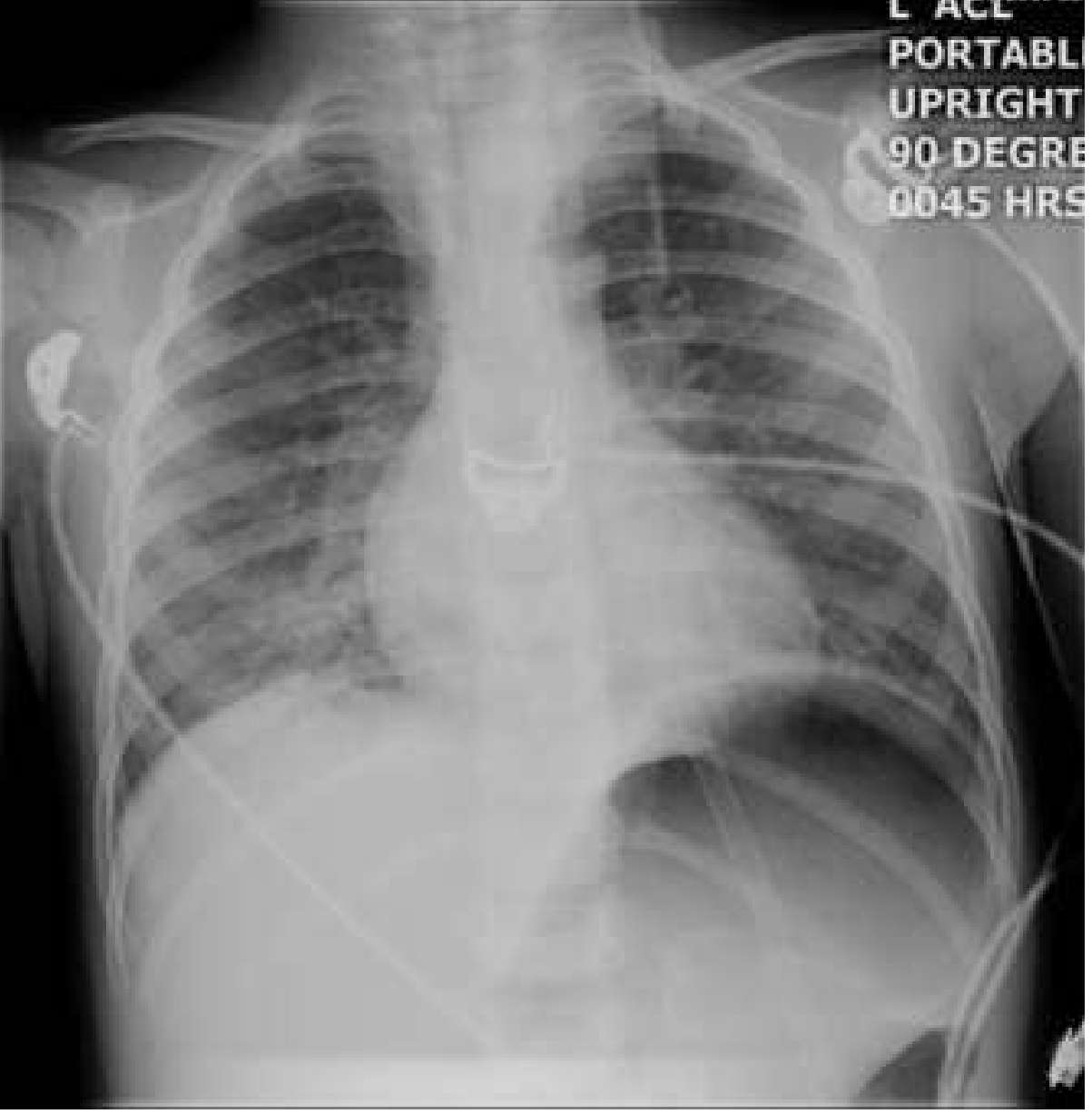
Typical pattern - diffuse, bilateral interstitial infiltrates (most common in primary viral pneumonia).
In children (Rosen's Emergency Medicine, p. 3192):
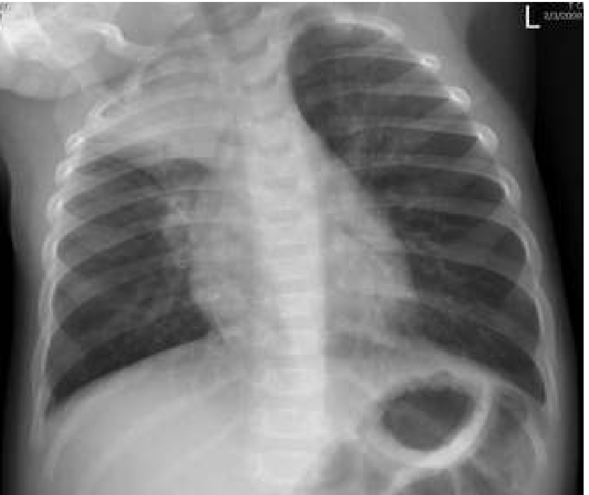
- Hyperinflation and peribronchial thickening
- Diffuse increase in interstitial markings
- Patchy consolidation (lobular atelectasis or alveolar pneumonia)
Note: lobar consolidation and pleural effusions, while possible, are more consistent with bacterial etiology. No radiographic pattern reliably differentiates bacterial from viral pneumonia.
Chest X-ray - bilateral interstitial infiltrates in viral pneumonia:
Risk Factors for Severe Disease
- Extremes of age (infants, elderly)
- Malnutrition and alcoholism
- Underlying cardiopulmonary disease (valvular heart disease, COPD)
- Pregnancy (especially 2nd and 3rd trimester; also postpartum) - 10% of 1957 influenza fatalities were pregnant women; 35% ICU admission and 7% mortality in H1N1 2009 pandemic
- Immunosuppression
- Comorbidities: diabetes, obesity, heart disease, immunosuppression (especially for influenza pandemics)
Complications
Bacterial Superinfection (Major Complication)
This is the most important complication and accounts for the majority of morbidity and mortality associated with viral pneumonia (Fishman's, p. 2220; Robbins, p. 664).
Classic presentation:
- Typical viral illness → partial or complete recovery → recurrence of fever + cough + dyspnea 2-14 days later
- CXR shows new lobar infiltrates
- Clinical course typical of bacterial pneumonia
Mechanisms facilitating bacterial superinfection:
- Disruption of epithelial barrier
- Loss of mucociliary clearance (markedly decreased, demonstrated with labeled particles in influenza)
- Virally-induced type I interferon response downregulates host antibacterial defenses
- Impaired innate immunity
Key pathogens: S. aureus (particularly common and often life-threatening post-influenza), S. pneumoniae, H. influenzae.
Other Complications
- ARDS (severe primary viral pneumonia)
- Dehydration (especially children)
- Bronchiolitis obliterans
- Apnea (infants < 3 months)
- Thromboembolic disease (especially COVID-19 in pregnancy)
Diagnosis
- Clinical - seasonal presentation, viral symptoms prodrome, typical age/epidemiology
- Children: do not require CXR or viral testing routinely in classic winter presentations with fever, cough, congestion, and wheezing (Rosen's EM, p. 3192)
- Rapid antigen tests (influenza, RSV)
- PCR-based respiratory panels (most sensitive; detect multiple viruses simultaneously)
- Sputum - scant; Gram stain shows minimal bacteria (helps distinguish from bacterial)
- Serology - retrospective, limited clinical utility
Treatment and Management
Most viral pneumonias resolve without specific therapy. Management is guided by pathogen and severity.
Outpatient Management (Goldman-Cecil Medicine, p. 3050)
| Pathogen | Treatment |
|---|---|
| SARS-CoV-2 | Antiviral therapy per COVID-19 guidelines (nirmatrelvir-ritonavir, remdesivir for outpatients at risk) |
| Influenza | Oseltamivir 75 mg twice daily × 5 days |
| Other viral pneumonia | Treat for possible bacterial coinfection |
Inpatient Management (Goldman-Cecil Medicine, p. 3085-3098)
| Pathogen | Treatment |
|---|---|
| SARS-CoV-2 | Per COVID-19 inpatient protocol |
| Influenza (inpatient) | Oseltamivir 75 mg BID × 5 days PLUS antibiotics for bacterial coinfection (beta-lactam ± macrolide or respiratory fluoroquinolone) |
| Other viral pneumonias | Treat for bacterial coinfection (same regimens as bacterial CAP) |
Specific Antiviral Therapies
- Influenza: Oseltamivir (oral), Zanamivir (inhaled), Baloxavir
- RSV: Supportive care; Ribavirin (severe/immunocompromised); Palivizumab (prophylaxis in high-risk infants)
- Varicella pneumonia: Acyclovir (reduces mortality, especially in pregnancy)
- CMV pneumonia (immunocompromised): Ganciclovir or Valganciclovir
- HSV pneumonia: Acyclovir
Children
- Most viral pneumonias resolve without therapy
- Consider antibiotics if severely ill or when bacterial co-infection cannot be excluded
- Supportive care: hydration, oxygen, respiratory support as needed
Special Populations
Pregnancy (Murray & Nadel's Respiratory Medicine, p. 3027)
- Viral pneumonia carries significantly higher maternal mortality
- 1918 pandemic: maternal mortality up to 50%
- 1957 epidemic: pregnant women = 10% of all fatalities
- H1N1 2009: 35% ICU admission, 7% mortality in infected pregnant women
- Early treatment with oseltamivir or zanamivir is beneficial
- Varicella pneumonia in pregnancy: 35% mortality (vs. 10% in other adults); treat with acyclovir
- Inactivated influenza vaccine is recommended in all trimesters; live attenuated vaccine is contraindicated
Immunocompromised
- Broader spectrum of pathogens (CMV, HSV, VZV, adenovirus)
- More severe disease; higher mortality
- Lower threshold for antiviral therapy and invasive diagnostics
Prevention
- Annual influenza vaccination (recommended for all ages > 6 months, all trimesters in pregnancy)
- COVID-19 vaccination
- RSV prophylaxis with palivizumab (high-risk infants; premature, BPD, congenital heart disease)
- Respiratory isolation, hand hygiene
- Varicella-zoster immune globulin within 96 hours of varicella exposure in susceptible pregnant women
Sources:
- Robbins, Cotran & Kumar - Pathologic Basis of Disease, pp. 663-664
- Fishman's Pulmonary Diseases and Disorders, pp. 2219-2222
- Murray & Nadel's Textbook of Respiratory Medicine, pp. 3027-3029
- Rosen's Emergency Medicine - Concepts and Clinical Practice, pp. 3192-3193
- Goldman-Cecil Medicine, pp. 3050-3098
Create a clinical reference guide for rapid identification & treatment of viral vs. bacterial pneumonia
Asking for Preferences
What format should the clinical reference guide be in? · Who is the primary audience?
Good. I'll pull targeted content from the medical library on bacterial pneumonia differentials, lab markers, and treatment alongside what we already have on viral pneumonia.
Finding Sources
Finding Sources
Finding Sources
Finding Sources
Reading File
Finding Sources
Finding Sources
Finding Sources
Reading File
Reading File
I now have all the material I need. Let me mark tasks complete and write the guide.---
🫁 Clinical Reference Guide: Rapid Identification & Treatment of Viral vs. Bacterial Pneumonia
For Medical Students | Based on Harrison's, Murray & Nadel, Robbins, Fishman's, Goldman-Cecil, Rosen's EM
SECTION 1 - First Glance: How to Start Thinking
When a patient presents with cough, fever, and respiratory symptoms, ask yourself three rapid questions:
- How did it start? - Abrupt onset favors bacterial or influenza; gradual/insidious favors atypical or viral
- What does the sputum look like? - Productive, purulent = bacterial; scant, non-purulent = viral/atypical
- What season is it, and what is the patient's age? - Winter + child = think RSV/parainfluenza; any season + elderly/comorbid = think bacterial until proven otherwise
SECTION 2 - Side-by-Side Comparison
2A. Clinical Features
| Feature | Bacterial | Viral |
|---|---|---|
| Onset | Often abrupt (pneumococcal) or subacute (atypical) | Gradual (most); abrupt with influenza |
| Cough | Productive, purulent (yellow/green/rusty) | Nonproductive; scant, watery sputum |
| Fever | High-grade (38.5-40°C), rigors with pneumococcal | Low-to-moderate grade; high with influenza |
| Pleuritic chest pain | Common (especially pneumococcal) | Uncommon |
| Myalgia/headache | Mild unless septic | Prominent (especially influenza) |
| Preceding URI symptoms | Uncommon with typical bacteria | Common - rhinitis, congestion, sore throat |
| Auscultation | Bronchial breathing, crackles, dullness to percussion | Diffuse crackles and wheezes; diffuse findings |
| Herpes labialis | May occur with pneumococcal | Absent |
| Relative bradycardia | Legionella, Mycoplasma, C. psittaci | Not typical |
| Extrapulmonary clues | Bullous myringitis (Mycoplasma); ecthyma gangrenosum (Pseudomonas) | Conjunctivitis, rhinitis (viral); rash (measles, varicella) |
Teaching pearl: Relative bradycardia means the pulse does NOT rise by the expected 10 bpm per 1°C of fever - this is a clue to atypical organisms (Legionella, Mycoplasma). - Murray & Nadel, p. 985
2B. Chest X-Ray (CXR) Patterns
| Finding | Favors Bacterial | Favors Viral |
|---|---|---|
| Lobar/segmental consolidation | ✔ Strong (especially pneumococcal) | Possible but less common |
| Air bronchograms | ✔ More common | Uncommon |
| Diffuse bilateral interstitial infiltrates | Less typical | ✔ Classic pattern |
| Peribronchial thickening | Uncommon | ✔ Common (children) |
| Hyperinflation | Uncommon | ✔ Common in children with RSV/viral |
| Pleural effusion | ✔ More common | Uncommon; if present, suggests bacterial |
| Cavitation | ✔ S. aureus, anaerobes, Klebsiella, GNB, TB | Rare |
| Bilateral patchy consolidation | Aspiration, S. aureus | ✔ Severe primary viral (ARDS pattern) |
| Round pneumonia | Hematogenous seeding | Uncommon |
CXR caveat: No radiographic pattern reliably confirms a specific pathogen. Lobar consolidation with air bronchograms is most characteristic of pneumococcal pneumonia but is not diagnostic. CXR sensitivity for pneumonia is only 44-78%. - Murray & Nadel, p. 985
Viral pneumonia CXR - bilateral interstitial infiltrates (child with RSV/viral):
Viral pneumonia CXR - hyperinflation + peribronchial thickening (3-year-old):
2C. Laboratory Findings
| Test | Bacterial | Viral | Clinical Notes |
|---|---|---|---|
| WBC/Leukocytes | Marked leukocytosis (esp. pneumococcal, S. aureus, H. influenzae, GNB) | Normal or mildly elevated; leukopenia in severe viral | Leukopenia with PVL-producing S. aureus or GNB = bad sign |
| Neutrophils | Neutrophilia | Lymphocytosis more common | |
| CRP | Elevated - higher values | Elevated - lower values | Raised to higher levels in bacterial than viral - Murray & Nadel, p. 984 |
| ESR | Elevated - higher values | Elevated - lower values | Same pattern as CRP |
| Procalcitonin (PCT) | Elevated (often >0.5 ng/mL) | Lower/normal | Variable sensitivity 38-91%; ATS/IDSA do not recommend PCT alone to decide whether to start antibiotics - Murray & Nadel, p. 984 |
| Sputum Gram stain | Numerous PMNs + organism | Minimal bacteria, few PMNs | Scant sputum with minimal bacteria = viral clue |
| Blood cultures | Positive in ~10-15% of hospitalized CAP | Negative | Always obtain in hospitalized patients |
| Urinary antigens | Legionella Ag, pneumococcal Ag | Negative | Useful for rapid detection |
| Rapid viral tests | Negative | Influenza Ag, RSV Ag, SARS-CoV-2 Ag | |
| Multiplex PCR panel | May detect bacteria + atypicals | Gold standard for viral ID | Rapidly replacing older diagnostics |
| SpO2 / ABG | Hypoxemia in moderate-severe disease | Prominent in viral (hypoxemia often out of proportion to CXR) |
Key teaching point: CRP, ESR, and PCT help trend direction, but no single lab test reliably differentiates viral from bacterial pneumonia. The Harriet Lane Handbook states directly: "CBC or inflammatory markers (CRP, ESR, procalcitonin) are not reliable to differentiate bacterial vs viral pneumonia."
2D. Common Pathogens by Category
Bacterial CAP - Typical
| Pathogen | Key Features |
|---|---|
| Streptococcus pneumoniae | Most common; rusty sputum, lobar consolidation, herpes labialis |
| Haemophilus influenzae | COPD patients; may cause lobar or bronchopneumonia |
| Staphylococcus aureus | Post-influenza superinfection; cavitation, pneumatoceles; PVL strains = necrotizing |
| Klebsiella pneumoniae | Alcoholics; upper lobe, "currant jelly" sputum; cavitation |
| Gram-negative bacilli | Aspiration-associated, nosocomial/healthcare-associated |
Bacterial CAP - Atypical ("walking pneumonia")
| Pathogen | Key Features |
|---|---|
| Mycoplasma pneumoniae | Young adults, gradual onset; bullous myringitis, erythema multiforme, cold agglutinins |
| Legionella pneumophila | Water exposure (AC units, hotels); relative bradycardia, hyponatremia, diarrhea; high mortality if untreated |
| Chlamydophila pneumoniae | Mild illness in young adults; pharyngitis prodrome |
Viral CAP
| Pathogen | Key Features |
|---|---|
| Influenza A/B | Abrupt onset; severe myalgia, high fever, then respiratory involvement |
| RSV | Most important cause in children < 2 years; bronchiolitis pattern; also severe in elderly |
| SARS-CoV-2 | Bilateral ground-glass opacities on CT; high risk in pregnancy, elderly, obese |
| Human Metapneumovirus (hMPV) | Similar to RSV clinically; spring predominance |
| Adenovirus | May mimic Mycoplasma or bacterial pneumonia radiographically |
| Varicella (VZV) | Rash + pneumonia; very dangerous in pregnancy (35% mortality) |
| CMV | Mainly immunocompromised patients |
| Measles | Children; giant cell pneumonia in measles-pneumonia |
SECTION 3 - Severity Assessment: Where Does This Patient Go?
CURB-65 Score (for hospitalization triage in CAP)
Award 1 point for each:
| Criterion | Score |
|---|---|
| C - Confusion (new disorientation) | 1 |
| U - Urea > 7 mmol/L (BUN > 20 mg/dL) | 1 |
| R - Respiratory rate ≥ 30 breaths/min | 1 |
| B - Blood pressure: systolic ≤ 90 or diastolic ≤ 60 mmHg | 1 |
| 65 - Age ≥ 65 years | 1 |
| Score | Mortality | Disposition |
|---|---|---|
| 0-1 | Low (<3%) | Outpatient treatment |
| 2 | Moderate (~9%) | Brief inpatient or supervised outpatient |
| 3-4 | High (15-40%) | Hospitalize |
| 5 | Very high (>50%) | Consider ICU |
CURB-65 is easy to use at the bedside. PSI is more accurate for predicting short-term mortality but requires more variables. Neither reliably identifies need for ICU - use clinical judgment + ATS/IDSA severe CAP criteria for ICU decisions. - Murray & Nadel, p. 907-913
SECTION 4 - Treatment
4A. Viral Pneumonia Treatment
| Pathogen | Setting | Treatment |
|---|---|---|
| Influenza | Outpatient | Oseltamivir 75 mg PO BID × 5 days |
| Influenza | Inpatient | Oseltamivir 75 mg PO BID × 5 days + antibiotics for bacterial coinfection |
| RSV | Most patients | Supportive care (O2, hydration) |
| RSV | Immunocompromised/severe | Inhaled ribavirin (specialist guidance) |
| Varicella (VZV) pneumonia | All | IV Acyclovir (reduces mortality, especially critical in pregnancy) |
| CMV pneumonia | Immunocompromised | Ganciclovir IV or Valganciclovir PO |
| SARS-CoV-2 | High-risk outpatient | Nirmatrelvir/ritonavir (Paxlovid) or remdesivir |
| SARS-CoV-2 | Inpatient, O2-requiring | Dexamethasone 6 mg daily × 10 days ± remdesivir ± baricitinib |
| Other viral pneumonia | Any | Supportive care; treat for possible bacterial coinfection |
Key rule: All inpatients with viral pneumonia should be treated for possible bacterial coinfection with standard CAP antibiotic regimens. - Goldman-Cecil Medicine, p. 3085-3098
4B. Bacterial Pneumonia Treatment
Outpatient CAP (no comorbidities)
- Amoxicillin 1 g TID × 5 days OR
- Doxycycline 100 mg BID × 5 days OR
- Azithromycin 500 mg day 1, then 250 mg days 2-5 (where macrolide resistance is low)
Outpatient CAP (comorbidities: COPD, DM, renal/hepatic disease, or recent antibiotics)
- Respiratory fluoroquinolone: Levofloxacin 750 mg OD × 5 days or Moxifloxacin 400 mg OD × 5 days OR
- Beta-lactam + macrolide combination
Inpatient CAP - Non-severe (no risk factors for MRSA or Pseudomonas)
- Beta-lactam + macrolide: ampicillin 1.5-3 g q6h OR ceftriaxone 1-2 g OD PLUS azithromycin 500 mg OD or clarithromycin 500 mg BID
- Monotherapy: Levofloxacin 750 mg OD or Moxifloxacin 400 mg OD
- If macrolide + fluoroquinolone contraindicated: beta-lactam + doxycycline 100 mg BID
- Newer agents: Omadacycline or Lefamulin (step-down capable)
Inpatient CAP - Severe
- Beta-lactam + macrolide OR beta-lactam + respiratory fluoroquinolone
- MRSA suspected: add vancomycin 15 mg/kg q12h (adjust by levels) or linezolid 600 mg q12h
- Pseudomonas suspected: piperacillin-tazobactam 4.5 g q6h, cefepime 2 g q8h, meropenem 1 g q8h
(Dosing from Goldman-Cecil Medicine, p. 3068-3083)
4C. Atypical Bacterial Pneumonia Treatment
| Pathogen | Drug of Choice | Alternative |
|---|---|---|
| Mycoplasma | Azithromycin or doxycycline | Respiratory fluoroquinolone |
| Legionella | Azithromycin or respiratory FQ | Doxycycline (less preferred) |
| Chlamydophila | Azithromycin or doxycycline | Respiratory FQ |
Note: Beta-lactams are NOT effective against atypical organisms (no cell wall).
SECTION 5 - Bacterial Superinfection After Viral Pneumonia
This is the most dangerous complication of viral pneumonia and accounts for the majority of deaths. Students must recognize the classic pattern:
Day 1-5: Viral illness (flu symptoms, low-grade fever, myalgia)
    ↓
Day 5-7: Apparent improvement
    ↓
Day 7-14: Sudden RECURRENCE of fever + productive cough + pleuritic pain
          New lobar infiltrates on CXR = BACTERIAL SUPERINFECTION
Mechanism: Viral destruction of ciliated epithelium + loss of mucociliary clearance + type I interferon response downregulating antibacterial defenses = open door for bacteria. - Fishman's, p. 2220
Most dangerous superinfecting pathogens:
- Staphylococcus aureus (including MRSA) - most common and most lethal post-influenza
- Streptococcus pneumoniae
- Haemophilus influenzae
Treatment: Full bacterial CAP regimen; MRSA coverage if signs of necrotizing disease (cavitation, rapidly worsening course).
SECTION 6 - Special Populations Quick Reference
| Population | Key Concern | Action |
|---|---|---|
| Pregnant (any trimester) | Influenza/varicella/COVID carry high maternal mortality | Early oseltamivir; IV acyclovir for varicella; inactivated flu vaccine; NO live vaccines |
| Children < 2 years | RSV is the major cause; may present with apnea only | Supportive care; antibiotics if bacterial co-infection suspected |
| Elderly | Higher mortality from all causes; RSV under-recognized | Lower threshold for admission; CURB-65 ≥ 2 = hospitalize |
| Immunocompromised | Broader spectrum: CMV, PCP, HSV, VZV, Aspergillus, Nocardia | Aggressive workup; BAL; ID consult |
| Alcoholics | Klebsiella, aspiration pneumonia, poor immunity | Cover for GNB + anaerobes; aspiration regimen |
| COPD | H. influenzae, Pseudomonas, Moraxella, encapsulated organisms | Cover for Gram-negatives if severe |
SECTION 7 - Diagnostic Algorithm (Step-by-Step)
Patient with fever + cough + respiratory symptoms
               │
    ┌──────────▼──────────────┐
    │   Clinical assessment   │
    │   Onset? Sputum? Season?│
    │   Comorbidities? Travel?│
    └──────────┬──────────────┘
               │
    ┌──────────▼──────────────────────────────────────────┐
    │  Chest X-Ray (always obtain if admitting or severe) │
    │  Lobar consolidation → likely bacterial             │
    │  Bilateral interstitial → likely viral              │
    │  Cavitation → S. aureus, anaerobes, Klebsiella, TB  │
    └──────────┬──────────────────────────────────────────┘
               │
    ┌──────────▼──────────────────────────────────────────┐
    │  Labs: CBC, CRP, PCT, blood cultures (if admitting) │
    │  Sputum Gram stain + culture                        │
    │  Rapid viral tests (flu, RSV, COVID)                │
    │  Urinary Legionella Ag + pneumococcal Ag            │
    │  Multiplex PCR if available                         │
    └──────────┬──────────────────────────────────────────┘
               │
    ┌──────────▼──────────────────────────────────────────┐
    │  CURB-65 → decide site of care                      │
    │  0-1: Outpatient   2: Supervised   ≥3: Admit        │
    └──────────┬──────────────────────────────────────────┘
               │
    ┌──────────▼────────────────────────────────────┐
    │  TREAT empirically - don't wait for cultures  │
    │  Viral + inpatient → ALWAYS add antibiotics   │
    │  for bacterial coinfection                    │
    └───────────────────────────────────────────────┘
SECTION 8 - High-Yield Memory Anchors
| "If you see/hear this..." | "Think of..." |
|---|---|
| Rusty/blood-tinged sputum + lobar consolidation | Streptococcus pneumoniae |
| Post-flu "got better, then got worse" | S. aureus superinfection |
| Relative bradycardia + hyponatremia + diarrhea | Legionella |
| Bullous myringitis + young adult + gradual onset | Mycoplasma |
| Rash (vesicular) + severe pneumonia + pregnant | Varicella - give IV acyclovir immediately |
| Diffuse bilateral interstitial + viral prodrome + no bacteria on sputum | Primary viral pneumonia |
| Child + winter + wheeze + low-grade fever | RSV / viral pneumonia |
| Cavitation + upper lobe + alcoholic | Klebsiella |
| Cavitation + rapid deterioration + post-influenza | MRSA (PVL-producing) |
| Immunocompromised + bilateral ground-glass on CT | CMV, PCP, or SARS-CoV-2 |
Sources
- Robbins, Cotran & Kumar - Pathologic Basis of Disease, pp. 663-664
- Fishman's Pulmonary Diseases and Disorders, pp. 2219-2222
- Murray & Nadel's Textbook of Respiratory Medicine, pp. 984-985, 907-913
- Rosen's Emergency Medicine - Concepts and Clinical Practice, pp. 3192-3193
- Goldman-Cecil Medicine, pp. 3050-3103
- The Harriet Lane Handbook (Johns Hopkins), Pulmonary section
This is a shared conversation. Sign in to Orris to start your own chat.