
Superolateral surface of brain
Reading File
The superolateral surface of the brain is the convex outer surface facing the skull, containing the major gyri and sulci. It is divided into the frontal, parietal, temporal, and occipital lobe regions, with key landmarks like the central sulcus, lateral sulcus (Sylvian fissure), and various functional areas.
Searching sources...
Finding Sources
Finding Sources
Finding Sources
Finding Sources
Finding Sources
Finding Sources
Finding Sources
Finding Sources
Finding Sources
Reading File
Reading File
Reading File
Reading File
Now let me fetch the brain diagram images to include in the answer.
Loading Image
Loading Image
Here is a complete, structured account of the superolateral (lateral) surface of the brain:
Superolateral Surface of the Brain
The superolateral surface is the large, convex outer surface of each cerebral hemisphere facing the calvarium. It is the most extensive of the three surfaces (the others being the medial and inferior surfaces). The surface is thrown into folds called gyri, separated by grooves called sulci (deeper ones are called fissures).
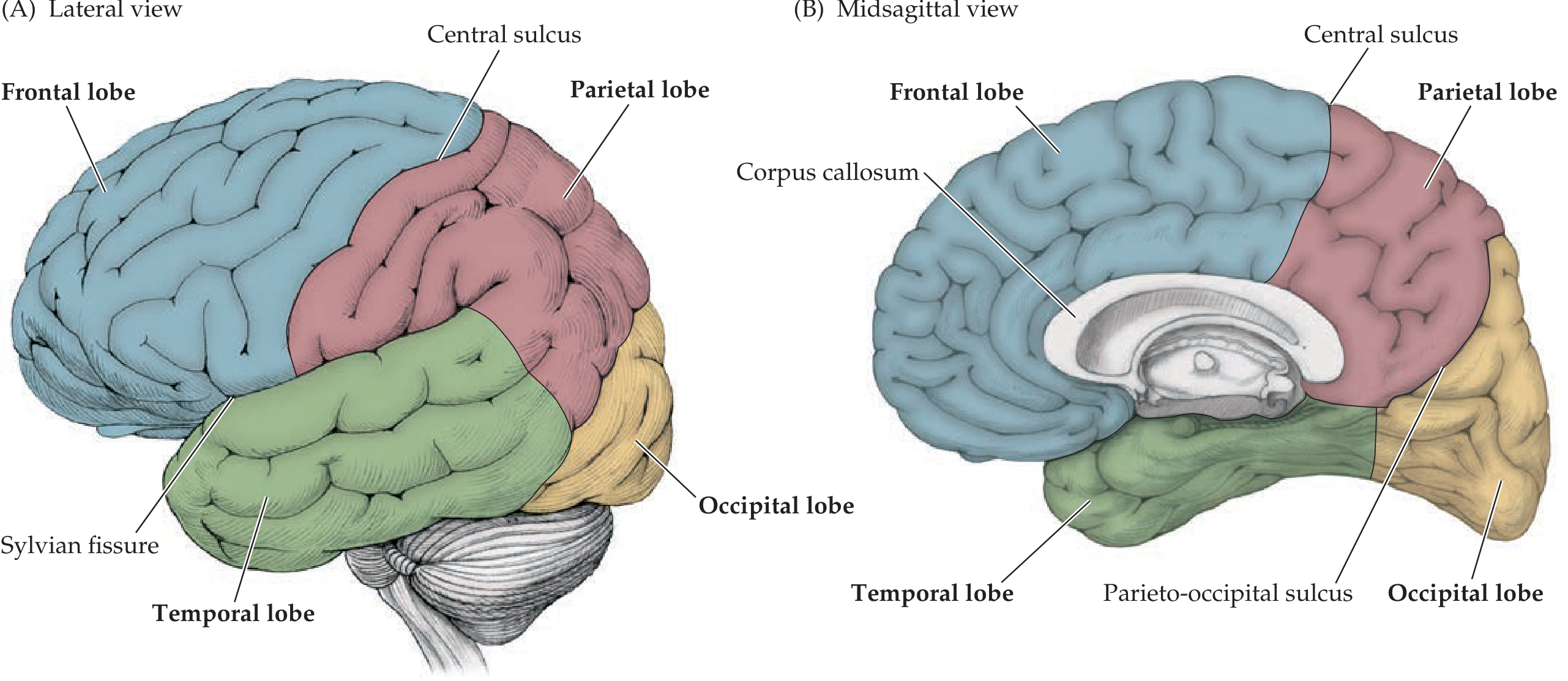
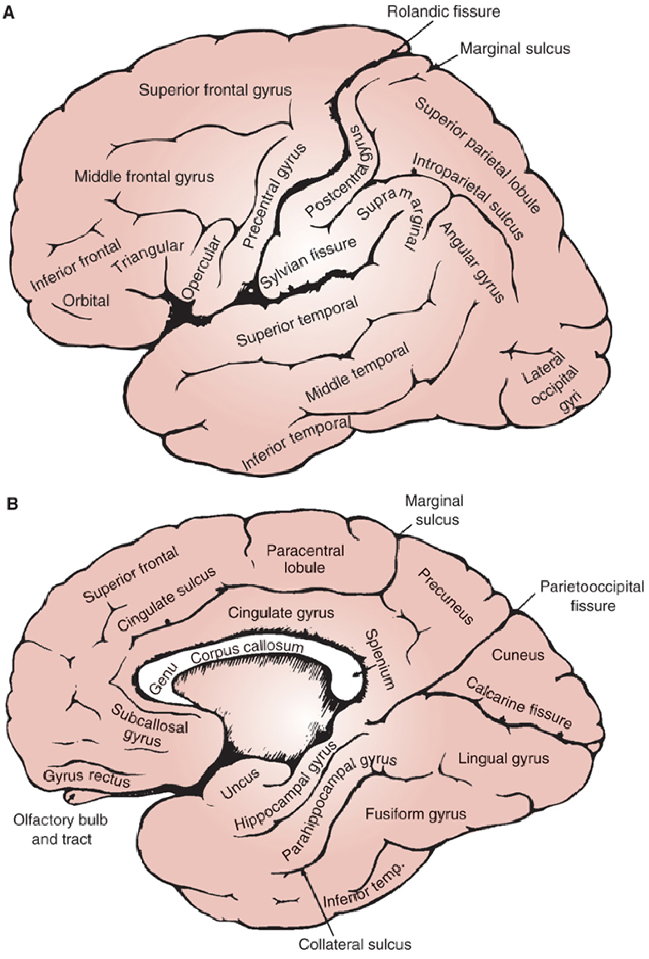
Key Fissures/Sulci Dividing the Surface
| Fissure / Sulcus | Separates |
|---|---|
| Central sulcus (of Rolando) | Frontal lobe (anterior) from parietal lobe (posterior) |
| Lateral sulcus (Sylvian fissure) | Frontal & parietal lobes (above) from temporal lobe (below) |
| Imaginary parieto-occipital line | Parietal & temporal lobes from occipital lobe laterally |
The insula lies buried in the depths of the Sylvian fissure, covered by the frontal operculum anteriorly and parietal operculum posteriorly.
Lobes on the Superolateral Surface
1. Frontal Lobe
- Bounded posteriorly by the central sulcus and inferolaterally by the lateral sulcus
- Precentral gyrus - lies immediately anterior to the central sulcus; contains the primary motor cortex (Brodmann area 4)
- The remaining lateral frontal surface is divided by the superior frontal sulcus and inferior frontal sulcus into three horizontal gyri:
- Superior frontal gyrus
- Middle frontal gyrus
- Inferior frontal gyrus - divided by the anterior ascending and horizontal limbs of the Sylvian fissure into three parts:
- Orbital part (pars orbitalis)
- Triangular part (pars triangularis)
- Opercular part (pars opercularis)
- The triangular + opercular parts of the left inferior frontal gyrus form Broca's area (Brodmann areas 44 & 45) - expressive speech
2. Parietal Lobe
- Bounded anteriorly by the central sulcus and antero-inferiorly by the Sylvian fissure
- Postcentral gyrus - immediately posterior to the central sulcus; contains the primary somatosensory cortex (areas 3, 1, 2)
- The intraparietal sulcus divides the lobe into:
- Superior parietal lobule (above intraparietal sulcus)
- Inferior parietal lobule (below), which consists of:
- Supramarginal gyrus - caps the upturned end of the Sylvian fissure
- Angular gyrus - caps the upturned end of the superior temporal sulcus
- The angular gyrus forms part of Wernicke's area (language comprehension)
3. Temporal Lobe
- Lies inferior to the Sylvian fissure
- Divided by the superior temporal sulcus and inferior temporal sulcus into:
- Superior temporal gyrus - its upper bank (planum temporale) contains Heschl's gyrus (primary auditory cortex, Brodmann area 41/42); the posterior portion of the superior temporal gyrus is part of Wernicke's area
- Middle temporal gyrus
- Inferior temporal gyrus
4. Occipital Lobe
- Forms the posterior pole of the hemisphere on the lateral surface
- Contains lateral occipital gyri; the primary visual cortex (calcarine cortex) is mostly on the medial surface but extends onto the lateral surface near the occipital pole
- No sharp lateral boundary separates it from parietal and temporal lobes; an imaginary line from the parieto-occipital sulcus to the preoccipital notch is used
Functional Areas (Lateral Surface)
| Area | Location | Function |
|---|---|---|
| Primary motor cortex | Precentral gyrus | Voluntary movement (contralateral body) |
| Primary somatosensory cortex | Postcentral gyrus | Contralateral sensation (touch, pain, proprioception) |
| Broca's area | Left inferior frontal gyrus (pars triangularis + opercularis) | Expressive/motor speech |
| Wernicke's area | Left posterior superior temporal gyrus + angular gyrus | Receptive/comprehension of speech |
| Primary auditory cortex | Heschl's gyrus (superior temporal gyrus, buried in Sylvian fissure) | Hearing |
| Premotor cortex | Anterior to precentral gyrus | Motor planning |
| Prefrontal cortex | Anterior frontal lobe | Executive function, working memory |
| Association cortex | Parietal, temporal, occipital junction | Multimodal sensory integration |
Clinical Relevance
- Lesion of precentral gyrus - contralateral spastic paresis (UMN pattern)
- Lesion of postcentral gyrus - contralateral cortical sensory loss (loss of discriminative touch, 2-point discrimination, graphesthesia)
- Broca's aphasia - non-fluent aphasia with intact comprehension; lesion in left inferior frontal gyrus
- Wernicke's aphasia - fluent aphasia with poor comprehension; lesion in left posterior superior temporal gyrus
- Supramarginal gyrus lesion - ideomotor apraxia, conduction aphasia
- Angular gyrus lesion - alexia, agraphia, acalculia, finger agnosia, left-right disorientation (Gerstmann's syndrome with involvement of left angular gyrus)
- Middle cerebral artery (MCA) supplies most of the superolateral surface; infarction causes contralateral hemiplegia, hemisensory loss, and aphasia (dominant hemisphere)
Sources: Neuroanatomy through Clinical Cases, 3rd Ed. | Localization in Clinical Neurology, 8th Ed.
This is a shared conversation. Sign in to Orris to start your own chat.