
stargards disease explain in detail
Stargardt disease
Stargardt disease National Eye Institute ABCA4 diagnosis treatment clinical trials
Stargardt disease fundus photograph yellow flecks bull's eye maculopathy
https://www.nei.nih.gov/learn-about-eye-health/eye-condition…
Stargardt disease treatment
https://www.nei.nih.gov/learn-about-eye-health/eye-condition…
PMID: 40725253
Stargardt disease, explained
What happens in the eye?
- Cones are concentrated in the macula and provide sharp central and color vision.
- Rods are more important for dim-light and peripheral vision.
- The retinal pigment epithelium (RPE) supports photoreceptors and handles waste from the visual cycle.
Symptoms
- Gradual blurring or loss of central vision in both eyes
- Difficulty reading, seeing classroom boards, or recognizing faces
- A central blind or blurry spot, called a central scotoma
- Reduced contrast sensitivity
- Reduced color discrimination
- Light sensitivity or slow adaptation after bright light
- Distortion of straight lines in some people
- Difficulty seeing in dim conditions in more extensive disease
What does the retina look like?
- Yellow-white deposits, called flecks, at the level of the RPE. They can look fish-tail shaped, termed pisciform flecks.
- Macular mottling or a “beaten-bronze” appearance.
- A ring-like area of atrophy called bull's-eye maculopathy.
- In later stages, more definite macular and RPE atrophy.
How is it diagnosed?
-
Dilated eye examination and color retinal photographs
Looks for macular change and flecks. -
Optical coherence tomography, OCT
This gives a cross-sectional retinal image. It can show outer-retinal, photoreceptor, and RPE damage, sometimes before obvious changes are visible on routine examination. -
Fundus autofluorescence, FAF
A particularly useful test for Stargardt disease. It maps lipofuscin-related signal and regions of RPE atrophy. It also helps track progression. -
Visual field testing or microperimetry
Identifies and monitors central scotomas. -
Fluorescein angiography, sometimes
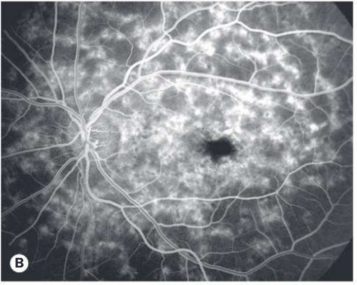
It may show a classic “dark” or “silent choroid”, because lipofuscin in the RPE blocks background choroidal fluorescence. OCT and FAF are now more commonly used for routine assessment. Kanski's Clinical Ophthalmology, 10th ed., p. 656. -
Electroretinography, ERG
It may be normal early on, particularly when disease is predominantly macular. It helps determine whether retinal dysfunction extends beyond the macula. -
Genetic testing
A retinal-dystrophy gene panel, including ABCA4, is important to confirm the diagnosis, distinguish similar disorders, clarify inheritance, provide family counseling, and determine trial eligibility. The National Eye Institute overview also lists retinal scans and genetic testing as parts of assessment.
Conditions that can look similar
- Cone or cone-rod dystrophy
- Best disease and other inherited macular dystrophies
- Pattern dystrophy
- Early-onset or atypical age-related macular degeneration
- Hydroxychloroquine or chloroquine retinal toxicity
- Fundus albipunctatus and retinitis punctata albescens
- Rarely, neurologic/metabolic conditions such as neuronal ceroid lipofuscinosis
Course and prognosis
- Central vision declines first.
- Peripheral vision remains relatively good for many people, especially early and middle stages.
- Some people keep useful reading vision for many years; others develop a larger central scotoma sooner.
- Advanced disease may involve widespread RPE and outer-retinal atrophy, which can affect more than just the center of vision.
Is there a cure or approved treatment?
- Therapies intended to reduce toxic vitamin-A byproducts or lipofuscin accumulation
- Visual-cycle modulation
- Gene-based treatments, including approaches that address the large ABCA4 gene
- RNA-based or gene-editing strategies
- Cell-based approaches
- Treatments targeting oxidative stress, inflammation, or retinal metabolism
Practical management
Regular review with a retina or inherited-retinal-disease specialist helps document change with visual acuity, OCT, FAF, and visual fields.
This often has the greatest day-to-day benefit. Options include:
- High-add reading glasses or magnifiers
- Electronic video magnifiers
- Large-print and high-contrast settings
- Phone accessibility features, screen readers, and text-to-speech
- Orientation and mobility training if needed
- School or workplace accommodations
Wear UV-blocking sunglasses and a hat outdoors. This is sensible light protection, although it should not be presented as a cure.
Because ABCA4-related disease involves vitamin-A-derived retinal byproducts, high-dose supplemental vitamin A is commonly avoided. This does not mean avoiding normal dietary vitamin A or starting an unproven restrictive diet. Discuss supplements with the eye specialist or primary clinician first. The Wills Eye Manual, p. 897.
This clarifies recurrence risk, carrier issues, testing of relatives, reproductive options, and trial eligibility.
Progressive central vision loss can affect education, employment, driving, independence, and mood. Low-vision services, rehabilitation specialists, and patient-support organizations can be useful early, not only after severe vision loss.
Family inheritance in the common ABCA4 form
- Usually, both parents are carriers.
- For each pregnancy of two carriers:
- 25% chance the child is affected
- 50% chance the child is an unaffected carrier
- 25% chance the child inherits neither altered copy
When urgent review is needed
Key takeaways
- Stargardt disease is an inherited macular degeneration, most often due to ABCA4 variants.
- It damages central vision, usually in both eyes, while peripheral vision is often relatively preserved.
- OCT, fundus autofluorescence, and genetic testing are central to diagnosis and follow-up.
- No approved therapy currently reverses the disease, but low-vision rehabilitation and genetic counseling provide real benefit.
- Multiple drug, gene, and cell-based treatments are in clinical research, but investigational treatments should not be assumed to work or replace standard specialist care.
honeycomb macular dystrophy explain in detail and dds and differentiationg points
Doyne honeycomb retinal dystrophy
Doyne honeycomb retinal dystrophy Malattia Leventinese EFEMP1 clinical features diagnosis differential
Doyne honeycomb retinal dystrophy Malattia Leventinese fundus photograph radial drusen
https://eyewiki.aao.org/Doyne_Honeycomb_Retinal_Dystrophy
PMID: 42569289
Honeycomb macular dystrophy
- Malattia Leventinese
- Familial dominant drusen
- Autosomal dominant radial drusen
Genetics and pathogenesis
Inheritance
- Autosomal dominant (AD) inheritance
- Caused by a pathogenic variant in EFEMP1 on chromosome 2p16
- Affected individuals have a 50% chance of passing the variant to each child.
- Expressivity is variable: members of the same family can have very different severity, and the two eyes of the same person may progress differently.
What happens anatomically?
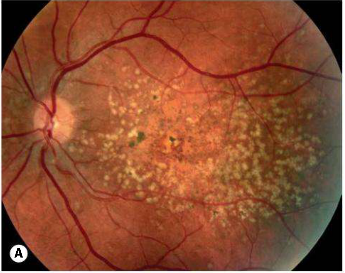
- Small radial drusen develop around the macula.
- The drusen enlarge, multiply, and coalesce.
- They form a dense honeycomb pattern.
- RPE dysfunction and atrophy may develop.
- A minority develop macular neovascularization, MNV, previously called choroidal neovascularization or CNV, with fluid, hemorrhage, scar formation, and sudden visual loss.
Clinical presentation
Age at onset
- Fundus lesions often appear in the second or third decade.
- Many patients are initially asymptomatic.
- Gradual symptomatic central visual impairment often occurs later.
- Severe central visual loss may appear in the seventh or eighth decade, usually due to RPE degeneration, macular atrophy, or MNV. Kanski's Clinical Ophthalmology, 10th ed., p. 664.
Symptoms
- Mild, gradual reduction of central vision
- Difficulty reading or seeing fine print
- Reduced contrast sensitivity
- Metamorphopsia, meaning straight lines look wavy
- Central blur or a central scotoma
- Poorer vision in dim illumination in some advanced cases
- Sudden distortion or vision loss if MNV, hemorrhage, or exudation occurs
Fundus findings
- Bilateral, generally symmetric disease, though severity can differ between eyes
- Multiple yellow-white drusen
- Lesions are often elongated and radially arranged, centered on the macula
- Drusen can extend toward and nasal to the optic disc, involving the disc margin
- With time, the drusen become more numerous, confluent, dense, and form a honeycomb pattern
- Late RPE atrophy, geographic atrophy, pigmentary change, and occasionally MNV
Young person with bilateral, radial macular drusen extending nasal to the optic disc, with an autosomal-dominant family history: think Doyne honeycomb retinal dystrophy.
Investigations
1. Color fundus photography
2. OCT
- RPE irregularity or atrophy
- Outer-retinal changes
- Subretinal or intraretinal fluid from MNV
- Pigment epithelial detachment
3. Fundus autofluorescence, FAF
- Hyperautofluorescence can indicate stressed RPE/lipofuscin-related change.
- Hypoautofluorescence usually indicates established RPE loss or atrophy.
4. OCT angiography, OCT-A
5. Fluorescein angiography, FA
6. Electrophysiology
- ERG: usually normal because this is mainly a macular/RPE disorder rather than a widespread rod-cone dystrophy.
- EOG: may become subnormal in advanced disease. Kanski's Clinical Ophthalmology, 10th ed., p. 664.
7. Genetic testing
- Distinguishing it from other early-onset drusen disorders
- Family screening
- Genetic counseling
- Trial eligibility
Differential diagnosis and differentiating points
High-yield comparison table
| Condition | Key differentiating points from DHRD |
|---|---|
| Age-related macular degeneration, AMD | Usually begins after age 50-60; multifactorial, not a clear autosomal-dominant EFEMP1 disorder; typical AMD risk factors such as age and smoking are more relevant. DHRD has onset of drusen in the second to third decade, radial distribution, frequent disc-margin involvement, and family history across generations. |
| Cuticular drusen / basal laminar drusen | Small, uniform yellow drusen, usually 25-75 μm, often numerous and clustered. FA classically shows a “stars in the sky” pattern. DHRD drusen are more elongated, radial, centered around the macula, may extend nasal to the disc, and evolve into a honeycomb pattern. Cuticular drusen are associated with CFH variants, not EFEMP1. Kanski's Clinical Ophthalmology, 10th ed., p. 600. |
| Stargardt disease / fundus flavimaculatus | Usually autosomal recessive, commonly ABCA4-related. Lesions are yellow flecks at RPE level, often pisciform or fish-tail shaped, with macular atrophy and a possible dark choroid on FA. Stargardt produces early central vision loss and outer-retinal photoreceptor degeneration, not classic radial drusen or a dominant EFEMP1 family history. |
| Sorsby fundus dystrophy | AD but caused by TIMP3, not EFEMP1. Often presents in the third decade with nyctalopia or in middle age with sudden visual loss due to MNV. Drusen-like deposits may occur along arcades and nasal to disc, but MNV and subretinal scarring occur earlier and more aggressively. Delayed rod dark adaptation is an important early clue. Kanski's Clinical Ophthalmology, 10th ed., p. 665. |
| Best disease / autosomal dominant vitelliform macular dystrophy | Yellow “egg-yolk” vitelliform lesion rather than multiple radial drusen. EOG is characteristically abnormal even when ERG is normal. Usually associated with BEST1, not EFEMP1. |
| Adult-onset foveomacular vitelliform dystrophy | Onset usually later, with a central solitary or bilateral yellow subfoveal vitelliform deposit and characteristic FAF hyperautofluorescence. No radial drusen pattern. |
| Pattern dystrophy | Often AD, typically PRPH2-related. Pigment/lipofuscin patterns such as butterfly, reticular, or fundus pulverulentus forms rather than true radial drusen. |
| North Carolina macular dystrophy | AD, often congenital or recognized early in life. Can show macular excavation, coloboma-like lesions, or atrophy. It is not characterized by classic radial drusen. |
| Central areolar choroidal dystrophy | Progressive central RPE and choriocapillaris atrophy with sharply circumscribed geographic atrophy. Drusen are not the dominant early finding. Often associated with PRPH2. |
| Membranoproliferative glomerulonephritis type II / dense deposit disease | Can cause bilateral diffuse drusen-like lesions in children or young adults. Ask about renal disease, hematuria, proteinuria, low complement, or nephrology follow-up. Kanski's Clinical Ophthalmology, 10th ed., p. 600. |
| Dominant drusen secondary to other inherited causes | Genetic-panel testing may reveal another retinal dystrophy gene. The hallmark of classical DHRD is an EFEMP1 pathogenic variant with radial macular and peripapillary drusen. |
DHRD versus cuticular drusen: an exam-focused distinction
| Feature | DHRD / Malattia Leventinese | Cuticular drusen |
|---|---|---|
| Gene | EFEMP1 | Often associated with CFH variants |
| Inheritance | Autosomal dominant, classically | Variable, often not a clear single-gene dominant pedigree |
| Shape | Elongated, radial drusen | Numerous small, round, uniform nodules |
| Distribution | Macula, often extends nasal to disc | Clustered, widely distributed small drusen |
| Fundus pattern | Radial then honeycomb | Grouped, “cuticular” |
| FA | Variable drusen fluorescence | Stars-in-the-sky hyperfluorescent pattern |
| Late problem | RPE atrophy, occasional MNV | PED, atrophy, MNV can occur |
Management
Regular monitoring
- Visual acuity and symptom review
- Dilated retinal examination
- OCT
- FAF where available
- OCT-A or FA if MNV is suspected
- Home monitoring for new metamorphopsia, for example with an Amsler grid, if advised by the retinal specialist
Treat MNV promptly
Low-vision rehabilitation
- Magnification devices
- High-contrast reading tools
- Electronic video magnifiers
- Accessibility functions and text-to-speech
- Occupational rehabilitation and workplace or educational accommodations
Genetics
- Genetic counseling
- Testing of at-risk relatives after the familial variant is identified
- Preconception and reproductive counseling when relevant
General retinal health
Prognosis
- Many patients retain good vision for many years.
- Drusen may remain asymptomatic early.
- Central vision may decline later due to RPE degeneration and geographic atrophy.
- MNV can cause a more abrupt deterioration but may respond to anti-VEGF therapy.
- The disorder generally does not produce early severe night blindness or widespread peripheral field loss, unlike many generalized retinal dystrophies.
Red flags needing urgent review
- New metamorphopsia
- Sudden central blur or scotoma
- A sharp decline in visual acuity
- New central dark patch
- New retinal hemorrhage noted on examination
One-line summary
brvo
branch retinal vein occlusion treatment
AAO branch retinal vein occlusion BRVO diagnosis treatment anti VEGF laser
branch retinal vein occlusion BRVO fundus photograph sectoral retinal hemorrhages
https://www.aao.org/education/preferred-practice-pattern/ret…
PMID: 42265442
BRVO: Branch Retinal Vein Occlusion
Pathogenesis
- The thickened, rigid retinal artery compresses the adjacent vein.
- Venous blood flow becomes turbulent and slow.
- Endothelial injury and venous thrombosis/occlusion occur.
- Venous pressure rises in the drainage territory.
- This causes sectoral hemorrhages, retinal edema, macular edema, ischemia, and sometimes neovascularization.
Arteriosclerotic artery compresses a retinal vein at an AV crossing, resulting in thrombosis and sectoral retinal venous obstruction.
Risk factors
Major systemic risk factors
- Hypertension: the most important association, especially for BRVO
- Older age
- Hyperlipidemia
- Diabetes mellitus
- Smoking
- Atherosclerotic cardiovascular disease
Ocular factors
- Retinal arterial sclerosis with AV nicking
- Glaucoma and raised intraocular pressure are more strongly linked to CRVO, but intraocular pressure should still be assessed in every retinal vein occlusion.
Consider in younger patients or unusual presentations
- Hyperhomocysteinemia
- Antiphospholipid syndrome
- Factor V Leiden and other thrombophilias
- Myeloproliferative disorders, such as polycythemia
- Hyperviscosity syndromes
- Autoimmune/inflammatory retinal vasculitis, for example Behçet disease or sarcoidosis
- Oral contraceptive use, pregnancy-related risk where relevant
- Severe dehydration
- Chronic kidney disease
Symptoms
- Sudden or subacute painless visual loss in one eye
- Blurred central vision, commonly due to macular edema
- Distortion of straight lines, metamorphopsia
- A localized field defect or blind spot
- Some peripheral BRVOs are asymptomatic and found incidentally
Fundus findings
- Flame-shaped and dot-blot hemorrhages in a retinal sector
- Dilated, tortuous branch vein
- Retinal edema
- Cotton-wool spots
- AV crossing changes, including arterial narrowing or venous compression
- Macular edema if the macula is involved
- Collateral vessels later
- Retinal or disc neovascularization in ischemic BRVO
- Vitreous hemorrhage if new vessels bleed
Key diagnostic point
In BRVO, hemorrhages lie along the involved vein and generally do not cross the horizontal raphe.
Classification
1. Major BRVO
- More extensive hemorrhage and ischemia
- Greater chance of macular edema and neovascularization
2. Macular BRVO
- Hemorrhages are localized near the macula
- May cause marked visual symptoms despite a smaller retinal area involved
3. Non-ischemic versus ischemic BRVO
| Feature | Non-ischemic BRVO | Ischemic BRVO |
|---|---|---|
| Capillary perfusion | Preserved or mildly reduced | Large areas of non-perfusion |
| Visual prognosis | Generally better | More guarded |
| Macular edema | May occur | May occur, often significant |
| Neovascularization risk | Low | Higher |
| Need for FA/OCT-A | Sometimes | Important to assess ischemia and MNV/NV risk |
Investigations
Ocular assessment
-
Visual acuity and refraction
-
Dilated fundus examination
-
Intraocular pressure measurement
-
OCT of the macula
- Detects and quantifies macular edema
- Shows intraretinal cysts, subretinal fluid, retinal thickness, and structural damage
- Used at follow-up to guide treatment response
-
Fluorescein angiography, FA
- Shows delayed venous filling
- Identifies macular ischemia
- Maps capillary non-perfusion
- Detects retinal neovascularization
- Often most useful once dense hemorrhages have cleared, unless neovascularization is already suspected
-
OCT angiography, OCT-A
- Can help assess macular perfusion and identify neovascularization without dye.
Systemic evaluation
- Blood pressure
- Fasting glucose or HbA1c
- Lipid profile
- Full blood count and platelet count
- Renal function where clinically appropriate
- Cardiovascular risk assessment
Treatment
1. Macular edema: anti-VEGF injections are first line
- Aflibercept
- Ranibizumab
- Bevacizumab, commonly used off-label
- Faricimab, depending on regulatory approval and local practice
2. Intravitreal corticosteroids
- May raise intraocular pressure
- Can accelerate cataract formation
- Requires monitoring for steroid response
3. Laser photocoagulation
4. Control systemic disease
- Strict blood pressure control
- Diabetes treatment
- Lipid reduction as indicated
- Smoking cessation
- Management of cardiovascular and renal risk factors
Follow-up
- Visual acuity
- OCT macula
- Dilated retinal examination
- Assessment for retinal/disc neovascularization
- Assessment for vitreous hemorrhage
Complications
- Macular edema: most common cause of reduced central vision
- Macular ischemia: limits visual recovery even after edema resolves
- Retinal neovascularization
- Vitreous hemorrhage from fragile new vessels
- Epiretinal membrane
- Tractional retinal detachment: uncommon, generally following untreated extensive neovascularization
- Chronic macular structural damage, including photoreceptor loss
Differential diagnosis
| Condition | Differentiating feature |
|---|---|
| CRVO | Hemorrhages, venous tortuosity, and edema involve all four quadrants rather than one sector. |
| Diabetic retinopathy | Usually bilateral; microaneurysms and dot-blot hemorrhages are diffuse and cross the horizontal raphe. |
| Hypertensive retinopathy | Usually bilateral; generalized arteriolar narrowing and hemorrhages are not confined to one venous drainage sector. |
| Ocular ischemic syndrome | Often has mid-peripheral hemorrhages, narrowed arteries, delayed arterial filling, carotid disease, and anterior-segment ischemic features. |
| Retinal vasculitis | May show perivascular sheathing, inflammatory cells, bilateral/asymmetric disease, and systemic inflammatory features. |
| Branch retinal artery occlusion | Sectoral retinal whitening rather than venous dilation and widespread sectoral hemorrhages. |
| Diabetic papillopathy/papillophlebitis | Disc swelling may occur, but papillophlebitis tends to affect younger people and requires distinction from true RVO. |
BRVO versus CRVO: rapid differentiation
| Feature | BRVO | CRVO |
|---|---|---|
| Site | Branch retinal vein | Central retinal vein |
| Hemorrhage distribution | One sector/quadrant | All four quadrants |
| Horizontal raphe | Usually does not cross it | Not confined by it |
| AV crossing compression | Classic mechanism | Not typical main site |
| Macular edema | Common | Common |
| Retinal neovascularization | Possible in ischemic cases | Higher overall ischemic complication burden |
| Anterior-segment neovascularization | Uncommon | More concerning in ischemic CRVO |
Prognosis
- The macula is not significantly involved
- Macular ischemia is limited
- Macular edema responds early to anti-VEGF treatment
- No retinal neovascularization develops