I now have comprehensive data from Gray's Anatomy, Neuroanatomy through Clinical Cases, Goldman-Cecil Medicine, and Adams & Victor's. I have all I need for a complete, well-organized answer with textbook diagrams.
Blood Supply of the Brain
The brain receives ~20% of cardiac output despite being only 2% of body weight. It is supplied by two systems that meet at the Circle of Willis.
Arterial Supply: Two Systems
70% from Internal Carotid Arteries (Anterior Circulation)
30% from Vertebral Arteries (Posterior Circulation)
Here are the anatomical diagrams from Neuroanatomy through Clinical Cases, 3rd Ed.:
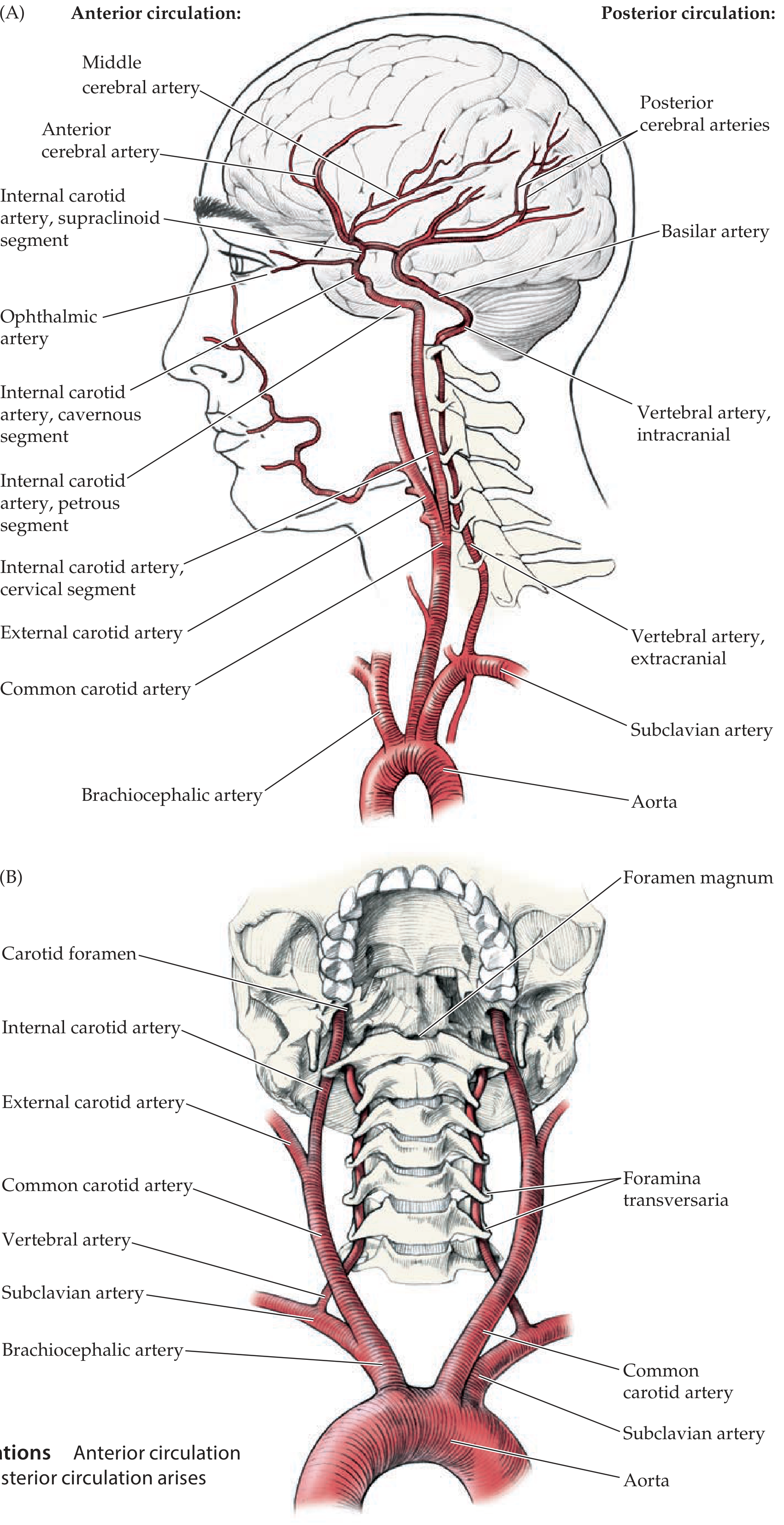
Fig. 10.2 - Anterior circulation (ICA) and posterior circulation (vertebral arteries), showing their origins from the aorta and course to the brain.
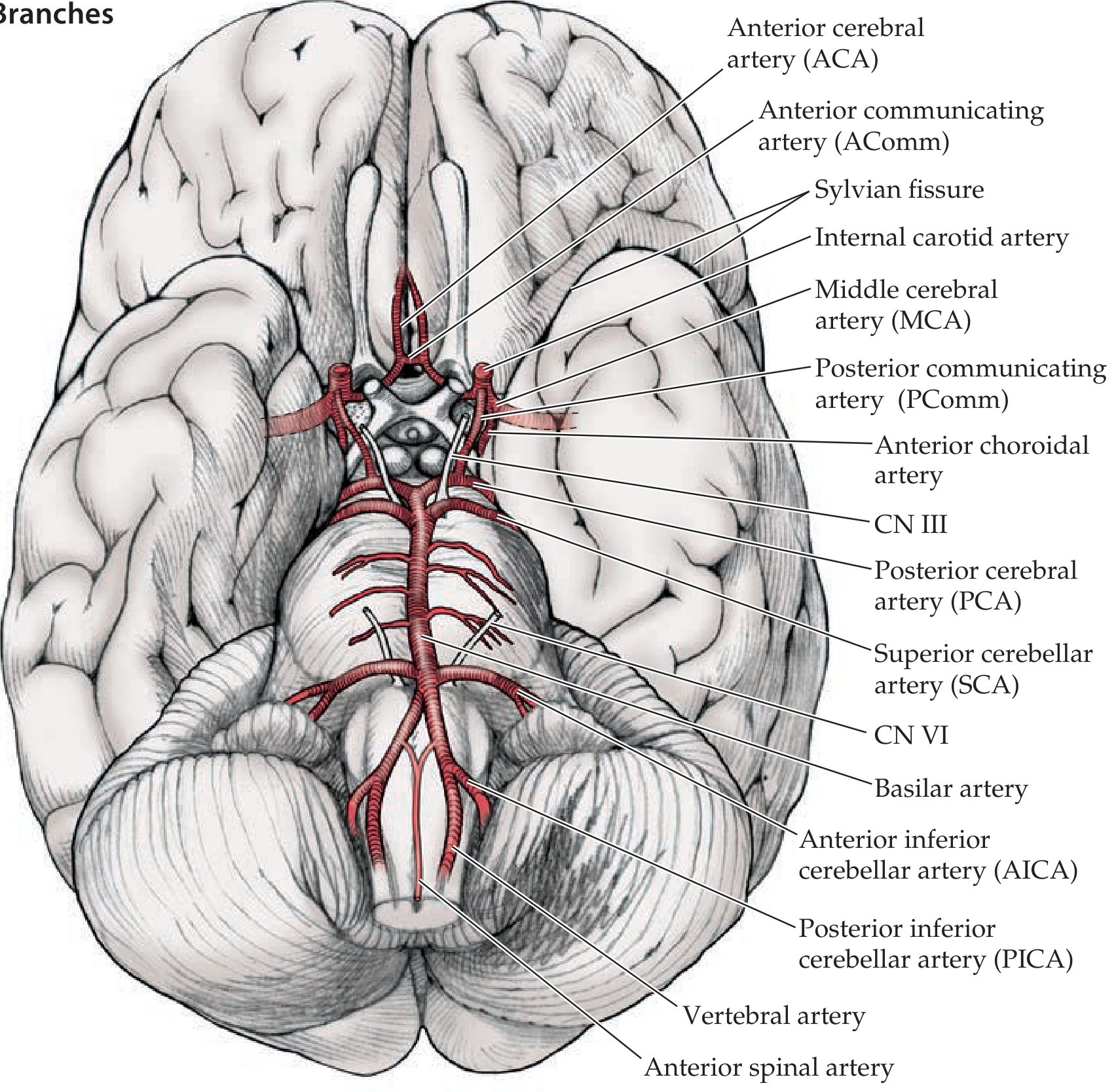
Fig. 10.3 - Inferior view of the brain showing the Circle of Willis and all major cerebral branches.
1. Internal Carotid Artery (ICA) - Anterior Circulation
The ICA arises from the common carotid artery at the carotid bifurcation and passes through four named segments:
| Segment | Location |
|---|
| Cervical | Neck - no branches |
| Petrous | Petrous canal of temporal bone |
| Cavernous | Cavernous sinus - gives off ophthalmic artery |
| Supraclinoid | Enters the dura - terminal branches |
Terminal branches of the ICA:
| Branch | Supplies |
|---|
| Ophthalmic artery | Retina, orbit (first branch after entering dura) |
| Anterior choroidal artery | Optic tract, posterior limb of internal capsule (inferior part), hippocampus, amygdala, geniculate body |
| Posterior communicating artery (PComm) | Links ICA to PCA; completes the Circle of Willis |
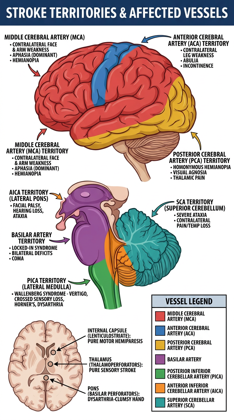
| Anterior cerebral artery (ACA) | Medial frontal and parietal lobes (leg area) |
| Middle cerebral artery (MCA) | Lateral frontal, parietal, temporal lobes (face + arm area) |
2. ACA - Anterior Cerebral Artery
- Travels anteriorly then turns posteriorly along the medial hemisphere
- Supplies medial frontal and parietal lobes, paracentral lobule, corpus callosum
- The A1 segment (before anterior communicating artery) gives off lenticulostriate branches
- The recurrent artery of Heubner (large medial striate branch) supplies: anterior internal capsule, head of caudate, putamen, anterior globus pallidus, hypothalamus
- Left and right ACAs connected by the anterior communicating artery (AComm)
3. MCA - Middle Cerebral Artery
- Largest branch of ICA; supplies the bulk of the lateral hemisphere
- The M1 segment gives rise to all lateral lenticulostriate arteries supplying the basal ganglia and internal capsule
- Bifurcates (20-30%) or trifurcates (70%) in the Sylvian fissure:
- Superior division - frontal and parietal lobes (Broca's area dominant hemisphere)
- Inferior division - lateral temporal lobe (Wernicke's area dominant hemisphere)
4. Vertebral Arteries - Posterior Circulation
- Arise from subclavian arteries
- Ascend through foramina transversaria of C6-C1 vertebrae
- Enter skull through foramen magnum
- Unite to form the basilar artery
- The fourth segment (V4) gives branches to brainstem and cerebellum
Branches of vertebral artery:
| Branch | Supplies |
|---|
| Posterior inferior cerebellar artery (PICA) | Lateral medulla + inferior cerebellum |
| Anterior spinal artery | Ventral medulla + upper cervical cord (pyramids, medial lemniscus, CN XII) |
5. Basilar Artery
Formed by the union of the two vertebral arteries. Runs along the ventral surface of the pons.
| Branch | Supplies |
|---|
| Anterior inferior cerebellar artery (AICA) | Lateral pontomedullary junction, anterior inferior cerebellum |
| Pontine perforators (paramedian + circumferential) | Basis pontis, MLF, pontine reticular nuclei, corticospinal tracts |
| Superior cerebellar artery (SCA) | Superior cerebellum, upper pons |
| Posterior cerebral artery (PCA) (terminal branch) | Occipital lobe, inferior temporal lobe, thalamus |
6. Circle of Willis
An anastomotic ring at the base of the brain providing collateral flow. Only a complete full-caliber ring is present in ~34% of individuals.
        ACA ─── AComm ─── ACA
         |                  |
        ICA                ICA
         |                  |
       PComm             PComm
         |                  |
        PCA ─── Basilar ─── PCA
                   |
              Vertebrals (x2)
Components:
- Anterior part: 2x ACA + anterior communicating artery (AComm)
- Posterior part: 2x PCA + 2x posterior communicating arteries (PComm)
- Connecting: 2x ICA (supraclinoid) joining anterior to posterior via PComm
Functional significance: In a complete circle, the entire intracranial circulation can theoretically be supplied by a single patent ICA or vertebral artery. Common variants (hypoplastic A1 or P1 segments) reduce this collateral capacity.
7. Deep Perforating Arteries (End Arteries)
These are small vessels with no collaterals - occlusion causes lacunar infarcts:
| Vessel | Structure Supplied | Source |
|---|
| Lateral lenticulostriate arteries | Putamen, caudate head/body, anterior internal capsule | MCA (M1) |
| Medial lenticulostriate arteries | Anterior internal capsule, caudate | ACA (A1) + Heubner |
| Thalamoperforators | Thalamus (VPL, VPM nuclei) | PCA (P1) |
| Thalamogeniculate arteries | Geniculate bodies, lateral thalamus | PCA |
| Basilar perforators (paramedian) | Ventral pons, corticospinal tracts | Basilar |
| Anterior choroidal artery | Posterior limb internal capsule (inferior), hippocampus | ICA |
8. Venous Drainage
Venous drainage is avalvular throughout. Blood flows: cortical veins → dural venous sinuses → internal jugular vein.
Dural Venous Sinuses (Gray's Anatomy for Students):
| Sinus | Location | Drains to |
|---|
| Superior sagittal | Superior edge of falx cerebri | Confluence of sinuses |
| Inferior sagittal | Inferior edge of falx cerebri | Straight sinus (via great vein of Galen) |
| Straight sinus | Junction of falx + tentorium | Confluence of sinuses |
| Confluence of sinuses | Internal occipital protuberance | Transverse sinuses bilaterally |
| Transverse sinuses | Along tentorium cerebelli | Sigmoid sinuses |
| Sigmoid sinuses | Groove of temporal/occipital bone | Internal jugular vein (via jugular foramen) |
| Cavernous sinuses | Lateral to sella turcica | Superior/inferior petrosal sinuses |
| Superior petrosal | Superior petrous ridge | Transverse sinus |
| Inferior petrosal | Petrous-occipital groove | Internal jugular vein |
Great vein of Galen drains deep structures (basal ganglia, deep white matter) → straight sinus.
Summary Diagram
AORTA
  ├── Brachiocephalic → Right Common Carotid → Right ICA
  ├── Left Common Carotid → Left ICA
  │       └── ICA branches: Ophthalmic, AChoroidal, PComm, ACA, MCA
  └── Subclavian (both sides) → Vertebral Arteries (x2)
            └── Unite at pons → BASILAR ARTERY
                    └── Branches: AICA, SCA, Pontine perforators, PCA (terminal)

CIRCLE OF WILLIS = ACA + AComm + ICA + PComm + PCA (basilar end)

VENOUS DRAINAGE:
  Cortical veins → Dural sinuses → Confluence → Transverse → Sigmoid → Internal Jugular Vein
  Deep veins → Great vein of Galen → Straight sinus → Confluence
Sources:
- Neuroanatomy through Clinical Cases, 3rd Ed. (Blumenfeld) - Figures 10.2 & 10.3, Circle of Willis anatomy
- Goldman-Cecil Medicine, International Ed. - Anterior choroidal, MCA, PCA, Circle of Willis variants
- Gray's Anatomy for Students - Venous drainage, dural venous sinuses (Table 8.3)
- Barash Clinical Anesthesia, 9th Ed. - 70%/30% supply proportions