
Components of blood transfusion and there indications
blood transfusion components packed red blood cells platelets plasma cryoprecipitate diagram
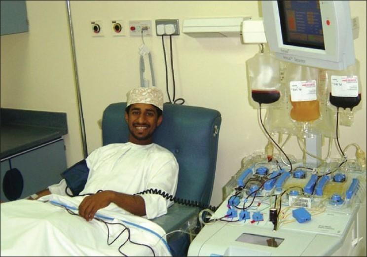
A clinical photograph depicting an apheresis procedure, specifically plateletapheresis, in a hospital or blood bank setting. A male donor is seated in a specialized blue reclining donor chair, connected to an automated blood cell separator (apheresis machine). The setup illustrates the extracorporeal circuit: blood is drawn through an intravenous line in the donor's arm, processed via a series of peristaltic pumps and a centrifugal separation chamber, and then returned. The machine features a monitoring screen and hangs several collection and infusion bags, containing red blood cells and amber-colored plasma or platelets, indicating the separation of blood components. A blood pressure cuff is visible on the donor's arm for monitoring hemodynamic stability during the procedure. This visual demonstrates the clinical application of medical technology in transfusion medicine for the collection of high-purity blood components like single-donor platelets.
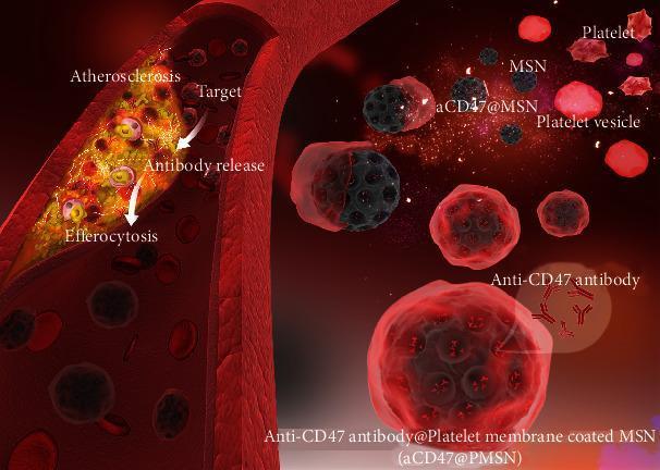
This medical illustration depicts a pathophysiology diagram of a therapeutic nanotechnology approach for treating atherosclerosis. The visual is divided into a longitudinal cross-section of a blood vessel and a detailed view of the microscopic components. The vessel shows a prominent atherosclerotic plaque characterized by a heterogeneous, lipid-rich core with internal cellular debris, causing significant luminal narrowing. Circulating within the vessel are red blood cells and various engineered nanoparticles. Key labeled components include mesoporous silica nanoparticles (MSN), shown as dark porous spheres; aCD47@MSN, representing MSN loaded with anti-CD47 antibody; and platelets/platelet vesicles. The primary therapeutic entity is the 'Anti-CD47 antibody@Platelet membrane coated MSN' (aCD47@PMSN), which consists of an antibody-loaded MSN core encapsulated in a semi-transparent red platelet membrane shell. Directional arrows illustrate the therapeutic mechanism: 'Targeting' of the plaque by aCD47@PMSN, 'Antibody release' into the lesion, and subsequent 'Efferocytosis' (clearance of necrotic cells). This diagram represents a biomimetic drug delivery system designed for cardiovascular intervention and immune-mediated plaque stabilization.
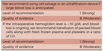
<table><tr><td>We recommend using cell salvage or an ultrafiltration device if large blood loss is anticipated.</td><td></td></tr><tr><td>Level of recommendation</td><td>1 (Strong)</td></tr><tr><td>Quality of evidence</td><td>B (Moderate)</td></tr><tr><td>If the intraoperative hemoglobin level is <10 g/dL and blood loss is ongoing, we recommend transfusion of packed blood cells along with fresh frozen plasma and platelets in a ratio of 1:1:1.</td><td></td></tr><tr><td>Level of recommendation</td><td>1 (Strong)</td></tr><tr><td>Quality of evidence</td><td>B (Moderate)</td></tr></table>
Components of Blood Transfusion and Their Indications
1. Packed Red Blood Cells (pRBCs)
- Symptomatic anemia (fatigue, dyspnea, angina) regardless of Hb level
- Restrictive threshold (general): Hb < 7 g/dL in most stable patients (supported by the landmark TRICC trial)
- Hb < 8 g/dL for patients with cardiovascular disease, cardiac surgery, or orthopedic surgery
- No transfusion benefit when Hb > 10 g/dL
- Acute blood loss with hemodynamic compromise (trauma, GI bleed, surgical hemorrhage)
- Hypoproliferative bone marrow disorders (aplastic anemia, myelodysplasia)
- Chronic hemolysis - sickle cell disease, thalassemia
- Preoperative optimization in symptomatic anemia
Note: In actively bleeding patients, Hb alone should not be the sole trigger - clinical status, hemodynamics, and rate of blood loss must be factored in.
- Tietz Textbook of Laboratory Medicine, 7th ed., p. 3743; Tintinalli's Emergency Medicine, p. 107
2. Fresh Frozen Plasma (FFP)
- Active bleeding with multiple coagulation factor deficiencies (DIC, massive hemorrhage, liver failure)
- Factor deficiency when specific factor concentrate is unavailable (e.g., Factor V or XI deficiency)
- Warfarin reversal with active bleeding or emergency surgery (where PCC is unavailable)
- Massive transfusion protocol - in a 1:1:1 ratio with pRBCs and platelets
- Thrombotic thrombocytopenic purpura (TTP) - as plasma exchange fluid
- Inherited or acquired coagulopathies
Important: Prophylactic FFP for mild coagulation abnormalities (INR < 1.5-2.0) in non-bleeding patients before procedures is not evidence-based and should be avoided.
- Tietz Textbook of Laboratory Medicine, 7th ed., p. 3748; Tintinalli's Emergency Medicine, p. 107
3. Platelets
- 1 apheresis unit (or 6 pooled random units) raises platelet count by ~50,000/mm³ in an adult
| Situation | Threshold |
|---|---|
| Prophylaxis - stable patients | < 10,000/mm³ |
| Prophylaxis - fever/infection/procedure | < 20,000/mm³ |
| Minor invasive procedures (e.g., LP, bone marrow biopsy) | < 50,000/mm³ |
| Major surgery or active bleeding | < 50,000-100,000/mm³ |
| Neurosurgery or ocular surgery | < 100,000/mm³ |
| Massive transfusion protocol | In 1:1:1 ratio |
- Qualitative platelet dysfunction (uremia, antiplatelet drugs) with active bleeding, regardless of count
- Dilutional thrombocytopenia during massive transfusion
Not indicated: In immune thrombocytopenic purpura (ITP) or TTP unless life-threatening bleeding, as transfused platelets are rapidly destroyed.
- Tietz Textbook of Laboratory Medicine, 7th ed.; Tintinalli's Emergency Medicine, p. 108
4. Cryoprecipitate (Cryo)
- Fibrinogen (~150-300 mg/unit)
- Factor VIII (~80-100 IU/unit)
- von Willebrand factor (vWF)
- Factor XIII
- Fibronectin
-
Hypofibrinogenemia (fibrinogen < 100-150 mg/dL) with bleeding - DIC, massive transfusion, obstetric hemorrhage
-
Hemophilia A (Factor VIII deficiency) when Factor VIII concentrate is unavailable
-
von Willebrand disease (types 1 and 2) when desmopressin (DDAVP) fails and specific concentrates are unavailable
-
Factor XIII deficiency with bleeding
-
Uremic bleeding (adjunct)
-
Topical fibrin sealant preparation
-
Tietz Textbook of Laboratory Medicine, 7th ed.; Fischer's Mastery of Surgery, 8th ed., p. 250
5. Granulocytes (Granulocyte Concentrates)
- Severe, prolonged neutropenia (ANC < 500/mm³) with life-threatening bacterial or fungal infection unresponsive to antibiotics/antifungals
- Neonatal sepsis with marrow failure
- Inherited neutrophil dysfunction (e.g., chronic granulomatous disease) with severe infection
6. Albumin
- Large-volume paracentesis (> 5L) - to prevent post-paracentesis circulatory dysfunction (8 g albumin per liter removed)
- Spontaneous bacterial peritonitis (SBP) - to reduce hepatorenal syndrome risk
- Hypoalbuminemia with hemodynamic instability (fluid resuscitation, severe burns)
- Hepatorenal syndrome as part of treatment protocols
7. Whole Blood
- Massive/combat trauma (military setting)
- Exchange transfusion in neonates
- Some massive transfusion protocols as an alternative to 1:1:1 component ratios
Massive Transfusion Protocol (MTP) Summary
- Ratio: pRBC : FFP : Platelets = 1:1:1
- Add cryoprecipitate when fibrinogen < 150 mg/dL
- Consider tranexamic acid (TXA) within 3 hours of injury
- Avoid crystalloid dilution
Key Points Summary
| Component | Main Contents | Key Indication | Storage |
|---|---|---|---|
| pRBCs | RBCs | Anemia, blood loss (Hb < 7-8 g/dL) | 42 days, 1-6°C |
| FFP | All clotting factors | Coagulopathy, DIC, massive transfusion | 1 year frozen |
| Platelets | Platelets | Thrombocytopenia < 10,000-50,000 | 5 days, 20-24°C |
| Cryoprecipitate | Fibrinogen, Factor VIII, vWF | Hypofibrinogenemia, Hemophilia A, vWD | 1 year frozen |
| Granulocytes | Neutrophils | Refractory severe infection with neutropenia | 24 hours |
| Albumin | Albumin | Large-volume paracentesis, SBP | Room temp |