
Can you make a powerpoint presentatiom on “malignant salivary gland neoplasm”
salivary gland carcinoma AND treatment
ESMO EURACAN salivary gland cancer clinical practice guideline 2022 diagnosis treatment follow up
https://pmc.ncbi.nlm.nih.gov/articles/PMC9808465
salivary gland carcinoma pathology histology mucoepidermoid adenoid cystic carcinoma
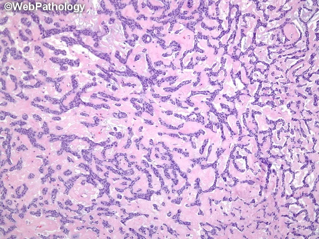
Imaging modality: Light microscopy of a hematoxylin and eosin stained histology slide from salivary gland tissue. The lesion demonstrates adenoid cystic carcinoma with classic cribriform architecture and multiple pseudocystic spaces. These gland-like spaces are variably sized and filled by dense, eosinophilic hyaline material representing duplicated basement membrane (basement lamina) produced by tumor cells. The basaloid neoplastic cells are arranged in small nests and narrow cords, with scant cytoplasm, oval to elongated (sometimes hyperchromatic) nuclei, and mild to moderate cytologic atypia. Delicate lace-like bands of stroma separate nests, creating a sieve-like, Swiss-cheese appearance. Overall tumor pattern may show admixtures of tubular and solid growth, but the cribriform component is prominent. The hyaline material within pseudocysts and the presence of basaloid cells support a diagnosis of adenoid cystic carcinoma. Clinically, ACC is associated with infiltrative growth and perineural invasion, contributing to local recurrence despite modest mitotic activity. This image is particularly relevant for educational purposes in surgical pathology, histology curricula, and diagnostic radiology correlations, aiding differentiation from polymorphous adenocarcinoma, mucoepidermoid carcinoma, and other salivary gland neoplasms. It provides a reference for recognizing the hallmark features in biopsy specimens and practicing pattern-based diagnosis.
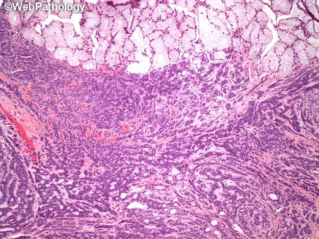
Low-power histology image of salivary gland tissue demonstrates polymorphic architectural patterns characteristic of polymorphous low-grade adenocarcinoma (PLGA). The section, stained with Hematoxylin and Eosin and viewed under light microscopy, shows residual benign minor salivary gland acini at the superior margin, providing an architectural reference for the tissue of origin. Tumor appears infiltrative within fibrous stroma, displaying a spectrum of growth patterns including tubular (glandular), cribriform, solid, papillary, trabecular, and single-file arrangements. The epithelial cells are relatively uniform, with small to moderate nuclei, inconspicuous nucleoli, and minimal cytologic atypia, consistent with a low-grade malignancy. Some areas exhibit a loosely arranged, hammock-like cellular nests that contribute to the polymorphous appearance. The overall patterning, combined with the intimate admixture of benign acini, supports a salivary gland neoplasm rather than a purely benign lesion. Diagnostic significance lies in recognizing architectural diversity as a hallmark of PLGA, which needs correlation with growth pattern and clinical behavior. Differential diagnoses include adenoid cystic carcinoma (cribriform pattern and perineural invasion), mucoepidermoid carcinoma, basal cell adenoma or carcinoma, and benign pleomorphic adenoma in the appropriate clinical context. This image is educational for pathology residents and clinicians learning salivary gland tumor histology and differential diagnosis.
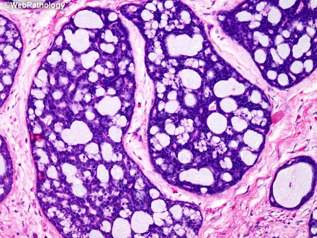
Adenoid cystic carcinoma of salivary gland on histology showing multiple nests with cribriform architecture, surrounded by a cuff of cellular myxoid stroma. Pseudoglandular lumina are filled with basophilic mucopolysaccharide-rich material that stains with Alcian blue, containing hyaluronic acid and sulfated acid mucins. The tumor cells are small, basaloid with uniform nuclei; perineural invasion may be present in some cases (not visible in this field). The pattern described—cribriform nests with mucoid, myxoid stroma—is classic for ACC and correlates with glandular origin from salivary tissue. Diagnostic significance lies in confirming ACC, guiding management toward surgical excision with wide margins and adjuvant radiotherapy, and anticipating potential perineural spread and recurrence. Differential considerations include mucoepidermoid carcinoma, basaloid adenocarcinoma, and polymorphous adenocarcinoma. Clinically, the image supports educational and diagnostic applications in head and neck pathology, pathology training, and research contexts. It is valuable for teaching histology of salivary gland neoplasms, pattern recognition of cribriform and myxoid components, and mucin staining interpretation. This image is valuable for diagnostic education, histopathology training, and multidisciplinary tumor boards.
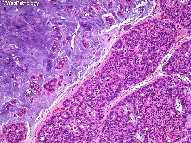
Imaging modality: light microscopy of hematoxylin and eosin stained paraffin-embedded salivary gland tissue from the parotid region. Anatomy: parotid gland tissue showing a classic biphasic neoplasm with epithelial elements intimately admixed with mesenchymal-like stroma. Description: Epithelial components form acinar and tubular structures, composed of duct‑forming cells and myoepithelial-like cells, set within a markedly hypocellular myxoid to chondromyxoid stroma. The epithelial nests are hypercellular in places and juxtaposed to expansive, basophilic stroma; the interface produces a biphasic appearance. The stroma often shows loose, lobulated myxoid material with mild chondroid differentiation, creating the distinctive mixed morphology. Overall, this pattern is typical of pleomorphic adenoma, a benign salivary gland tumor with epithelial–mesenchymal differentiation and typically a well circumscribed capsule. Diagnostic significance: the combination of acinar/tubular epithelial differentiation within a myxoid/chondromyxoid stroma strongly supports pleomorphic adenoma; differentiates from malignant salivary neoplasms. Differential considerations: mucoepidermoid carcinoma, adenoid cystic carcinoma, basal cell adenoma. Clinical correlation: presents as a slow-growing, painless parotid mass in adults; treatment usually involves complete surgical excision with facial nerve preservation and careful follow-up for recurrence. Potential applications: educational histology exemplar, pathology teaching repository, and radiology-pathology correlation studies. This image exemplifies classic pleomorphic adenoma histology suitable for teaching and diagnostic training in pathology education.
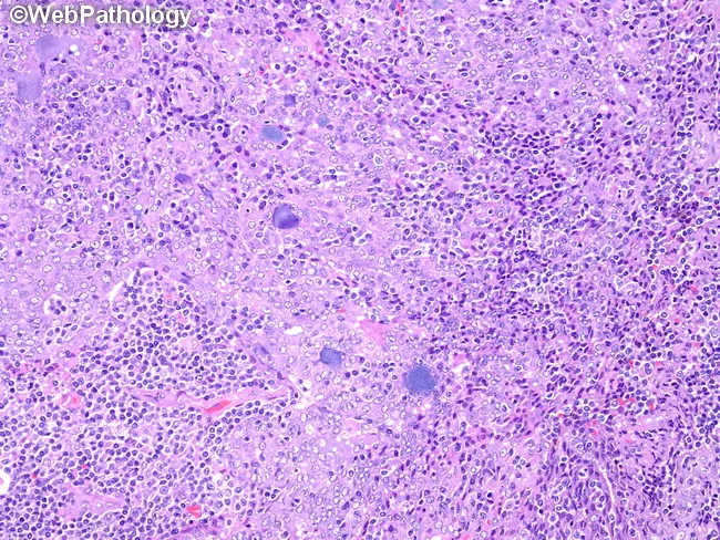
Imaging modality and technique: Light microscopy of a hematoxylin and eosin (H&E) stained salivary gland tissue section analyzed at high magnification, illustrating pleomorphic adenoma features. Anatomic location: parotid or salivary gland region; primary subject and tissue: salivary gland epithelium with myoepithelial components embedded in a myxoid/chondromyxoid stroma. Visual features: epithelial nests and tubules with cuboidal to columnar lining, intermingled with plasmacytoid or spindle-shaped myoepithelial cells; the background matrix is myxoid, frequently with mucopolysaccharide-rich stroma and occasional cartilaginous differentiation. The lesion is typically well circumscribed and shows a biphasic pattern comprising duct-forming epithelial elements and myoepithelial/myxoid components, without conspicuous cytologic atypia or invasion in benign cases. Pathological significance: this classic histology supports a diagnosis of pleomorphic (mixed) adenoma of the salivary gland, a benign tumor with rare malignant transformation and potential for recurrence if incompletely excised. Differential diagnosis: adenoid cystic carcinoma, mucoepidermoid carcinoma, basal cell adenoma, canalicular adenoma; clinical correlation: common presentation as a slow-growing painless parotid or minor salivary gland mass in middle adulthood; relevance to surgical planning and prognosis. Use cases: educational resource, tumor biology study, differential diagnosis training, and pathology exam prep. The image highlights the characteristic biphasic architecture and the myxoid stromal milieu essential for recognizing this entity.
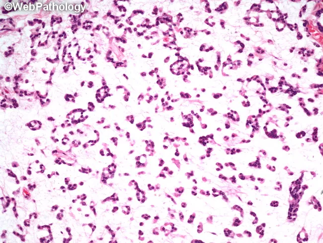
Comprehensive description: Light microscopy of a salivary gland lesion stained with Hematoxylin and Eosin (H&E). The specimen demonstrates a malignant salivary gland neoplasm, most consistent with acinic cell carcinoma. Tumor cells are arranged in sheets and minor acinar differentiation is evident, with a minority of cells showing conventional non-specific glandular morphology. The acinar cells have basophilic cytoplasm rich in coarse granules; cytoplasmic borders are crisp, and nuclei are relatively uniform with mild atypia. The tumor is embedded in a fibrous stroma with mild desmoplasia. An accompanying mild to moderate lymphoplasmacytic infiltrate surrounds tumor nests, and scattered plasma cells are present in the peritumoral milieu. No obvious necrosis is seen. Ductal differentiation is limited, supporting acinic lineage. Mitotic activity is low. Overall, the architecture shows lobulocentric arrangement with cohesive nests of mucin-poor, basophilic cells, consistent with acinic cell differentiation rather than serous acinar cells of normal gland. Clinically, this histology has diagnostic significance for salivary gland neoplasms and helps distinguish from mucoepidermoid carcinoma and adenoid cystic carcinoma; immunohistochemical staining for amylase, DOG1, or S-100 may aid confirmation. This image is suitable for educational pathology, differential diagnosis training, and correlates with radiographic parotid-region masses undergoing parotidectomy. Correlative molecular data may refine classification.
~/
mkdir -p "/tmp/workspace/malignant-salivary-gland-neoplasm/src" "/tmp/workspace/malignant-salivary-gland-neoplasm/output" "/tmp/workspace/malignant-salivary-gland-neoplasm/assets" && python /tmp/skills/shared/scripts/fetch_images.py "https://cdn.orris.care/cdss_images/Pathology_1760046781317_59561d95-cb12-4e9f-91cc-30a79824827f.jpg" > "/tmp/workspace/malignant-salivary-gland-neoplasm/assets/image_fetch.json"
~/malignant-salivary-gland-neoplasm/
python -c "import json,base64; d=json.load(open('/tmp/workspace/malignant-salivary-gland-neoplasm/assets/image_fetch.json'))[0]['base64'].split(',',1)[1]; open('/tmp/workspace/malignant-salivary-gland-neoplasm/assets/adenoid_cystic_histology.webp','wb').write(base64.b64decode(d))"
python - <<'PY' from PIL import Image p='/tmp/workspace/malignant-salivary-gland-neoplasm/assets/adenoid_cystic_histology.webp' im=Image.open(p); print(im.format, im.size) im.convert('RGB').save('/tmp/workspace/malignant-salivary-gland-neoplasm/assets/adenoid_cystic_histology.jpg') PY
python - <<'PY' from pathlib import Path p=Path('/tmp/workspace/malignant-salivary-gland-neoplasm/src/create_deck.py') p.write_text(r'''from pptx import Presentation from pptx.util import Inches, Pt from pptx.dml.color import RGBColor from pptx.enum.text import PP_ALIGN, MSO_ANCHOR from pptx.enum.shapes import MSO_SHAPE, MSO_CONNECTOR from pptx.enum.dml import MSO_THEME_COLOR from pptx.enum.text import MSO_AUTO_SIZE OUT='/tmp/workspace/malignant-salivary-gland-neoplasm/output/Malignant_Salivary_Gland_Neoplasm.pptx' IMG='/tmp/workspace/malignant-salivary-gland-neoplasm/assets/adenoid_cystic_histology.jpg' prs=Presentation(); prs.slide_width=Inches(13.333); prs.slide_height=Inches(7.5) blank=prs.slide_layouts[6] NAVY=RGBColor(13,37,63); TEAL=RGBColor(0,124,140); CYAN=RGBColor(93,203,214); PALE=RGBColor(231,247,247); WHITE=RGBColor(255,255,255); INK=RGBColor(28,42,55); GREY=RGBColor(92,108,121); LINE=RGBColor(205,220,224); RED=RGBColor(181,56,60); GOLD=RGBColor(232,178,57); LIGHT=RGBColor(247,250,251) def rect(sl,x,y,w,h,color, radius=False, line=None): sh=sl.shapes.add_shape(MSO_SHAPE.ROUNDED_RECTANGLE if radius else MSO_SHAPE.RECTANGLE, Inches(x), Inches(y), Inches(w), Inches(h)); sh.fill.solid(); sh.fill.fore_color.rgb=color; sh.line.color.rgb=line or color if radius: sh.adjustments[0]=0.12 return sh def text(sl,x,y,w,h,txt,size=18,color=INK,bold=False,align=None,font='Aptos',val=None): tb=sl.shapes.add_textbox(Inches(x),Inches(y), Inches(w),Inches(h)); tf=tb.text_frame; tf.clear(); tf.word_wrap=True; tf.margin_left=Inches(.04); tf.margin_right=Inches(.04); tf.margin_top=Inches(.02); tf.margin_bottom=Inches(.02) p=tf.paragraphs[0]; p.text=txt; p.font.name=font; p.font.size=Pt(size); p.font.bold=bold; p.font.color.rgb=color if align: p.alignment=align if val: tf.vertical_anchor=val return tb def rich(sl,x,y,w,h,items, size=17): tb=sl.shapes.add_textbox(Inches(x),Inches(y), Inches(w), Inches(h)); tf=tb.text_frame; tf.clear(); tf.word_wrap=True; tf.margin_left=0; tf.margin_right=0; tf.margin_top=0; tf.margin_bottom=0 for i,(head,body) in enumerate(items): p=tf.paragraphs[0] if i==0 else tf.add_paragraph(); p.space_after=Pt(10); p.level=0 r=p.add_run(); r.text=head; r.font.name='Aptos'; r.font.size=Pt(size); r.font.bold=True; r.font.color.rgb=NAVY r=p.add_run(); r.text=body; r.font.name='Aptos'; r.font.size=Pt(size); r.font.color.rgb=INK return tb def bullet(sl,x,y,w,h,arr,size=17): tb=sl.shapes.add_textbox(Inches(x), Inches(y), Inches(w), Inches(h)); tf=tb.text_frame; tf.clear(); tf.word_wrap=True; tf.margin_left=Inches(.10); tf.margin_right=0; tf.margin_top=0; tf.margin_bottom=0 for i,s in enumerate(arr): p=tf.paragraphs[0] if i==0 else tf.add_paragraph(); p.text=s; p.level=0; p.font.name='Aptos'; p.font.size=Pt(size); p.font.color.rgb=INK; p.space_after=Pt(9); p.bullet=True return tb def header(sl,title,kicker, n): rect(sl,0,0,13.333,.24,TEAL); text(sl,.55,.42,11.9,.25,kicker.upper(),10,TEAL,True); text(sl,.55,.7,11.9,.55,title,28,NAVY,True); rect(sl,.55,1.35,12.2,.02,LINE) text(sl,.55,7.12,11.8,.18,'Malignant salivary gland neoplasms',9,GREY); text(sl,12.25,7.12,.5,.18,f'{n:02d}',9,TEAL,True,PP_ALIGN.RIGHT) def foot(sl,src): text(sl,.55,6.86,12.1,.16,src,7.4,GREY) def add_note(sl,txt): ts=sl.notes_slide.notes_text_frame; ts.text=txt def badge(sl,x,y,label, color=TEAL): rect(sl,x,y,1.25,.34,color,True); text(sl,x,y+.05,1.25,.18,label,9,WHITE,True,PP_ALIGN.CENTER) def card(sl,x,y,w,h,title,body,accent=TEAL): rect(sl,x,y,w,h,WHITE,True,LINE); rect(sl,x,y,.10,h,accent); text(sl,x+.28,y+.22,w-.45,.25,title,15,NAVY,True); text(sl,x+.28,y+.62,w-.45,h-.75,body,13.4,INK) # 1 s=prs.slides.add_slide(blank); rect(s,0,0,13.333,7.5,NAVY); rect(s,0,0,13.333,.22,TEAL); rect(s,.65,1.02,.10,4.85,TEAL); text(s,1.0,1.12,10.9,.35,'HEAD & NECK ONCOLOGY',13,CYAN,True); text(s,1.0,1.78,10.9,1.4,'Malignant salivary\ngland neoplasms',38,WHITE,True); text(s,1.02,3.48,9.7,.6,'A practical framework for classification, diagnosis, treatment, and follow-up',19,RGBColor(214,231,235)); for x,y,r,c in [(11.25,1.15,.45,CYAN),(10.72,2.18,.24,GOLD),(11.95,2.68,.32,TEAL),(10.93,3.35,.18,WHITE),(11.45,4.05,.6,RGBColor(31,72,98))]: sh=s.shapes.add_shape(MSO_SHAPE.OVAL,Inches(x),Inches(y), Inches(r), Inches(r)); sh.fill.solid(); sh.fill.fore_color.rgb=c; sh.line.color.rgb=c text(s,1.02,6.43,9.0,.25,'Educational presentation | Adapt local multidisciplinary protocols',11,RGBColor(181,210,216)); text(s,.65,7.05,12,.2,'Sources: Cummings Otolaryngology; ESMO-EURACAN Guideline (2022)',8,RGBColor(181,210,216)); add_note(s,'Opening slide. Explain that salivary gland carcinoma is a heterogeneous group, not one disease.') # 2 s=prs.slides.add_slide(blank); header(s,'Why this topic matters','Context',2) card(s,.65,1.75,3.85,1.6,'Rare but diverse','Many distinct epithelial malignancies arise in major and minor salivary glands. Histologic subtype changes behavior and management.',TEAL) card(s,4.74,1.75,3.85,1.6,'Site predicts risk','Parotid masses are most common, but a smaller proportion are malignant. Submandibular, sublingual, and minor-gland lesions carry higher malignant proportions.',GOLD) card(s,8.83,1.75,3.85,1.6,'Long natural history','Adenoid cystic carcinoma may relapse or metastasize many years after primary treatment, requiring prolonged surveillance.',RED) text(s,.72,3.85,11.6,.33,'Clinical implication',18,NAVY,True); rect(s,.72,4.32,11.6,1.25,PALE,True); text(s,1.0,4.62,11.0,.55,'A painless lump is not reassuring when it is fixed, rapidly enlarging, associated with pain, skin involvement, cervical nodes, or facial nerve dysfunction.',19,NAVY,True,PP_ALIGN.CENTER, val=MSO_ANCHOR.MIDDLE) foot(s,'Cummings Otolaryngology Head and Neck Surgery, Salivary Gland Tumors, key points and physical examination.'); add_note(s,'Set the clinical context. Emphasize heterogeneity and the need for specialist pathology and tumor-board review.') # 3 s=prs.slides.add_slide(blank); header(s,'Where do these tumors arise?','Anatomy & epidemiology',3) # gland schematic text(s,.7,1.72,5.3,.28,'Major glands',18,NAVY,True) for y,label,sub,col in [(2.2,'Parotid','Preauricular / retromandibular',TEAL),(3.55,'Submandibular','Submandibular triangle',GOLD),(4.9,'Sublingual','Floor of mouth',RED)]: sh=s.shapes.add_shape(MSO_SHAPE.OVAL, Inches(.82), Inches(y), Inches(.86), Inches(.7)); sh.fill.solid(); sh.fill.fore_color.rgb=col; sh.line.color.rgb=col text(s,1.98,y+.02,3.9,.25,label,17,NAVY,True); text(s,1.98,y+.34,3.9,.2,sub,12,GREY) rect(s,6.25,1.65,.02,4.6,LINE); text(s,6.72,1.72,5.55,.28,'Pattern by site',18,NAVY,True) rich(s,6.72,2.2,5.45,3.5,[('Parotid: ','largest share of all salivary tumors; in one large review, 73% arose here and about 15% were malignant.\n'),('Minor glands: ','less common overall but a higher malignant proportion, especially palate lesions.\n'),('Clinical rule: ','the probability of malignancy rises as one moves from parotid toward submandibular, sublingual, and minor glands.')],17) foot(s,'Cummings Otolaryngology Head and Neck Surgery, Salivary Gland Tumors, key points (review of 2,410 cases).'); add_note(s,'Explain the clinical heuristic: site informs pre-test probability but does not replace tissue diagnosis.') # 4 s=prs.slides.add_slide(blank); header(s,'Classification: a family of carcinomas','Histopathology',4) text(s,.7,1.67,11.8,.3,'The WHO framework recognizes numerous entities. In clinical practice, subtype, grade, stage, and biomarkers drive decisions.',16,GREY) cols=[('Common entities',['Mucoepidermoid carcinoma','Adenoid cystic carcinoma','Acinic cell carcinoma','Secretory carcinoma'],TEAL),('Aggressive / high-grade',['Salivary duct carcinoma','Carcinoma ex pleomorphic adenoma','High-grade adenocarcinoma','Poorly differentiated carcinoma'],RED),('Other important entities',['Polymorphous adenocarcinoma','Epithelial-myoepithelial carcinoma','Myoepithelial carcinoma','Clear cell carcinoma'],GOLD)] for i,(title,items,col) in enumerate(cols): x=.65+i*4.15; rect(s,x,2.25,3.72,3.35,WHITE,True,LINE); rect(s,x,2.25,3.72,.48,col,True); text(s,x+.18,2.36,3.34,.2,title,13,WHITE,True); bullet(s,x+.28,3.0,3.1,2.25,items,15) rect(s,.65,5.93,12.0,.55,PALE,True); text(s,.92,6.09,11.45,.18,'Do not treat “salivary gland cancer” as a single entity: prognosis and systemic options are histology-specific.',14,NAVY,True,PP_ALIGN.CENTER) foot(s,'Cummings Otolaryngology Head and Neck Surgery, Malignant Salivary Gland Tumors; WHO classification referenced by ESMO-EURACAN guideline.'); add_note(s,'Mention that a final histologic classification should come from experienced head and neck pathology review.') #5 s=prs.slides.add_slide(blank); header(s,'Clinical presentation: recognize red flags','Assessment',5) text(s,.7,1.65,11.5,.32,'Malignant lesions may be indolent or rapidly progressive. Symptoms reflect local invasion and neural involvement.',16,GREY) for i,(t,b,c) in enumerate([('Mass behavior','Rapid growth, firmness, fixation, ulceration or skin tethering',RED),('Neural symptoms','Pain, paresthesia, trismus, facial weakness or paralysis',RED),('Nodal disease','Cervical lymphadenopathy, especially in high-grade tumors',GOLD),('Minor gland lesions','Palatal, buccal, lip, or sinonasal submucosal mass',TEAL)]): x=.7+(i%2)*6.05; y=2.25+(i//2)*1.55; card(s,x,y,5.55,1.18,t,b,c) rect(s,.7,5.62,11.65,.68,NAVY,True); text(s,1.0,5.83,11.05,.25,'Facial nerve weakness in a parotid mass is a major warning sign for malignancy and should expedite specialist evaluation.',15,WHITE,True,PP_ALIGN.CENTER) foot(s,'Cummings Otolaryngology Head and Neck Surgery, Physical Examination, malignant salivary gland neoplasms.'); add_note(s,'Facial weakness and pain are particularly important. Do not biopsy a parotid mass via open incision before proper imaging and FNA/core planning.') #6 s=prs.slides.add_slide(blank); header(s,'Diagnostic work-up: image, sample, stage','Diagnosis',6) steps=[('1','Focused assessment','History, cranial nerve examination, oral cavity and neck examination.'),('2','Imaging','MRI is preferred when malignancy is suspected. CT is useful when MRI is contraindicated or for bone assessment.'),('3','Tissue diagnosis','Fine-needle aspiration (FNA) for screening. If inadequate or more architecture is needed, use core needle biopsy.'),('4','Stage & plan','Neck evaluation in all patients. Consider FDG-PET/CT for high-grade disease and distant-metastasis assessment.')] for i,(n,t,b) in enumerate(steps): y=1.72+i*1.16; sh=s.shapes.add_shape(MSO_SHAPE.OVAL,Inches(.75), Inches(y+.05), Inches(.55), Inches(.55)); sh.fill.solid(); sh.fill.fore_color.rgb=TEAL; sh.line.color.rgb=TEAL; text(s,.75,y+.19,.55,.14,n,12,WHITE,True,PP_ALIGN.CENTER) text(s,1.55,y,3.1,.22,t,16,NAVY,True); text(s,4.45,y,7.75,.48,b,15,INK) foot(s,'ESMO-EURACAN Clinical Practice Guideline for salivary gland cancer (2022): diagnosis and staging recommendations.'); add_note(s,'Mention the Milan system for FNA reporting. For minor salivary tumors, biopsy approach differs and should include tumor and surrounding stroma.') #7 s=prs.slides.add_slide(blank); header(s,'Pathology should answer more than “malignant”','Diagnostic pathology',7) s.shapes.add_picture(IMG, Inches(.72), Inches(1.7), width=Inches(5.0), height=Inches(3.75)); rect(s,.72,5.45,5.0,.48,NAVY,True); text(s,.87,5.59,4.7,.14,'Adenoid cystic carcinoma: cribriform “Swiss-cheese” pattern',10.5,WHITE,True,PP_ALIGN.CENTER) text(s,6.23,1.78,5.6,.3,'Essential pathology report elements',18,NAVY,True) bullet(s,6.25,2.25,5.55,3.0,['Histologic entity and grade','Tumor size and extent; margins','Lymphovascular and perineural invasion','Nodal status and extranodal extension, if dissected','Ancillary immunohistochemistry and molecular findings when relevant'],16) rect(s,6.25,5.5,5.55,.66,PALE,True); text(s,6.5,5.69,5.05,.22,'Perineural invasion is a characteristic concern in adenoid cystic carcinoma.',14,NAVY,True,PP_ALIGN.CENTER) foot(s,'Image: educational histology, adenoid cystic carcinoma. Cummings Otolaryngology; ESMO-EURACAN guideline.'); add_note(s,'Use the image to introduce the cribriform architecture of ACC. Perineural invasion helps explain pain and local recurrence risk.') #8 s=prs.slides.add_slide(blank); header(s,'Staging and risk stratification','Prognostic factors',8) text(s,.7,1.68,11.5,.28,'AJCC TNM staging for major salivary gland tumors is combined with histologic and patient-specific risk factors.',16,GREY) for i,(t,b,c) in enumerate([('Tumor factors','High grade, large T category, positive/close margin, lymphovascular invasion, perineural invasion',RED),('Nodal factors','Clinical or pathologic nodal metastases and extranodal extension',GOLD),('Histology / biology','Subtype-specific behavior, HER2 or androgen receptor expression, NTRK fusion, other actionable alterations',TEAL)]): card(s,.72+i*4.08,2.3,3.7,2.25,t,b,c) rect(s,.72,5.17,11.86,.85,NAVY,True); text(s,1.0,5.42,11.28,.25,'Risk is not defined by stage alone: a small high-grade salivary duct carcinoma and an adenoid cystic carcinoma demand different planning and surveillance.',15,WHITE,True,PP_ALIGN.CENTER) foot(s,'ESMO-EURACAN guideline (2022); Cummings Otolaryngology, malignant salivary gland tumors.'); add_note(s,'Avoid relying on a single factor. This is why multidisciplinary discussion is useful before and after surgery.') #9 s=prs.slides.add_slide(blank); header(s,'Localized disease: surgery is the foundation','Management',9) rect(s,.7,1.72,12.0,.7,PALE,True); text(s,.98,1.94,11.4,.22,'Goal: complete resection with negative margins while preserving function when oncologically safe.',17,NAVY,True,PP_ALIGN.CENTER) for i,(t,b,c) in enumerate([('Primary resection','Appropriate parotidectomy, submandibular resection, or wide local excision for minor gland primaries. Preserve facial nerve if uninvolved.',TEAL),('Neck management','Treat the neck therapeutically for clinically positive nodes. Consider elective treatment when occult-nodal risk is meaningful, particularly in high-grade disease.',GOLD),('Reconstruction & rehabilitation','Plan facial nerve, soft-tissue, oral function, and cosmetic reconstruction early. Supportive care is part of definitive treatment.',RED)]): card(s,.72+i*4.08,2.82,3.7,2.7,t,b,c) foot(s,'ESMO-EURACAN guideline (2022): local and regional management; Cummings Otolaryngology.'); add_note(s,'The extent of parotid surgery depends on disease location and involvement. Facial nerve sacrifice is reserved for direct tumor invasion, not proximity alone.') #10 s=prs.slides.add_slide(blank); header(s,'When is postoperative radiotherapy considered?','Adjuvant treatment',10) text(s,.7,1.67,11.5,.32,'Radiotherapy is commonly integrated after surgery for features associated with local or regional recurrence risk.',16,GREY) for i,(t,items,c) in enumerate([('Typical indications',['High-grade histology','T3-T4 primary','Positive or close margins','Perineural invasion','Nodal disease'],RED),('Treatment principles',['Cover primary bed and indicated nodal volumes','Account for named-nerve pathways when perineural spread is present','Use modern conformal planning to limit organ-at-risk dose'],TEAL)]): x=.72+i*5.95; rect(s,x,2.35,5.4,3.28,WHITE,True,LINE); rect(s,x,2.35,5.4,.48,c,True); text(s,x+.22,2.47,4.95,.18,t,14,WHITE,True); bullet(s,x+.35,3.03,4.7,2.2,items,15) rect(s,.72,5.98,11.35,.42,RGBColor(255,246,229),True); text(s,.92,6.1,10.96,.16,'Concurrent chemotherapy with postoperative radiation is not routine for all salivary gland carcinomas; individualize in a specialist team.',12.5,INK,True,PP_ALIGN.CENTER) foot(s,'ESMO-EURACAN guideline (2022): postoperative radiotherapy recommendations.'); add_note(s,'Avoid describing chemoradiotherapy as standard for all cases. Evidence is limited and differs from squamous cell carcinoma of the head and neck.') #11 s=prs.slides.add_slide(blank); header(s,'Recurrent or metastatic disease: subtype and biomarkers matter','Systemic management',11) text(s,.7,1.64,11.7,.28,'First ask: can limited recurrence be treated locally? If systemic therapy is needed, match therapy to histology and molecular findings.',16,GREY) for i,(t,b,c) in enumerate([('Local options','Selected re-resection, re-irradiation, stereotactic approaches, or ablation may benefit oligometastatic or locoregional recurrence.',TEAL),('Salivary duct carcinoma','Test androgen receptor and HER2 in distant metastatic disease. Androgen deprivation or HER2-directed regimens may be considered when positive.',RED),('Secretory carcinoma','Test for NTRK fusion. TRK inhibitors can be effective in fusion-positive disease.',GOLD),('Other histologies','Cytotoxic chemotherapy, targeted agents, or clinical trials are selected by disease tempo, symptoms, prior therapy, and actionable findings.',TEAL)]): x=.7+(i%2)*6.05; y=2.22+(i//2)*1.68; card(s,x,y,5.55,1.32,t,b,c) foot(s,'ESMO-EURACAN guideline (2022); Prost et al. ESMO Open 2024, systematic review of systemic treatments in recurrent/metastatic salivary gland cancer (PMID 39368417).'); add_note(s,'Stress that evidence is limited and subtype-specific. Molecular testing can directly change options in salivary duct and secretory carcinoma.') #12 s=prs.slides.add_slide(blank); header(s,'Surveillance and survivorship','Follow-up',12) text(s,.72,1.67,11.5,.3,'Follow-up is tailored to subtype, recurrence risk, anatomy, treatment effects, and whether measurable disease remains.',16,GREY) for i,(t,b,c) in enumerate([('Clinical review','History, cranial nerve and head-and-neck examination; evaluate swallowing, speech, shoulder function, pain, and xerostomia.',TEAL),('Imaging','MRI is useful for local and perineural assessment. Chest CT is especially important in adenoid cystic carcinoma because late pulmonary metastases may occur.',GOLD),('Suggested cadence','For adenoid cystic carcinoma, guideline-based imaging is more frequent early, then continues long-term. Other types are often imaged every 6-12 months initially, then less often.',RED)]): card(s,.72+i*4.08,2.35,3.7,2.85,t,b,c) rect(s,.72,5.65,11.9,.52,PALE,True); text(s,.95,5.83,11.45,.16,'Late relapse is a defining issue in adenoid cystic carcinoma. Follow-up should not stop after a short disease-free interval.',13.5,NAVY,True,PP_ALIGN.CENTER) foot(s,'ESMO-EURACAN guideline (2022): follow-up recommendations.'); add_note(s,'Follow-up should include functional rehabilitation and management of radiation-related xerostomia, dental health, and psychosocial needs.') #13 s=prs.slides.add_slide(blank); header(s,'Take-home messages','Summary',13) for i,(num,t,b,c) in enumerate([('01','Think “heterogeneous”','Histology and site change prognosis, local treatment, molecular testing, and surveillance.'),('02','Investigate systematically','MRI, appropriate needle biopsy, pathology review, and neck/distant staging are the core sequence.'),('03','Aim for durable local control','Surgery plus risk-adapted radiotherapy remains central for most resectable malignancies.'),('04','Use biomarkers intelligently','AR/HER2 and NTRK testing can open subtype-specific options in recurrent or metastatic disease.')]): y=1.68+i*1.18; text(s,.8,y,.7,.35,num,22,c,True); text(s,1.72,y,3.6,.26,t,16,NAVY,True); text(s,5.05,y,6.85,.44,b,14.5,INK); rect(s,.8,y+.72,11.6,.012,LINE) foot(s,'Teaching summary based on Cummings Otolaryngology and ESMO-EURACAN clinical practice guideline (2022).'); add_note(s,'Conclude with the need for multidisciplinary care and personalized, subtype-specific treatment.') #14 s=prs.slides.add_slide(blank); header(s,'Selected references','References',14) refs=[ 'van Herpen C, et al. Salivary gland cancer: ESMO-EURACAN Clinical Practice Guideline for diagnosis, treatment and follow-up. ESMO Open. 2022;7:100602. PMID: 36567082.', 'Cummings Otolaryngology Head and Neck Surgery. Salivary gland tumors and malignant salivary gland tumors. Elsevier.', 'Prost D, et al. Systemic treatments in recurrent or metastatic salivary gland cancer: a systematic review. ESMO Open. 2024. PMID: 39368417.', 'WHO Classification of Head and Neck Tumours. 5th ed. International Agency for Research on Cancer.', 'Image used on slide 7: educational H&E histology of adenoid cystic carcinoma from the indexed medical image library.' ] bullet(s,.75,1.75,11.7,4.55,refs,15) rect(s,.75,6.1,11.8,.5,NAVY,True); text(s,1,6.26,11.25,.18,'For education only. Clinical decisions require local protocols and multidisciplinary specialist assessment.',12.5,WHITE,True,PP_ALIGN.CENTER) prs.save(OUT) print(OUT) ''') PY python "/tmp/workspace/malignant-salivary-gland-neoplasm/src/create_deck.py"
~/malignant-salivary-gland-neoplasm/src/create_deck.py
Successfully replaced 1 instance(s) of the string in '/tmp/workspace/malignant-salivary-gland-neoplasm/src/create_deck.py'
python "/tmp/workspace/malignant-salivary-gland-neoplasm/src/create_deck.py" && python - <<'PY' from pptx import Presentation p='/tmp/workspace/malignant-salivary-gland-neoplasm/output/Malignant_Salivary_Gland_Neoplasm.pptx' prs=Presentation(p) print('slides=',len(prs.slides)) for i,s in enumerate(prs.slides,1): texts=[sh.text.strip().replace('\n',' | ') for sh in s.shapes if getattr(sh,'has_text_frame',False) and sh.text.strip()] print(i, ' :: '.join(texts[:3])) PY
python - <<'PY' from pptx import Presentation from pathlib import Path p=Path('/tmp/workspace/malignant-salivary-gland-neoplasm/output/Malignant_Salivary_Gland_Neoplasm.pptx') prs=Presentation(str(p)) assert len(prs.slides)==14 assert p.stat().st_size > 50000 for i,slide in enumerate(prs.slides,1): assert any(getattr(shape,'has_text_frame',False) and shape.text.strip() for shape in slide.shapes), f'empty slide {i}' print(f'Validated: {len(prs.slides)} slides, {p.stat().st_size:,} bytes') PY
Create a one-page PDF quick reference
salivary gland carcinoma AND treatment
ESMO EURACAN salivary gland cancer guideline diagnosis treatment follow-up 2022
~/
python - <<'PY' from pathlib import Path p=Path('/tmp/workspace/malignant-salivary-gland-neoplasm/src/create_quick_reference.py') p.write_text(r'''from reportlab.pdfgen import canvas from reportlab.lib.pagesizes import A4 from reportlab.lib.colors import HexColor, white from reportlab.pdfbase.pdfmetrics import stringWidth from reportlab.lib.utils import simpleSplit from reportlab.pdfbase.ttfonts import TTFont from reportlab.pdfbase import pdfmetrics from reportlab.lib.colors import Color OUT='/tmp/workspace/malignant-salivary-gland-neoplasm/output/Malignant_Salivary_Gland_Neoplasm_Quick_Reference.pdf' W,H=A4 NAVY=HexColor('#0D253F'); TEAL=HexColor('#007C8C'); CYAN=HexColor('#5BCBD6'); PALE=HexColor('#E7F7F7'); INK=HexColor('#1C2A37'); GREY=HexColor('#596C79'); LINE=HexColor('#CEDCE0'); RED=HexColor('#B5383C'); GOLD=HexColor('#D99E25'); LIGHT=HexColor('#F7FAFB'); WHITE=white c=canvas.Canvas(OUT,pagesize=A4); c.setTitle('Malignant Salivary Gland Neoplasm - Quick Reference') def rect(x,y,w,h,fill,stroke=None,r=0): c.setFillColor(fill); c.setStrokeColor(stroke or fill) if r: c.roundRect(x,y,w,h,r,fill=1,stroke=1 if stroke else 0) else: c.rect(x,y,w,h,fill=1,stroke=1 if stroke else 0) def txt(x,y,s,size=9,color=INK,bold=False): c.setFillColor(color); c.setFont('Helvetica-Bold' if bold else 'Helvetica',size); c.drawString(x,y,s) def wrap(x,y,w,s,size=8.3,color=INK,leading=None,bold=False): leading=leading or size*1.22; c.setFillColor(color); c.setFont('Helvetica-Bold' if bold else 'Helvetica',size) lines=simpleSplit(s,'Helvetica-Bold' if bold else 'Helvetica',size,w) for line in lines: c.drawString(x,y,line); y-=leading return y def bullet(x,y,w,s,size=8.1,color=INK): lines=simpleSplit(s,'Helvetica',size,w-11); c.setFillColor(TEAL); c.circle(x+2.5,y+2,1.6,fill=1,stroke=0); c.setFillColor(color); c.setFont('Helvetica',size) for j,line in enumerate(lines): c.drawString(x+10,y-j*10,line) return y-len(lines)*10-3 def section(x,y,w,h,title,accent): rect(x,y,w,h,WHITE,LINE,6); rect(x,y+h-21,w,21,accent,None,6); c.setFillColor(WHITE); c.setFont('Helvetica-Bold',10); c.drawString(x+9,y+h-14,title) # header rect(0,H-61,W,61,NAVY); rect(0,H-8,W,8,TEAL) txt(33,H-32,'MALIGNANT SALIVARY GLAND NEOPLASM',19,WHITE,True) txt(34,H-47,'One-page clinical quick reference | Adult head and neck oncology',8.5,HexColor('#C9E8EB')) # warning band rect(33,H-88,W-66,18,PALE,None,5); txt(42,H-81,'Clinical suspicion: rapidly enlarging, fixed, painful mass; facial weakness; skin involvement; or cervical lymphadenopathy.',8.4,NAVY,True) # columns L=33; gap=12; col=(W-66-gap)/2; R=L+col+gap # left top section(L,H-236,col,134,'1. CLASSIFY & RECOGNIZE',TEAL) y=H-124 for s in ['Major sites: parotid (most common), submandibular, sublingual. Minor-gland tumors frequently involve palate.','Common entities: mucoepidermoid carcinoma, adenoid cystic carcinoma (ACC), acinic cell carcinoma, salivary duct carcinoma, carcinoma ex pleomorphic adenoma.','Risk clue: malignancy proportion increases outside the parotid; sublingual masses deserve particular concern.']: y=bullet(L+10,y,col-20,s) # right top section(R,H-236,col,134,'2. DIAGNOSTIC SEQUENCE',GOLD) y=H-124 for s in ['Focused examination: cranial nerves, facial nerve, oral cavity, skin, and both necks.','Imaging: MRI preferred when malignancy is suspected. CT if MRI is contraindicated or bony anatomy is key.','Tissue: FNA for initial sampling. Core needle biopsy if FNA is inadequate or architecture is needed.','Staging: assess neck; consider FDG-PET/CT for high-grade disease or distant spread.']: y=bullet(R+10,y,col-20,s) # middle left section(L,H-406,col,150,'3. PATHOLOGY & RISK',RED) y=H-254 for s in ['Report: histologic entity, grade, size/extent, margins, lymphovascular invasion, perineural invasion, nodes and extranodal extension.','ACC: neural invasion, local recurrence, and late lung metastases are characteristic clinical concerns.','Biomarkers: in recurrent/metastatic disease, test AR and HER2 in salivary duct carcinoma; test for NTRK fusion in secretory carcinoma.']: y=bullet(L+10,y,col-20,s) # middle right section(R,H-406,col,150,'4. LOCALIZED DISEASE',TEAL) y=H-254 for s in ['Surgery is the core treatment: pursue complete resection with negative margins while preserving function when safe.','Neck: therapeutic nodal management for clinically positive nodes; elective treatment is considered for meaningful occult-node risk.','Postoperative radiotherapy is generally considered for high grade, T3-T4 tumors, close/positive margins, perineural invasion, or nodal disease.','Concurrent chemotherapy is not routine for every salivary gland carcinoma.']: y=bullet(R+10,y,col-20,s) # lower wide section(L,92,W-66,130,'5. RECURRENT / METASTATIC DISEASE & FOLLOW-UP',NAVY) # use two mini cols x1=L+11; x2=L+col+7; y=183 txt(x1,y,'Management principles',9,NAVY,True); y-=15 for s in ['Reassess for local therapy: selected re-resection, re-irradiation, stereotactic treatment or ablation may help limited recurrence.','Systemic treatment is subtype- and biomarker-directed. Consider AR-targeted treatment or HER2-directed therapy in appropriately selected salivary duct carcinoma; TRK inhibitor in NTRK fusion-positive secretory carcinoma.','Discuss clinical trials when feasible.']: y=bullet(x1,y,col-20,s,7.8) y=183; txt(x2,y,'Surveillance',9,NAVY,True); y-=15 for s in ['Clinical review plus tailored imaging. MRI is useful for local and perineural disease; chest CT is important for ACC.','ACC needs long-term follow-up because recurrence and metastases can occur late.','Supportive care: xerostomia, dental care, swallowing, facial function, pain, nutrition, and psychosocial needs.']: y=bullet(x2,y,col-20,s,7.8) # bottom key actions rect(33,57,W-66,23,NAVY,None,5); txt(43,66,'BOTTOM LINE',8.5,CYAN,True); txt(112,66,'Image appropriately, obtain high-quality tissue, stage accurately, and treat in a multidisciplinary specialist team.',8.4,WHITE,True) # footer c.setStrokeColor(LINE); c.line(33,44,W-33,44) txt(33,32,'Sources: ESMO-EURACAN Salivary Gland Cancer Clinical Practice Guideline (2022, PMID 36567082);',6.8,GREY) txt(33,23,'Cummings Otolaryngology Head and Neck Surgery; Prost et al., ESMO Open 2024 (PMID 39368417). Educational use only.',6.8,GREY) c.save(); print(OUT) ''') PY python "/tmp/workspace/malignant-salivary-gland-neoplasm/src/create_quick_reference.py" && python - <<'PY' from pypdf import PdfReader p='/tmp/workspace/malignant-salivary-gland-neoplasm/output/Malignant_Salivary_Gland_Neoplasm_Quick_Reference.pdf' r=PdfReader(p) print('pages',len(r.pages),'bytes',__import__('os').path.getsize(p)) print(r.pages[0].extract_text()[:500]) PY