
I
It looks like your message got cut off - I only see "I" with no actual question or request. Could you let me know what you'd like help with?
Make notes on topic-“ anagen telogen ratio” also tell its. Clinical implications
Reading File
Finding Sources
Finding Sources
Searching Images
anagen telogen hair cycle diagram trichogram
Searching the Web
anagen telogen ratio clinical significance telogen effluvium trichogram
Reading File
Anagen-Telogen Ratio
The Hair Cycle Background
Each hair follicle cycles independently through three phases:
- Anagen - active growth phase (~2-6 years for scalp hair; grows at ~0.4 mm/day)
- Catagen - brief regression/transition phase (~2-3 weeks)
- Telogen - resting phase, ends with the hair (a "club hair") being shed (~3 months)
At any given time, hair follicles on the scalp are distributed across these phases, not synchronized (in adult humans). The anagen-telogen ratio (A:T ratio) describes the proportion of hairs in the growth phase versus the resting phase in a given scalp sample, and it is a key marker of overall follicular activity.
Normal Values
- Normal scalp: ~85-90% anagen, ~1% catagen, ~10-13% telogen (telogen 13%, range 4-20%, per Fitzpatrick's Dermatology).
- Expressed as an anagen:telogen ratio, this is roughly 9:1 to 14:1 in normal scalp.
How It Is Measured
- Trichogram - 50-100 hairs are forcibly plucked (patient avoids washing hair for 3-5 days beforehand) and root morphology is examined microscopically to classify each as anagen, catagen, or telogen. This is the classic method for obtaining the A:T ratio directly (Fitzpatrick's Dermatology, TRICHOGRAM).
- Hair pull test - a gentler bedside screen; pulling out more than 5-6 hairs from 2+ areas suggests increased telogen shedding, but a negative test does not exclude pathology.
- Phototrichogram / TrichoScan - sequential photography of a shaved/trimmed scalp area; since only anagen hairs elongate, comparing growth over days estimates the A:T ratio non-invasively and reproducibly.
- Scalp biopsy (histology) - horizontal sectioning of a 4 mm punch biopsy at the reticular dermis/subcutis junction allows quantification of the telogen:anagen ratio plus terminal:vellus hair ratio (the latter indicating miniaturization).
- Trichoscopy (dermoscopy) - indirect assessment via signs like empty follicular openings, short vellus hairs, black dots, or hair diameter diversity.
Clinical Implications
1. Telogen Effluvium (TE) - the ratio shifts strongly toward telogen due to premature termination of anagen and synchronous entry of follicles into telogen after a triggering stressor (illness, childbirth, surgery, drugs, severe dietary restriction, psychological stress).
- Normal ~90:10 (anagen:telogen) shifts to roughly 70:30 in TE.
- Trichogram showing >25% telogen hairs strongly supports the diagnosis of acute TE.
- On biopsy, >15-25% follicles in telogen phase confirms significant shift; total hair count/density remains normal (distinguishing TE from androgenetic alopecia).
- Chronic TE: biopsy shows an anagen:telogen ratio of about 8:1, compared with ~14:1 in normal scalp.
2. Anagen Effluvium - by contrast, hair loss here occurs from abrupt interruption of the anagen phase itself (chemotherapy, radiation, toxins, severe alopecia areata), so the anagen:telogen ratio typically remains normal, but shed hairs show tapered/broken "pencil point" tips rather than the club-shaped root of telogen hair. A normal A:T ratio with acute diffuse hair loss points toward anagen effluvium rather than TE.
3. Androgenetic Alopecia (AGA) - the primary abnormality is follicular miniaturization rather than a dramatically altered A:T ratio; the key discriminator is the terminal:vellus hair ratio (a ratio <4:1 is considered pathognomonic for AGA), while telogen percentage is often only mildly elevated. This distinguishes AGA histologically from chronic TE, where terminal:vellus ratio stays >8:1 but the A:T ratio is more markedly shifted.
4. Monitoring treatment response - serial trichograms or phototrichogram/TrichoScan measurements of the A:T ratio are used in clinical trials and practice to track recovery (return toward normal anagen predominance) in TE, AGA on minoxidil/finasteride, or after treating an underlying trigger (thyroid disease, iron deficiency, postpartum state).
5. Distinguishing diffuse alopecias generally - because a hair pull test or trichogram directly measures the A:T ratio, it is a first-line, low-cost tool to differentiate TE, AGA, and diffuse alopecia areata before proceeding to biopsy in ambiguous or prolonged (>6 months) cases.
Summary Table
| Condition | A:T ratio change | Hair count/density | Distinguishing biopsy feature |
|---|---|---|---|
| Normal scalp | ~9:1 to 14:1 | Normal | - |
| Acute Telogen Effluvium | Shifts to ~70:30 (anagen:telogen); telogen >25% | Normal total count | No miniaturization |
| Chronic Telogen Effluvium | ~8:1 (vs 14:1 normal) | Normal to mildly reduced | Terminal:vellus ratio still >8:1 |
| Androgenetic Alopecia | Mildly increased telogen | Reduced terminal hair density | Terminal:vellus ratio <4:1 (miniaturization) |
| Anagen Effluvium | Normal A:T ratio | Reduced (acute shedding) | Tapered/broken hair shafts, no club roots |
Sources: Fitzpatrick's Dermatology (9th ed.), sections on Telogen Effluvium, Trichogram, and Histology of hair disorders; Andrews' Diseases of the Skin, "Diseases of the Hair"; corroborated with the IJDVL review on telogen effluvium and DermNet NZ.
A brief evidence check via recent systematic reviews/meta-analyses on this topic found no updates contradicting these established diagnostic thresholds - the core reference remains the Cureus review "Telogen Effluvium: A Review of the Literature," which aligns with the textbook figures above.


IMG_4039.png
+2 more
Based on information in these images make notes on blood supply of face ank its clinical implications also add this diagram showing arteries position
analyse_media
analyse_media
analyse_media
Finding Sources
Blood Supply of the Face — Notes
Overview
The face is supplied predominantly by branches of the external carotid artery (facial artery, transverse facial artery, superficial temporal artery), with a small but critical central "mask-like" region (eyes, upper nose, central forehead) supplied by the ophthalmic artery — a branch of the internal carotid artery. This dual supply and its anastomoses are the anatomical basis of most filler-related vascular complications.
Major Arteries and Their Course
| Artery | Origin | Course / Distribution |
|---|---|---|
| Facial artery | External carotid artery, just posteromedial to the angle of the mandible | Crosses the mandible, courses tortuously across the cheek toward the medial canthus; gives off the labial, angular, and nasal branches; the "major vessel of the face" |
| Superior labial artery | Facial artery, at the angle of the mouth | Runs between mucosa and orbicularis oris, anastomoses in the midline with the contralateral vessel; supplies the upper lip, with terminal branches to nasal alae and septum |
| Inferior labial artery | Facial artery, at the angle of the mouth | Supplies the lower lip and part of the superior chin |
| Angular artery | Terminal continuation of the facial artery | Runs along the medial cheek/nasolabial fold up to the medial canthus; supplies the nasal ala, sidewall, and dorsum; anastomoses with the dorsal nasal artery — the key link between external and internal carotid systems |
| Supratrochlear artery | Ophthalmic artery (internal carotid system) | Exits the medial orbit, ascends to supply the nasal root and inferior central forehead |
| Supraorbital artery | Ophthalmic artery | Supplies the lateral forehead and scalp |
| Dorsal nasal artery | Terminal branch of ophthalmic artery | Supplies the nasal dorsum; anastomoses with the angular artery |
| Infraorbital artery | Maxillary artery (via infraorbital foramen) | Supplies mid-face, lower eyelid, and upper lip region |
| Transverse facial artery | Superficial temporal artery | Crosses the cheek over the masseter, supplying the parotid region and cheek |
| Lateral/external nasal arteries | Facial artery / angular artery | Supply the nasal sidewall and tip |
Facial Vasculature Diagram

Figure 2-21 (as labeled in the source text): shows the vertical relationship of the supraorbital and supratrochlear arteries at the forehead, the dorsal nasal and angular arteries converging at the medial canthus, and the facial and transverse facial arteries coursing across the lower face — illustrating the anastomotic network between the internal carotid (ophthalmic) and external carotid (facial, superficial temporal) systems.
Layered (Plexus) Blood Supply of the Skin
Facial skin is nourished by a stack of horizontal vascular planes:
- Fascial plexus — at the level of deep muscle fascia, fed by septocutaneous/musculocutaneous perforators
- Subcutaneous plexus — at the level of the SMAS (superficial musculoaponeurotic system)
- Subdermal (cutaneous) plexus — the most significant horizontal layer, at the junction of reticular dermis and subcutaneous fat; responsible for distributing blood across regions (clinically corresponds to the "dermal bleeding" seen at flap edges)
- Dermal plexus and subepidermal plexus — provide thermoregulation and nutrient supply to skin respectively
The Angiosome Concept
An angiosome (Taylor and Palmer, 1987) is a composite block of skin, muscle, nerve, and bone supplied by a single source vessel. Most facial skin is supplied by the external carotid system, except the central "mask" region (eyes, upper nose, central forehead), which is supplied by the ophthalmic artery (internal carotid system) — this central zone is the anastomotic watershed between the two systems and explains why it is the highest-risk area for vascular complications.
Sensory Correlation (Trigeminal Dermatomes)
Though not arterial, the three trigeminal divisions map onto the same facial regions relevant to vascular danger zones:
- Ophthalmic (V1) — forehead, upper nose, cornea, eyelids (supratrochlear/supraorbital artery territory)
- Maxillary (V2) — mid-face, cheek, upper lip, nasal ala (infraorbital/angular artery territory)
- Mandibular (V3) — lower lip, chin, jaw, temporal region (inferior labial/facial artery territory)
Clinical Implications
1. Dermal filler-induced vascular occlusion — the central concern in cosmetic injection practice:
- Nose and glabella are highest-risk because of limited collateral circulation and the very superficial course of the supratrochlear and supraorbital arteries there.
- Arterial embolization/occlusion: immediate skin blanching in a geographic distribution, pain ranging from severe to minimal, followed by dusky/violaceous discoloration, then eschar and skin necrosis if untreated.
- Venous occlusion: presents with persistent dull aching pain and swelling with violaceous discoloration; pain is out of proportion to simple bruising.
- Retrograde embolization into the ophthalmic artery via the supratrochlear/angular artery anastomosis can cause retinal and cerebral embolization — vision loss or blindness (rare but reported), particularly with glabellar injections.
- Angular artery injection risk: because it anastomoses with the dorsal nasal artery (linking external and internal carotid systems), intravascular injection during nasolabial fold or nasal tip augmentation can propagate emboli centrally.
- Labial artery injury: risk during perioral/lip filler augmentation, given their submucosal course crossing the midline.
2. Preventive technique — inject small volumes slowly, place filler superficially (deep dermis/high subcutaneous fat) rather than deep near named vessels, aspirate before injection (reflex aspiration test), and use blunt cannulas over sharp needles in high-risk zones (glabella, nasal region, nasolabial fold).
3. Alar groove vulnerability — embryonic fusion planes at the alar groove limit tissue distension, so even modest filler volume there can cause vascular compromise/necrosis, presenting as violaceous or reticulated discoloration of the upper lip, nasolabial fold, alar groove, and nose.
4. Flap surgery — understanding the fascial, subcutaneous, and subdermal plexuses guides fasciocutaneous flap design; including the fascial plexus in flap elevation improves tissue survival. The angiosome concept underlies safe flap design so that a flap based on one source vessel can safely incorporate adjacent angiosome tissue via collateral channels.
5. Regional anesthesia/nerve blocks — while distinct from arterial supply, the facial artery and its branches (e.g., crossing the masseter) must be avoided during injections (e.g., botulinum toxin for masseter hypertrophy) to prevent hematoma, and awareness of vessel course alongside the facial nerve branches reduces complication risk during regional blocks.
Sources: ACSI Textbook (Applied Anatomy of Facial Vasculature, Chapter 2 — Applied Surgical Anatomy in Relation to Facial Rejuvenation), cross-referenced with Gray's Anatomy for Students and Fitzpatrick's Dermatology (Vascular Anatomy sections).


IMG_4041.png
+2 more
Make notes on nerve supply of face
Nerve Supply of the Face — Notes
The face has a dual nerve supply: sensory innervation from the trigeminal nerve (and upper cervical nerves), and motor innervation from the facial nerve (CN VII). Both are clinically critical in dermatologic surgery, nerve blocks, and cosmetic procedures.
A. Sensory Innervation — Trigeminal Nerve (CN V)
The trigeminal nerve divides into three sensory branches: Ophthalmic (V1), Maxillary (V2), Mandibular (V3).
.png)
1. Ophthalmic Nerve (V1)
Supplies the cornea, iris, lacrimal gland/conjunctiva, eyelids, eyebrow, forehead, and upper lateral nose. Divides into three branches: frontal, nasociliary, lacrimal.
- Supraorbital nerve — terminal branch of the frontal nerve; exits the supraorbital notch (palpable ~27 mm lateral to the glabellar midline), traverses the corrugator muscles, splits into medial and lateral branches supplying the forehead and anterior scalp.
- Supratrochlear nerve — exits a foramen ~17 mm from the midline; supplies the mid-forehead; found under the medial centimeter of the eyebrow.
- Infratrochlear nerve — branch of the nasociliary nerve; runs along the medial orbital wall, exits below the trochlea; supplies the medial eyelids, side of the nose above the medial canthus, conjunctiva, and lacrimal apparatus.
2. Maxillary Nerve (V2)
Appears at the infraorbital foramen as the infraorbital nerve.
- Infraorbital nerve — foramen located on a line dropped from the medial limbus of the iris, 4-7 mm below the orbital rim. Terminal branches supply the nasal ala, lower eyelid, and upper lip. Alveolar branch supplies anterior maxillary gingiva/teeth.
- Zygomaticotemporal nerve — supplies skin over the side of the forehead/temple (fan-shaped area posterior to the lateral orbital rim, extending into the hairline).
- Zygomaticofacial nerve — exits at the inferolateral zygoma; supplies skin over the prominence of the cheek.
3. Mandibular Nerve (V3)
Supplies the teeth/gums of the mandible, temporal region, part of the external ear, lower lip, and chin.
- Inferior alveolar nerve — the largest branch; traverses the mandibular canal, exits at the mental foramen, dividing into incisive and mental nerves.
- Mental nerve — exits below the apex of the second bicuspid (variable 6-10 mm anterior/posterior); divides into 2-3 branches supplying the pink lip, vermillion-to-labiomental fold, and skin of the chin.
- Buccal branch (sensory, from V3) — supplies skin over the buccinator muscle.
B. Motor Innervation — Facial Nerve (CN VII)
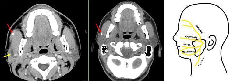
The facial nerve supplies all muscles of facial expression. After exiting the parotid gland, it divides into five terminal branches, classically called the "pes anserinus" (goose's foot):
- Temporal (frontal) branch — crosses the zygomatic arch; supplies frontalis, orbicularis oculi (upper), corrugator supercilii. Highly susceptible to surgical injury as it crosses the arch.
- Zygomatic branch — supplies orbicularis oculi (lower) and muscles of the midface.
- Buccal branch — supplies buccinator, orbicularis oris, and other perioral muscles; often has multiple interconnecting rami with the zygomatic branch.
- Marginal mandibular branch — runs along the lower border of the mandible; supplies depressor anguli oris, depressor labii inferioris, and mentalis. Second most commonly injured branch; has the least collateral interconnection, so injury causes a very visible, uncompensated deficit.
- Cervical branch — supplies platysma.
Clinical Implications
1. Regional nerve blocks for facial esthetic surgery — accurate blocks of the supraorbital/supratrochlear, infratrochlear, infraorbital, zygomaticotemporal, zygomaticofacial, and mental nerves (the "seven blocks") can anesthetize the entire face without general anesthesia, allowing dermabrasion, chemical peels, laser resurfacing, and filler injections to be performed safely and comfortably. Advantages include avoiding general anesthesia/sedation, thorough and long-lasting anesthesia; disadvantage is longer procedure time with multiple injections.
2. Anatomical landmarks reduce complication risk:
- Infraorbital nerve block via the transcutaneous nasolabial approach avoids inadvertent entry into the orbit; palpating the rim before injecting prevents superior misplacement.
- Mental nerve is often visible/palpable submucosally near the lower canine (~85% of the time), guiding safe needle placement.
- Supraorbital notch and zygomaticofrontal suture are reliable palpable landmarks for their respective blocks.
3. Iatrogenic facial nerve injury during surgery — the temporal branch (crossing the zygomatic arch) and the marginal mandibular branch (crossing the mandible near the facial artery/vein) are the two branches most vulnerable during dermatologic and cosmetic surgery (e.g., rhytidectomy, submandibular excisions, masseter botulinum toxin injection). Injury causes:
- Temporal branch damage → inability to raise the eyebrow (frontalis paralysis)
- Marginal mandibular damage → asymmetric smile, drooping of the lower lip corner (most visible deformity due to minimal cross-innervation with adjacent branches)
4. Botulinum toxin injection safety — knowledge of facial nerve branch course (crossing the masseter, zygomatic/buccal/marginal mandibular branches) guides safe injection depth and site to avoid diffusion-related paralysis of unintended muscles.
5. Dermal filler placement guided by sensory nerve anatomy — knowing where sensory nerves emerge (e.g., zygomaticotemporal nerve foramen for temporal volumization, zygomaticofacial nerve area for cheek augmentation, infraorbital nerve area for nasolabial/lip work) allows targeted anesthesia before filler procedures in the temples, cheek, lips, and nasolabial folds.
6. Diagnostic and localizing value — since each sensory branch maps a discrete facial dermatome (V1 forehead/nose, V2 midface, V3 lower face/jaw), and facial nerve branches map discrete motor zones, lesions (trauma, tumor, Bell's palsy, herpes zoster) can be localized based on the pattern of sensory loss or motor weakness observed clinically.
Sources: ACSI Textbook, Chapter 2 - "Applied Anatomy of Nerve Supply to the Face in Relation to Nerve Blocks" and "Surgical Anatomy of the Midface" (pp. 26-28); cross-referenced with Gray's Anatomy for Students (Motor innervation of face - facial nerve branches) and Fitzpatrick's Dermatology (Facial Motor Nerves section) for the five facial nerve branches and their surgical vulnerability.
This is a shared conversation. Sign in to Orris to start your own chat.