
Triangles of neck
triangles of the neck anterior and posterior triangle diagram
"neck"[MeSH Terms] AND "anatomy"[MeSH Terms] AND triangle
Triangles of the Neck
1. Anterior triangle
- Laterally: the anterior border of sternocleidomastoid.
- Superiorly: the inferior border of the mandible.
- Medially: the midline of the neck.
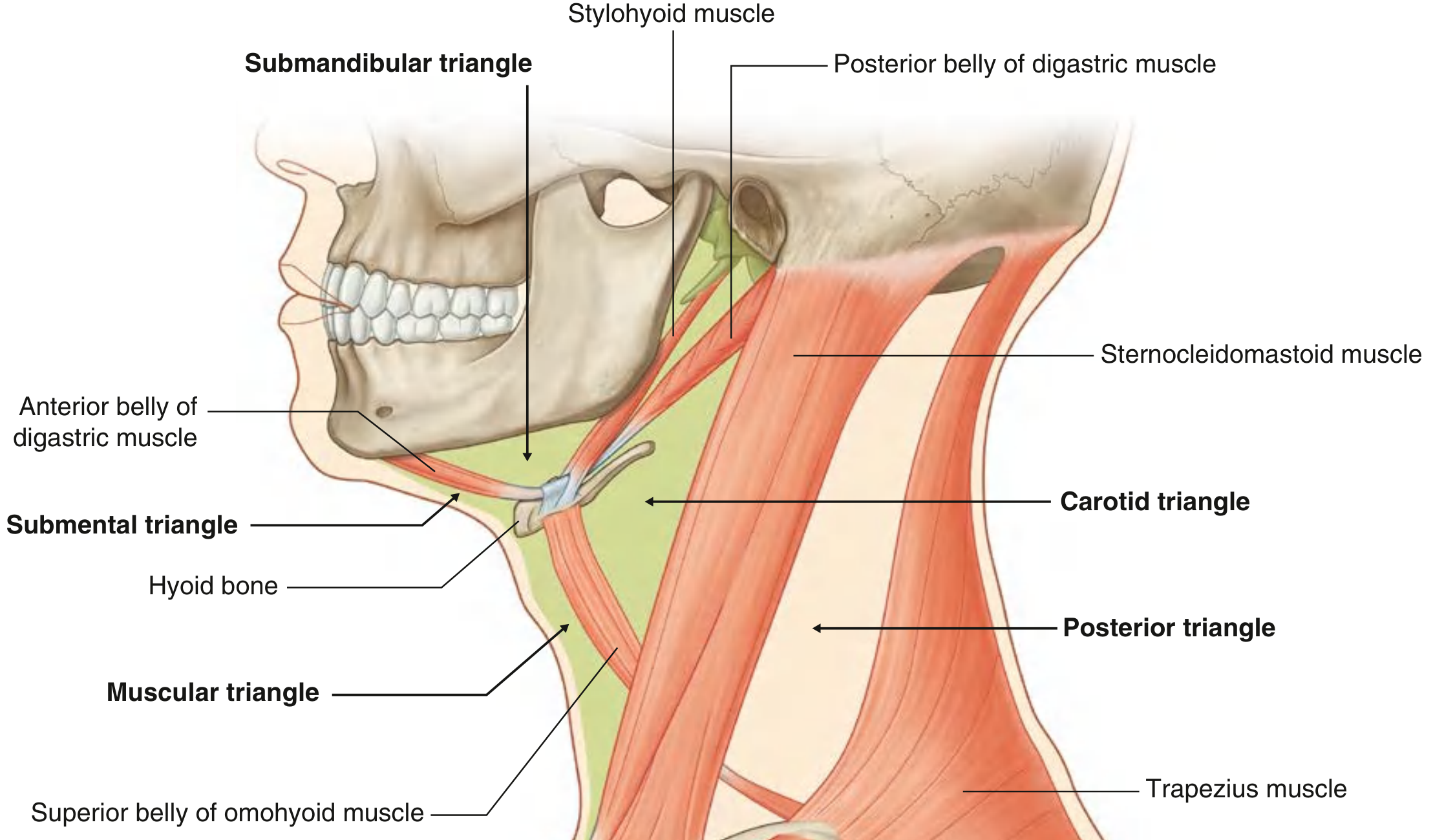
| Triangle | Boundaries | Main contents |
|---|---|---|
| Submental (single, unpaired) | Hyoid body (inferior), anterior bellies of digastric (lateral), mandibular symphysis (apex) | Submental lymph nodes; tributaries forming the anterior jugular vein |
| Submandibular (paired) | Lower border of mandible, anterior and posterior bellies of digastric | Submandibular gland and nodes; hypoglossal nerve; nerve to mylohyoid; facial artery and vein |
| Carotid (paired) | Superior belly of omohyoid (anteroinferior), stylohyoid and posterior belly of digastric (superior), anterior border of SCM (posterior) | Common carotid and its bifurcation; internal jugular vein; vagus, hypoglossal and accessory nerves; ansa cervicalis roots; branches of the external carotid artery (superior thyroid, ascending pharyngeal, lingual, facial, occipital) |
| Muscular (paired) | Hyoid (superior), superior belly of omohyoid, anterior border of SCM, midline | Infrahyoid (strap) muscles: sternohyoid, omohyoid, sternothyroid, thyrohyoid; thyroid and parathyroid glands; pharynx |
2. Posterior triangle
- Anteriorly: the posterior border of SCM.
- Posteriorly: the anterior border of trapezius.
- Base: the middle third of the clavicle.
- Apex: the occipital bone, behind the mastoid process, where SCM and trapezius meet.
- splenius capitis
- levator scapulae
- posterior, middle and anterior scalenes
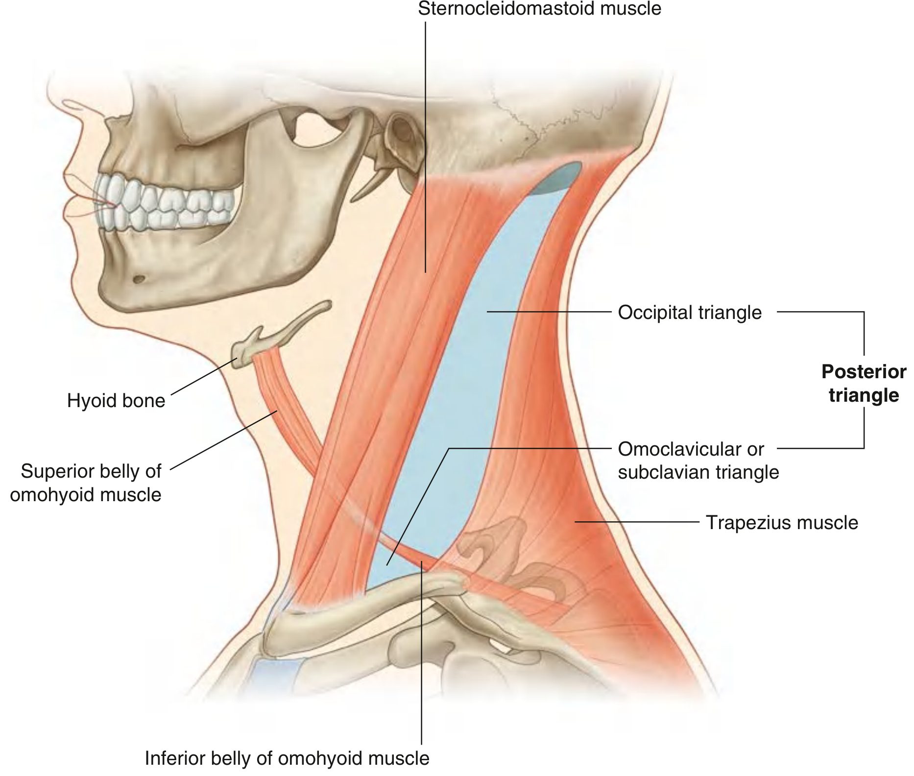
- a large occipital triangle above
- a small omoclavicular (subclavian) triangle below
- Nerves:
- Spinal accessory nerve (XI). It runs superficially in the roof and is vulnerable in lymph node biopsy.
- Cervical plexus branches (lesser occipital, great auricular, transverse cervical, supraclavicular).
- Roots and trunks of the brachial plexus.
- Phrenic nerve, on the anterior surface of the anterior scalene.
- Nerve to serratus anterior and dorsal scapular nerve.
- Vessels:
- External jugular vein, which crosses the roof, pierces the fascia and drains into the subclavian vein.
- Third part of the subclavian artery.
- Transverse cervical and suprascapular arteries.
- Lymph nodes: the occipital and supraclavicular groups.
Clinical points
- Spinal accessory nerve injury: it is superficial in the posterior triangle. Injury, for example during node biopsy, causes trapezius weakness and shoulder drop. A 2024 systematic review (PMID 37767816) examined the anatomy of this nerve in the posterior triangle. I did not review it in detail.
- Carotid triangle: this is the site for carotid endarterectomy, carotid pulse palpation and carotid sinus massage.
- Submandibular triangle: the site of submandibular gland excision. The marginal mandibular, lingual and hypoglossal nerves are at risk.
- Muscular triangle: the site of thyroidectomy and tracheostomy approaches.
- Posterior triangle landmarks:
- Erb's point, where the cervical plexus branches emerge, is at the midpoint of the posterior border of SCM.
- The subclavian artery and the lower brachial plexus trunk lie in the omoclavicular triangle.
- Subclavian vein and brachial plexus blocks are done in this region.
Quick memory aid
- Anterior triangle: Submental, Submandibular, Carotid, Muscular. All are bounded by the midline, mandible and SCM.
- Posterior triangle: Occipital (large) and Omoclavicular (small).

Mark it
ls -la /tmp/workspace/attachments/0ee15e0e-d4d3-446c-96d7-3f35027123ed/ && python3 -c "from PIL import Image; im=Image.open('/tmp/workspace/attachments/0ee15e0e-d4d3-446c-96d7-3f35027123ed/photo-1793776737074.jpg'); print(im.size)"
mkdir -p /tmp/workspace/neck-triangles && cat > /tmp/workspace/neck-triangles/mark.py <<'EOF' from PIL import Image, ImageDraw, ImageFont src='/tmp/workspace/attachments/0ee15e0e-d4d3-446c-96d7-3f35027123ed/photo-1793776737074.jpg' im=Image.open(src).convert('RGBA') ov=Image.new('RGBA',im.size,(0,0,0,0)); d=ImageDraw.Draw(ov) f=ImageFont.truetype('/usr/share/fonts/truetype/dejavu/DejaVuSans-Bold.ttf',30) fs=ImageFont.truetype('/usr/share/fonts/truetype/dejavu/DejaVuSans-Bold.ttf',22) polys={ 'Occipital (posterior)':([(335,520),(440,680),(510,830),(560,960),(500,985),(400,1040),(415,900),(410,780),(370,650)],(0,120,255),(430,780)), 'Omoclavicular':([(400,1040),(500,985),(560,960),(600,1120),(395,1120)],(160,0,220),(470,1085)), 'Submandibular':([(585,480),(880,520),(790,600),(650,570)],(0,200,0),(700,530)), 'Carotid':([(620,560),(745,610),(700,790),(640,700)],(255,230,0),(675,670)), 'Muscular':([(750,620),(850,650),(845,940),(800,1040),(745,880),(715,800)],(0,230,230),(790,830)), } for n,(p,c,_) in polys.items(): d.polygon(p,fill=c+(70,)); d.line(p+[p[0]],fill=c+(255,),width=5) im=Image.alpha_composite(im,ov); d=ImageDraw.Draw(im) def lab(t,xy,fill,font=fs): x,y=xy for dx in(-2,2): for dy in(-2,2): d.text((x+dx,y+dy),t,font=font,fill='black') d.text(xy,t,font=font,fill=fill) lab('POSTERIOR TRIANGLE',(40,500),(120,190,255),f) lab('Occipital',(380,760),(120,190,255)) lab('Omoclavicular',(420,1060),(220,150,255)) lab('ANTERIOR TRIANGLE',(640,330),(255,255,255),f) lab('Submandibular',(640,495),(120,255,120)) lab('Carotid',(655,650),(255,240,60)) lab('Muscular',(745,980),(100,255,255)) lab('Submental (at midline,',(860,420),(255,170,170)); lab('cut off by model edge)',(860,446),(255,170,170)) lab('SCM (11)',(560,800),'white') d.line([(600,830),(580,830)],fill='white',width=1) im.convert('RGB').save('/tmp/workspace/neck-triangles/marked_neck_triangles.jpg',quality=92) EOF python3 /tmp/workspace/neck-triangles/mark.py
python3 -c " import matplotlib, os print(os.path.join(os.path.dirname(matplotlib.__file__),'mpl-data/fonts/ttf'))"; ls $(python3 -c "import matplotlib,os;print(os.path.join(os.path.dirname(matplotlib.__file__),'mpl-data/fonts/ttf'))") | grep -i bold | head
cd /tmp/workspace/neck-triangles && sed -i "s#/usr/share/fonts/truetype/dejavu/DejaVuSans-Bold.ttf#/opt/amazon/genesis1p-tools/venv/lib64/python3.12/site-packages/matplotlib/mpl-data/fonts/ttf/DejaVuSans-Bold.ttf#g" mark.py && python3 mark.py && ls -la
cd /tmp/workspace/neck-triangles && python3 -c " from PIL import Image im=Image.open('marked_neck_triangles.jpg'); im.thumbnail((900,1200)); im.save('preview.jpg',quality=80)" && ls -la
marked neck triangles
Image · JPG

- Occipital (blue): this is the large upper part. It is bounded by the back edge of the SCM, the front edge of the trapezius (the muscle strip on the left) and the inferior belly of omohyoid below.
- Omoclavicular (purple): this is the small lower part, above the clavicle. The small slanting muscle labelled 10 is, I think, the inferior belly of omohyoid. It separates the two parts.
- Submandibular (green): below the mandible, between the two bellies of digastric.
- Carotid (yellow): bounded by the front edge of the SCM, the posterior belly of digastric and the superior belly of omohyoid. The orange area at its lower part is probably the neurovascular region.
- Muscular (cyan): from the midline to the front edge of the SCM, over the strap muscles (labelled 7, 8 and 9).
- Submental: I did not outline this one. It lies at the midline under the chin, and the model is cut off at the midline.
- The outlines are approximate. I placed them by eye, because the image tool could not open the file.
- I'm unsure what the small numbers 1 to 14 stand for. There is no key in the photo, so I don't name muscles from those numbers, apart from the SCM (11) and the probable omohyoid (10).
- If you have the key sheet for model "2", send it. I can then correct the borders against the muscle labels.
Label this diagram
cd /tmp/workspace/neck-triangles && python3 -c " from PIL import Image im=Image.open('/tmp/workspace/attachments/0ee15e0e-d4d3-446c-96d7-3f35027123ed/photo-1793776737074.jpg') im.crop((380,380,980,700)).resize((1200,640)).save('c1.jpg') im.crop((350,560,900,1180)).save('c2.jpg') "
cd /tmp/workspace/neck-triangles && python3 -c " from PIL import Image im=Image.open('/tmp/workspace/attachments/0ee15e0e-d4d3-446c-96d7-3f35027123ed/photo-1793776737074.jpg') im.crop((380,380,980,700)).resize((1200,640)).save('c1.jpg') im.crop((350,560,900,1180)).save('c2.jpg') "; ls -la
cd /tmp/workspace/neck-triangles && cat > label.py <<'EOF' from PIL import Image, ImageDraw, ImageFont src='/tmp/workspace/attachments/0ee15e0e-d4d3-446c-96d7-3f35027123ed/photo-1793776737074.jpg' FP='/opt/amazon/genesis1p-tools/venv/lib64/python3.12/site-packages/matplotlib/mpl-data/fonts/ttf/DejaVuSans-Bold.ttf' ph=Image.open(src).convert('RGB') W,H=1750,1950 cv=Image.new('RGB',(W,H),(245,245,245)); cv.paste(ph,(0,0)) d=ImageDraw.Draw(cv) F=ImageFont.truetype(FP,27) def txt(xy,t,fill=(0,0,0)): d.text(xy,t,font=F,fill=fill) def ring(p,c): x,y=p; d.ellipse((x-17,y-17,x+17,y+17),outline=c,width=5) def call(tag,pts,label,c,lxy): ring(tag,c) d.line([tag]+pts,fill=c,width=4) txt(lxy,label,c) # right margin R=1120 items=[ ((843,466),[(1000,400),(R-10,400)],'3 Digastric, anterior belly',(0,110,200),(R,384)), ((885,503),[(1000,470),(R-10,470)],'5 Geniohyoid',(150,0,170),(R,454)), ((712,613),[(1000,560),(R-10,560)],'6 Thyrohyoid',(0,130,60),(R,544)), ((733,740),[(1000,670),(R-10,670)],'9 Omohyoid, superior belly',(200,90,0),(R,654)), ((792,755),[(1000,760),(R-10,760)],'8 Sternohyoid',(190,0,0),(R,744)), ((735,843),[(1000,860),(R-10,860)],'7 Sternothyroid',(0,120,120),(R,844)), ] for t,p,l,c,x in items: call(t,p,l,c,x) # top labels on board d.rectangle((0,0,W,200),fill=(245,245,245)) call((772,460),[(772,262)],'4 Mylohyoid',(0,100,0),(790,236)) call((565,468),[(500,262)],'1 Stylohyoid',(160,60,0),(380,215)) call((512,491),[(200,262)],'2 Digastric, posterior belly',(0,60,160),(20,215)) # SCM direct ring((570,885),(0,0,0)); d.rectangle((590,868,590+330,908),fill=(255,255,255)); txt((596,872),'11 Sternocleidomastoid',(0,0,0)) # bottom rows=[((560,1068),'14 Anterior scalene',(190,0,100),1640), ((493,1070),'13 Middle scalene',(0,110,0),1710), ((480,1010),'10 Omohyoid, inferior belly',(200,90,0),1780), ((418,1100),'12 Posterior scalene',(0,70,170),1850)] for tag,l,c,y in rows: ring(tag,c); d.line([tag,(tag[0],y)],fill=c,width=4); txt((tag[0]+10,y-10),l,c) d.text((20,1600),'Right lateral view of the neck (model "2")',font=ImageFont.truetype(FP,24),fill=(60,60,60)) cv.save('labelled_neck_model.jpg',quality=92) p=cv.copy(); p.thumbnail((1000,1200)); p.save('labelled_preview.jpg',quality=80) EOF python3 label.py
cd /tmp/workspace/neck-triangles && python3 - <<'EOF' s=open('label.py').read() s=s.replace("call((512,491),[(200,262)],'2 Digastric, posterior belly',(0,60,160),(20,215))","call((512,491),[(200,262),(200,170)],'2 Digastric, posterior belly',(0,60,160),(210,150))") s=s.replace("d.text((20,1600),","d.text((20,30),") open('label.py','w').write(s) EOF python3 label.py
labelled neck model
Image · JPG

| No. | Structure | Group |
|---|---|---|
| 1 | Stylohyoid | Suprahyoid |
| 2 | Digastric, posterior belly | Suprahyoid |
| 3 | Digastric, anterior belly | Suprahyoid |
| 4 | Mylohyoid | Suprahyoid |
| 5 | Geniohyoid | Suprahyoid |
| 6 | Thyrohyoid | Infrahyoid |
| 7 | Sternothyroid | Infrahyoid |
| 8 | Sternohyoid | Infrahyoid |
| 9 | Omohyoid, superior belly | Infrahyoid |
| 10 | Omohyoid, inferior belly | Crosses the posterior triangle |
| 11 | Sternocleidomastoid | Divides the neck into anterior and posterior triangles |
| 12 | Posterior scalene | Floor of the posterior triangle |
| 13 | Middle scalene | Floor of the posterior triangle |
| 14 | Anterior scalene | Floor of the posterior triangle |
- Most reliable: 7 to 11. The strap muscles, the SCM and the omohyoid are clear from their course and relations. The inferior belly (10) is the one that splits the posterior triangle.
- Scalenes (12, 13, 14): these are numbered from posterior to anterior, so 14, next to the SCM, is the anterior scalene. This follows the usual order and is probably right.
- Least certain: 3, 4, 5 and 6. They sit near the chin and hyoid on a cut edge of the model, and 6's tag is hard to read. In particular, 3 (anterior belly of digastric) and 5 (geniohyoid) could be swapped.
- Tag 2: it is smudged in the photo. I read it as the posterior belly of digastric, which lies just below the stylohyoid (1).
- The digastric bellies (2 and 3) form the borders of the submandibular and submental triangles. The posterior belly (2) also bounds the carotid triangle.
- The superior belly of omohyoid (9) bounds the carotid and muscular triangles.
- The inferior belly of omohyoid (10) splits the posterior triangle into the occipital and omoclavicular parts.
- The scalenes (12 to 14) form part of the floor of the posterior triangle.