
First aid in a suspected cervical spine injury
Reading File
Now let me search the medical library and web in parallel for first aid in cervical spine injury.
Finding Sources
Finding Sources
Searching the Web
first aid suspected cervical spine injury management 2024 guidelines
Good results. Let me now read the most relevant textbook sections in parallel.
Reading File
Reading File
Finding Sources
Reading Web Page
https://www.anzcor.org/home/first-aid/guideline-9-1-6-manage…
Now let me read the key textbook sections on catastrophic cervical spine injury and the Roberts & Hedges section on background/procedures:
Reading File
Reading File
I now have excellent textbook and guideline data. Let me read the remaining portions of the on-field assessment section:
Reading File
I now have comprehensive information from multiple authoritative sources. Let me compile the full answer.
First Aid in Suspected Cervical Spine Injury
When to Suspect Cervical Spine Injury
Suspect cervical spine injury in any patient with:
- Significant neck or spine pain after trauma
- Diminished level of consciousness or unconsciousness after trauma
- Neurologic deficits (weakness, numbness, tingling in limbs)
- High-energy mechanism: motor vehicle collision, fall from height, axial loading (diving, "spear tackling" in football), direct blow to head/neck
- Any unconscious trauma patient - assume unstable cervical spine until proven otherwise
Step-by-Step First Aid Principles
1. Scene Safety & Call for Help
- Ensure the scene is safe before approaching
- Call emergency services (EMS) immediately
- Do not attempt to move the patient unless there is immediate life-threatening danger (fire, drowning, airway obstruction)
2. Initial Assessment - ABCs First
- Airway, Breathing, Circulation (ABCs) take priority over spinal precautions
- Assess level of consciousness
- If the patient is unconscious, assume an unstable cervical spine injury
3. Manual Inline Spinal Motion Restriction (SMR) - Key Modern Concept
The current evidence-based approach (ILCOR 2024, ANZCOR 2025) has shifted from "full immobilization" to spinal motion restriction (SMR):
- Position yourself at the top of the patient's head
- Place your hands on the patient's shoulders, thumbs extending anteriorly over the clavicles, fingers behind the neck
- The head is immobilised between your forearms - this is the preferred manual inline restriction technique
- Alternatively, hold the head between both hands on either side of the skull
- Keep the head in a neutral position (not flexed or extended), pointing in the same direction as the rest of the body
- In a conscious patient, this should be the most comfortable position - do not force the head into a neutral position if doing so causes pain
- Maintain this position until EMS arrives
4. Airway Management
- If the patient is breathing normally, maintain manual SMR and do not move them
- If the patient is NOT breathing or airway is obstructed: use the jaw thrust maneuver (NOT head-tilt/chin-lift) to open the airway - this opens the airway while keeping the cervical spine stable
- If jaw thrust fails to open the airway and the patient is in cardiac arrest, a head-tilt/chin-lift is acceptable - airway takes absolute priority
- If the patient vomits and risks aspiration, log-roll them with the team while maintaining spinal alignment
5. Cervical Collars - Revised Guidance
- Soft cervical collars only indicate a possibility of spinal injury - they provide minimal motion restriction. Use only as a marker, not a primary protective device (ANZCOR 2025)
- Semi-rigid (hard) collars may be used when manual SMR is impossible, for extrication/transport, but only for the shortest time necessary by trained providers
- Head blocks that restrict only the head more than the body can actually increase cervical spine motion when the body moves and should NOT be used by first aiders
- Do NOT rely on any device to replace manual inline restriction in an unconscious patient
6. Do NOT Move the Patient Unless Necessary
- Do not attempt to move, straighten, or position the patient unless there is immediate danger or airway compromise
- If transport is needed before EMS, use a 6-person lift or log-roll technique onto a spine board, with one designated person controlling the head and neck throughout
7. Helmet Considerations (Sports Injuries)
- In football/hockey players wearing shoulder pads: leave the helmet on - removing it causes the head to hyperextend (shoulder pads elevate the torso, helmet maintains neutral alignment)
- The face mask should be removed as early as possible to allow airway access, but the helmet and chin strap should remain in place
- If shoulder pads AND helmet must both be removed, they must be removed simultaneously by a coordinated team
8. Transport
- Transport should be to a trauma center with diagnostic (CT/MRI) and surgical spinal capabilities
- Do not place the patient back on a long spine board longer than necessary - prolonged use causes pressure injuries and respiratory compromise
- Continue monitoring neurologic status: limb movement, sensation, respiratory pattern
What NOT to Do
| Avoid | Reason |
|---|---|
| Rotating, flexing, or extending the neck | Risk of cord injury from unstable fracture |
| Removing a football helmet without also removing shoulder pads | Causes neck hyperextension |
| Using head blocks alone | Can shear the spine when the torso moves |
| Assuming a conscious, ambulatory patient cannot have a fracture | Incomplete neurological injury can still be mobile |
| Prolonged routine full immobilization on a rigid long board | Causes pressure ulcers, respiratory compromise, raised ICP without proven neurological benefit |
| Applying cervical spine restrictions to penetrating neck trauma without neurological signs | May delay transport and worsen outcomes |
Red Flags Requiring Immediate EMS Response
- Bilateral limb weakness or paralysis
- Sensory loss below neck level
- Inability to control bladder or bowel
- Priapism (male patients) - a sign of spinal shock
- Diaphragmatic breathing with absent chest wall movement
- Rapidly deteriorating consciousness after neck trauma
Key Conceptual Shift: "Motion Restriction" Not "Immobilization"
Current guidelines (ILCOR CoSTR Dec 2024, updated March 2025) reflect a major shift in thinking. True spinal immobilization is not achievable in the prehospital setting, and rigid immobilization has known complications. The goal is now spinal cord protection (SCP) through motion restriction - reducing unnecessary movement while prioritizing airway and circulation. Selective, clinical-decision-rule-guided restriction is preferred over blanket immobilization for every trauma patient. - Roberts and Hedges' Clinical Procedures in Emergency Medicine; ANZCOR Guideline 9.1.6
Sources: Textbook of Family Medicine 9e (Catastrophic Cervical Spine Injuries); Roberts and Hedges' Clinical Procedures in Emergency Medicine; Rosen's Emergency Medicine; ANZCOR Guideline 9.1.6 (ILCOR CoSTR Dec 2024, updated March 2025); Wilderness Medical Society 2024 Spinal Care Guidelines
Blunt abdominal trauma
Blunt abdominal trauma is a major cause of morbidity/mortality, most commonly from MVCs, falls, and assaults. The spleen is the most commonly injured solid organ, followed by the liver. Key priorities are rapid assessment, identifying occult hemorrhage, and determining operative vs. non-operative management. FAST ultrasound has become the primary bedside tool.
Searching sources...
Reading File
Finding Sources
Finding Sources
Searching the Web
blunt abdominal trauma management guidelines 2024 FAST ATLS
Excellent. Now let me read the key textbook sections in parallel - Rosen's, Tintinalli's, Sabiston, and Current Surgical Therapy:
Reading File
Reading File
Reading File
Excellent content. Now let me get more detail - the specific organ injury management, grading systems, and Rosen's management algorithm sections:
Reading File
Reading File
Finding Sources
Let me get the AAST grading tables, NOM criteria, and specific organ sections:
Reading File
Reading File
Reading File
Excellent - I now have comprehensive data including AAST grading tables. Let me get the AAST grading tables from Current Surgical Therapy and any remaining key details on hollow viscus and pancreatic injuries:
Reading File
Finding Sources
I now have all the material I need for a comprehensive, well-structured answer. Let me also quickly fetch the BAT algorithm image from Rosen's:
Loading Image
I now have all the material needed for a comprehensive, authoritative answer. Let me compile it:
Blunt Abdominal Trauma (BAT)
Epidemiology & Common Mechanisms
Motor vehicle collisions account for the majority of cases. Falls from height, direct blows, sports injuries, and blast injuries make up the rest. The abdomen is one of the most vulnerable anatomic regions in blunt trauma, and injuries are frequently occult - physical examination alone is unreliable, especially in patients with altered mental status, intoxication, distracting injuries, or spinal cord injury. - Rosen's Emergency Medicine
Most commonly injured organs:
- Spleen - most common; isolated in ~2/3 of splenic injuries
- Liver - second most common
- Kidney - third
- Small bowel / hollow viscus - less common (1-5% of BAT admissions) but frequently missed
Initial Assessment - ATLS Framework
The primary approach follows the xABCDE pattern (updated ATLS):
- xA - eXsanguinating hemorrhage control first
- A - Airway
- B - Breathing
- C - Circulation (two large-bore IV lines, blood transfusion if needed)
- D - Disability (GCS, pupils)
- E - Exposure/Environment (undress completely, logroll for back)
The hemodynamic status is the single most important driver of the subsequent workup and management pathway. Everything diverges at this fork.
Diagnostic Tools
1. FAST / eFAST (First-Line Bedside Tool)
FAST (Focused Assessment with Sonography for Trauma) is now the standard adjunct to the primary survey. It is performed bedside, simultaneously with resuscitation, and evaluates four windows:
| Window | Structure Examined |
|---|---|
| Subxiphoid | Pericardium / cardiac tamponade |
| Right subcostal | Hepatorenal fossa (Morrison's pouch) |
| Left subcostal | Splenorenal fossa |
| Suprapubic | Pelvis / pouch of Douglas |
Extended FAST (eFAST) adds bilateral thoracic views to detect hemothorax and pneumothorax.
- Sensitivity: 73-88% | Specificity: 98-100% | Accuracy: 96-98%
- Requires ≥200 mL of free fluid for detection
- Cannot evaluate the retroperitoneum - a major limitation
- Can be repeated serially with any hemodynamic change
- Positive FAST in an unstable patient → immediate laparotomy
- Positive FAST in a stable patient → proceed to CT for characterization
Current practice is trending toward "Complete Abdominal Sonography for Trauma" (CAST) in stable patients to allow more selective CT use (BJS 2025).
2. CT Abdomen/Pelvis with IV Contrast (Definitive Imaging in Stable Patients)
CT has essentially become the primary method for comprehensive evaluation of hemodynamically stable BAT patients. Key points:
- Performed with IV contrast in arterial and portal venous phases - best demonstrates solid organ perfusion and active bleeding
- Negative predictive value up to 99.63% for significant intraabdominal injury
- Identifies solid organ injury (spleen, liver, kidney), retroperitoneal hematoma, and active extravasation ("vascular blush")
- Less sensitive for hollow viscus / bowel injury - CT signs include bowel wall thickening, mesenteric stranding, free fluid without solid organ injury, pneumoperitoneum
- A hemodynamically unstable patient should not be taken to CT unless resuscitative measures can continue simultaneously and the facility can provide true "scoop and scan"
3. Diagnostic Peritoneal Lavage / Aspirate (DPL/DPA)
DPL has been largely supplanted by FAST and CT. Remaining indications:
- FAST unavailable or technically inadequate
- Suspected occult bowel injury not detectable by CT
- Rapid triage when other modalities unavailable
DPL positive criteria:
| Finding | Threshold |
|---|---|
| Gross blood on aspiration | >10 mL |
| RBCs in lavage effluent | >100,000/mL |
| WBCs in lavage effluent | >500/mL |
| Bacteria, bile, or GI contents | Any amount |
Pitfalls: high false-positive rate, cannot evaluate retroperitoneum, risk of iatrogenic injury. Always place Foley catheter and NGT first. Use a supraumbilical approach in pelvic fracture or pregnancy.
Management Algorithm
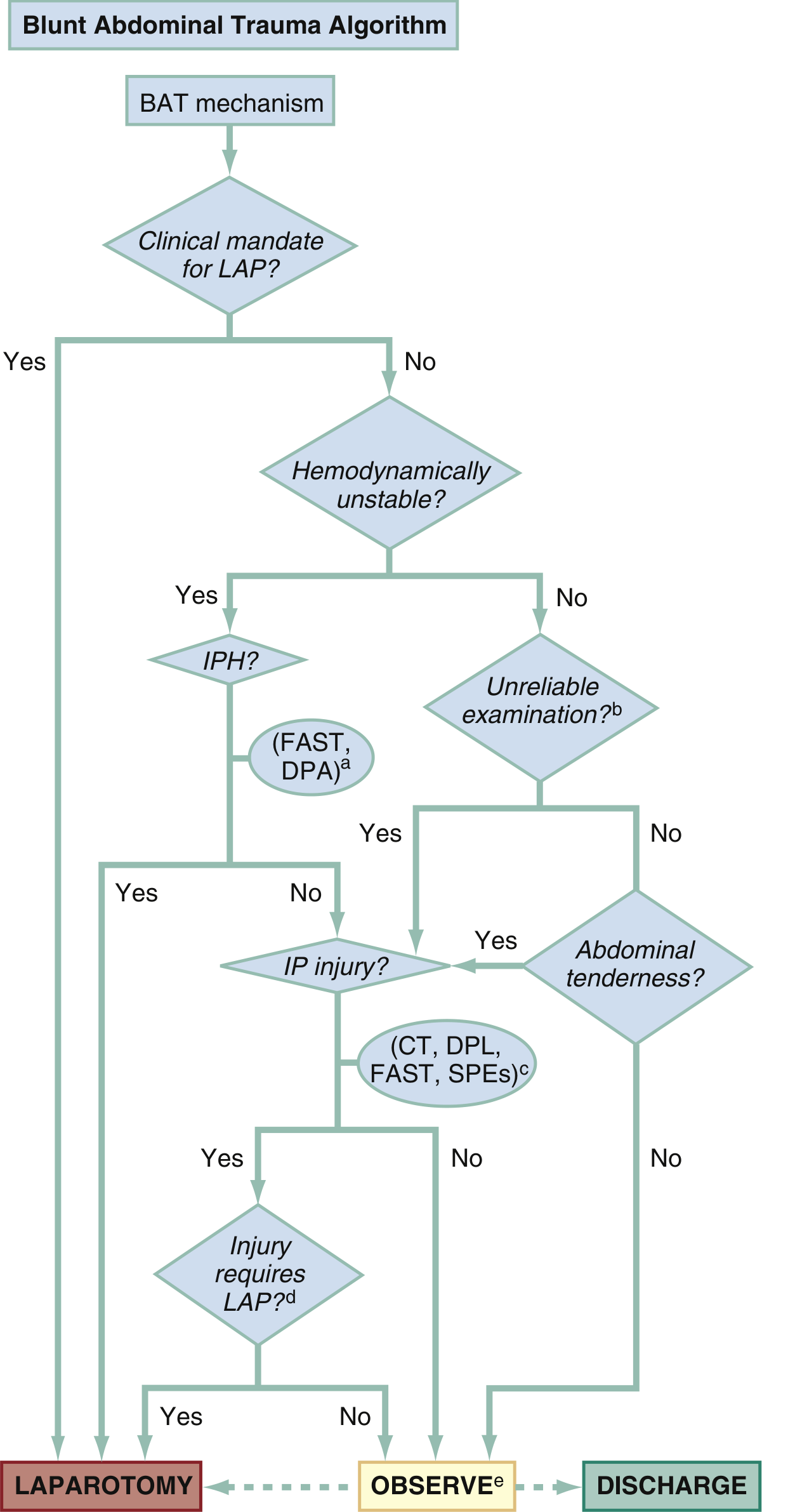
Fig. 38.8 - Rosen's Emergency Medicine: BAT Algorithm. IPH = intraperitoneal hemorrhage; DPA = diagnostic peritoneal aspirate; SPEs = serial physical examinations.
Unstable Patient + Positive FAST/DPA → Immediate Laparotomy
Clinical indications for immediate laparotomy after blunt trauma (Rosen's, Table 38.2):
| Indication | Pitfall |
|---|---|
| Unstable vitals with suspected abdominal injury | Alternate shock sources (pelvic fracture, tension pneumothorax) |
| Unequivocal peritoneal irritation | May be unreliable |
| Pneumoperitoneum | Can come from non-abdominal sources |
| Evidence of diaphragmatic injury | Nonspecific |
| Significant GI bleeding | Uncommon |
Stable Patient → CT then Selective NOM or Surgery
Organ-Specific Management
Spleen
The most commonly injured organ. Clinical signs: LUQ pain, LUQ tenderness, hypotension, Kehr's sign (referred pain to the left shoulder on deep inspiration from diaphragmatic irritation by splenic hematoma).
AAST Spleen Injury Scale:
| Grade | Description |
|---|---|
| I | Subcapsular hematoma <10% surface area; capsular tear <1 cm depth |
| II | Subcapsular hematoma 10-50%; intraparenchymal <5 cm; laceration 1-3 cm depth |
| III | Subcapsular hematoma >50% or expanding; laceration >3 cm or involving trabecular vessels |
| IV | Laceration involving segmental/hilar vessels with >25% devascularization |
| V | Shattered spleen; hilar vascular injury devascularizing entire spleen |
(Advance one grade for multiple injuries, up to Grade III)
NOM (Nonoperative Management): ~60-80% of blunt splenic injuries can be managed nonoperatively at Level I/II trauma centers. Success rate ~95% when criteria are met.
Criteria for NOM:
- Hemodynamic stability
- No peritoneal signs
- No hollow viscus injury
- No free extravasation of IV contrast from splenic parenchyma
Risk factors for NOM failure:
- Grade III-V injury
- Age >55 years
- Moderate-large hemoperitoneum
- Subcapsular hematoma (risk of delayed rupture at 6-8 days)
- Portal hypertension / cirrhosis
- Contrast "blush" on CT (pseudoaneurysm) - present in ~2/3 of NOM failures
Angioembolization: Used for contained contrast blush (pseudoaneurysm) to selectively occlude splenic arterial branches; reduces NOM failure rate. About 20% of patients initially on NOM eventually require intervention.
Operative management: Splenorrhaphy (repair) preferred when possible at surgery. If splenectomy is unavoidable: post-splenectomy vaccines (pneumococcus, meningococcus, Hib) and prophylactic antibiotics to prevent Overwhelming Post-Splenectomy Infection (OPSI).
Liver
Second most commonly injured organ. The right lobe is injured more frequently due to its larger size.
AAST Liver Injury Scale:
| Grade | Description |
|---|---|
| I | Subcapsular hematoma <10%; capsular tear <1 cm depth |
| II | Subcapsular hematoma 10-50%; intraparenchymal <10 cm; laceration 1-3 cm depth, <10 cm length |
| III | Subcapsular hematoma >50% or expanding; laceration >3 cm depth |
| IV | Parenchymal disruption 25-75% of hepatic lobe or 1-3 Couinaud segments |
| V | Disruption >75% hepatic lobe or >3 Couinaud segments; juxtahepatic venous injury |
| VI | Hepatic avulsion |
NOM: Selective NOM is the standard of care for hemodynamically stable patients. Similar to the spleen: NOM of liver and spleen combined has a 95% success rate at centers with adequate resources.
- Angiographic embolization has similarly reduced morbidity and mortality for arterial liver injuries
- Complications of NOM: delayed hemorrhage, biloma, abscess, hemobilia
Operative management: Damage control surgery (packing + temporary closure) for Grade IV-V or hemodynamically unstable. Hepatorrhaphy or partial hepatectomy for definitive repair.
Kidney
AAST Kidney Injury Scale: Grades I-V (contusion/hematoma through vascular pedicle injury).
- CT with IV contrast is the imaging modality of choice for renal injury evaluation
- Hematuria (macro or micro) is a common but not universal finding
- Most renal injuries (Grades I-III) are managed nonoperatively with bed rest and monitoring
- Grade IV-V: often require angioembolization or operative repair/nephrectomy
- Retroperitoneal hematoma from renal injury may not be visible on FAST
Hollow Viscus (Bowel & Mesentery)
Incidence: 1-5% of BAT admissions - the diagnosis most likely to be missed.
High-risk mechanisms/signs:
- Lap belt / seatbelt sign (abdominal wall ecchymosis) - highly suspicious
- Handlebar injury
- "Chance fracture" of lumbar spine (flexion-distraction) - associated with bowel injury in ~50%
CT findings (often subtle):
- Overt: pneumoperitoneum, extravasation of oral contrast
- Subtle: bowel wall thickening, mesenteric fat stranding, free fluid without solid organ injury
Management: Any perforation requires surgical repair. Serial abdominal examinations are a critical adjunct for patients in whom exam is reliable. DPL can evaluate for occult bowel injury when CT is non-diagnostic. Hollow viscus injury must be ruled out before committing to NOM of solid organ injuries.
Pancreas
- Rare (2-12% of BAT), but frequently missed - CT sensitivity for pancreatic injury is lower
- Suspect with epigastric pain, rising amylase/lipase after upper abdominal blow
- MRCP or ERCP may be needed to evaluate ductal injury
- Isolated contusions: NOM with pancreatic rest
- Ductal disruption (Grade III+): surgical drainage or resection
Special Scenarios
Pelvic Fracture + BAT
Pelvic fractures complicate management by creating an additional retroperitoneal hemorrhage source not visible on FAST. Algorithm:
- Unstable + FAST positive for hemoperitoneum → laparotomy (pelvic packing if needed)
- Unstable + FAST negative → pelvic angioembolization or preperitoneal pelvic packing
- Stable → CT to differentiate intraperitoneal vs retroperitoneal hemorrhage
Head Injury + BAT
Closed head trauma is the scenario most vulnerable to delayed diagnosis of bowel perforation - impaired neurologic exam masks evolving peritonitis. Serial imaging and DPL have a higher threshold of use in this population.
Seatbelt Sign
Patients with visible abdominal wall ecchymosis from a lap belt should be admitted for observation and serial examination. Detection of intraperitoneal fluid by FAST or CT mandates DPL to determine the nature of the fluid, or exploratory laparotomy.
Key Laboratory Studies
- CBC - baseline Hgb/Hct (may be normal initially due to redistribution)
- Comprehensive metabolic panel - liver enzymes (ALT/AST elevated in liver injury), BUN/Cr
- Amylase/Lipase - pancreatic injury screen (low sensitivity alone)
- Urinalysis - hematuria suggests genitourinary injury
- Coagulation studies (PT/PTT/INR, fibrinogen) - guides damage control resuscitation
- Type and Screen / Crossmatch
- Lactate / Base deficit - severity of shock and resuscitation endpoint
Damage Control Surgery (DCS) Principles
For the hemodynamically unstable patient who goes to the OR:
- Hemorrhage control - packing, vascular clamps
- Contamination control - bowel clamps, temporary closure
- ICU resuscitation - correction of "lethal triad" (hypothermia, acidosis, coagulopathy)
- Definitive repair - return to OR at 24-72 hours once physiology normalized
Modern damage control resuscitation uses balanced transfusion (1:1:1 ratio of packed RBCs : FFP : platelets) and avoids large-volume crystalloid which worsens coagulopathy and abdominal compartment syndrome.
Sources: Rosen's Emergency Medicine (8th ed., Chapter 38); Current Surgical Therapy 14e; Sabiston Textbook of Surgery; Maingot's Abdominal Operations; BJS 2024 Management Review; EAST Practice Management Guidelines
This is a shared conversation. Sign in to Orris to start your own chat.