
Intestinal obstruction
intestinal obstruction bowel X-ray radiology
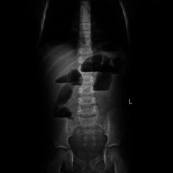
This diagnostic image is an erect anterior-posterior (AP) abdominal X-ray (radiography) demonstrating classic features of an acute intestinal obstruction. The primary radiographic findings include multiple dilated loops of small bowel, most prominent in the left upper quadrant and central abdomen, with diameters reaching approximately 4 cm. Distinctive horizontal air-fluid levels are visible within these distended loops, creating a 'stepladder' appearance characteristic of a small bowel obstruction (SBO). There is a notable paucity of distal colonic gas, supporting the diagnosis of a high-grade or complete mechanical obstruction. The image highlights key educational concepts such as the '3-6-9 rule' for bowel diameter and the utility of plain film imaging in the initial triage of patients presenting with acute abdominal pain and vomiting. This clinical photograph is highly relevant for medical education in radiology, emergency medicine, and general surgery for identifying signs of ileus or mechanical obstruction, such as intussusception.
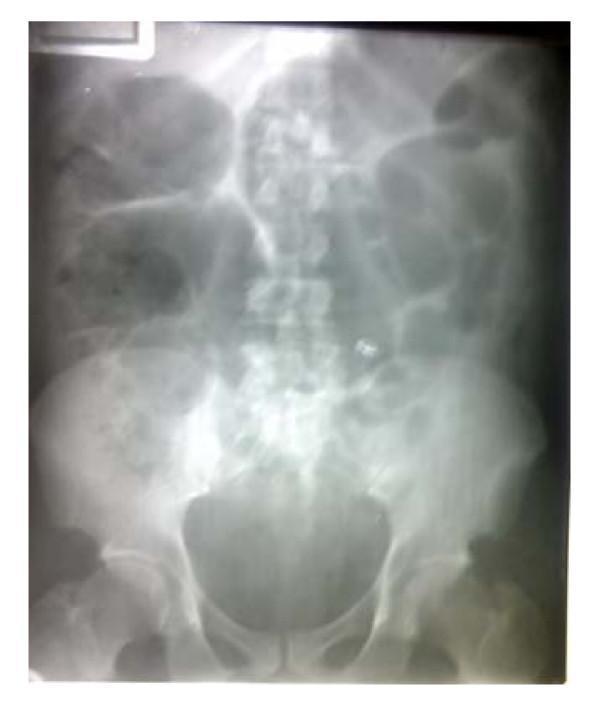
This diagnostic image is a supine abdominal X-ray (radiography) demonstrating classic features of intestinal obstruction. The radiograph reveals significant gaseous distension of multiple small bowel loops, primarily located in the upper and mid-abdomen. There is a notable paucity of gas in the distal colon and pelvic region, a finding highly suggestive of a mechanical obstruction rather than an adynamic ileus. The distended loops appear stacked and show increased caliber, exceeding the normal anatomical limits for small bowel diameter. Visible air-fluid levels indicate trapped gas and liquid within the dilated segments. This clinical imaging finding is essential for the diagnosis of acute abdominal conditions, such as small bowel obstruction (SBO), and serves as an important educational example of intestinal gas pattern analysis in emergency radiology and general surgery.
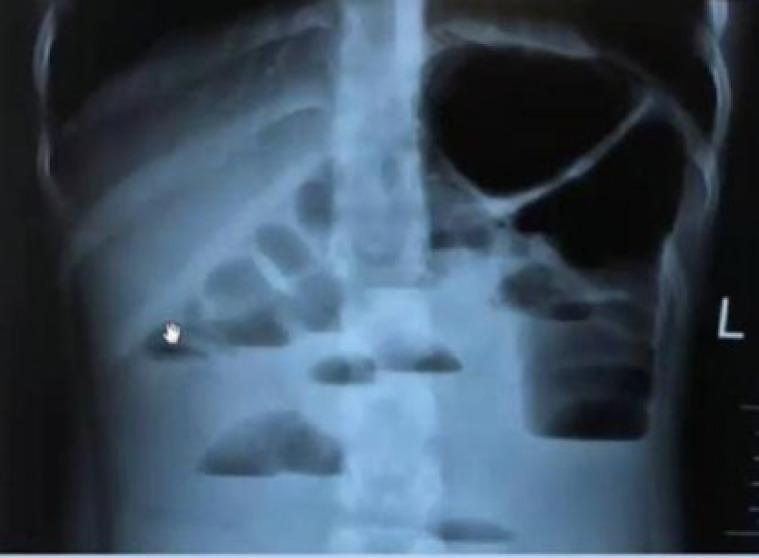
This diagnostic image is an erect abdominal plain radiograph (X-ray) demonstrating signs of intestinal obstruction. The primary finding is the presence of multiple, dilated loops of bowel containing prominent horizontal air-fluid levels. These levels appear as sharp linear interfaces between superior radiolucent gas and inferior radiopaque fluid. The distribution involves several small bowel loops in the central abdomen and a larger, more dilated gas-filled structure in the upper left quadrant. The radiographic appearance is characteristic of a mechanical bowel obstruction or paralytic ileus. Anatomical landmarks include the visible lumbar spine, lower rib cage, and a lateral 'L' orientation marker. The study illustrates key diagnostic features used in emergency medicine and radiology to evaluate acute abdominal pain and suspected colonic or small bowel transit issues.
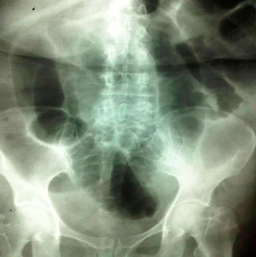
This diagnostic image is an anteroposterior (AP) abdominal X-ray (radiography) demonstrating classic radiological signs of a small bowel obstruction. The image reveals multiple centrally located, air-filled, and markedly dilated loops of small intestine. These distended loops occupy a significant portion of the abdominal cavity, showing a disorganized bowel gas pattern with loss of normal caliber. Key anatomical landmarks visible include the lumbar spine, pelvis, and proximal femurs. The visible pathology is highly suggestive of a mechanical obstruction, which clinically correlated with ileo-ileal intussusception. The image serves as an educational example of obstructive bowel patterns in emergency radiology, highlighting the use of plain film radiography in diagnosing acute abdominal distension and intestinal blockage.
Intestinal Obstruction
Definition and Classification
- Mechanical (dynamic) - a physical barrier to flow (most common)
- Functional (adynamic/paralytic ileus) - failure of peristalsis without a physical block
- Vascular - occlusion of the intestinal blood supply
- Complete (absolute constipation - no flatus or faeces)
- Partial/Incomplete (some gas may still pass)
- Small bowel obstruction (SBO) - high or low
- Large bowel obstruction (LBO)
- Simple - blood supply intact
- Strangulated - blood supply compromised
Causes
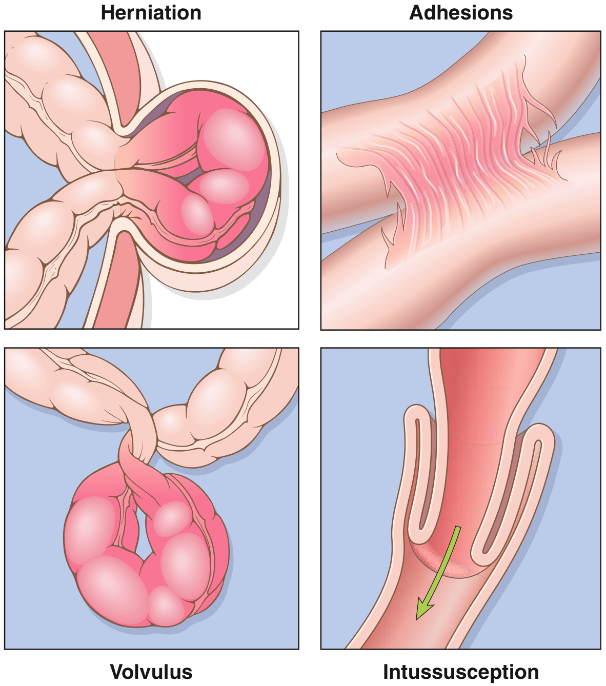
Small Bowel Obstruction (Adults)
| Cause | Notes |
|---|---|
| Adhesions (>60%) | Most common overall; especially post-pelvic surgery (gynecologic, appendectomy, colorectal) |
| Hernias (~10%) | Inguinal, femoral, umbilical, ventral, internal (post-op mesenteric defects) |
| Neoplastic | Carcinomatosis, extraintestinal neoplasms |
| Intraabdominal abscess | External compression |
- Congenital: malrotation, duplications
- Inflammatory: Crohn disease, tuberculosis, diverticulitis, actinomycosis
- Neoplastic: primary or metastatic tumours
- Traumatic: haematoma
- Miscellaneous: intussusception, endometriosis, radiation stricture
- Gallstone ileus, bezoars, swallowed foreign bodies, meconium (neonates)
Large Bowel Obstruction
- Carcinoma of the colon - most common cause
- Sigmoid volvulus - especially in elderly, institutionalised patients
- Diverticular stricture
- Caecal volvulus
- Sabiston Textbook of Surgery, Box 91.1; Bailey and Love's Short Practice of Surgery 28th ed., Ch. 78
Pathophysiology
- Fluid and electrolyte loss - up to 6 litres of salt-rich fluid secreted daily into the small intestine normally gets reabsorbed by the colon; obstruction prevents this reabsorption. Vomiting and luminal sequestration cause profound hypovolaemia
- Bowel distension - swallowed air and gas from bacterial fermentation accumulate proximal to the obstruction
- Bacterial overgrowth - intraluminal proliferation leads to faeculant vomitus and, with mucosal compromise, translocation and sepsis
- Strangulation - venous congestion first, then arterial occlusion, leading to ischaemia, gangrene, and perforation
- Pye's Surgical Handicraft 22nd ed.
Clinical Features
- Abdominal pain - sudden onset, colicky, periumbilical (SBO) or lower abdomen (LBO)
- Abdominal distension - greater the more distal the obstruction
- Vomiting - onset earlier and more profuse in high SBO; later and faeculant in distal obstruction
- Absolute constipation - no passage of flatus or faeces
Variations by Level
| Feature | High SBO | Low SBO | LBO |
|---|---|---|---|
| Vomiting | Early, profuse, rapid dehydration | Later | Late |
| Distension | Minimal | Central, prominent | Early, pronounced |
| Pain | Colicky, periumbilical | Colicky, central | Less severe |
| Radiograph | Little dilated SB; no colonic gas | Multiple dilated SB loops | Dilated colon; ± SB if ileocaecal valve incompetent |
Exceptions to Absolute Constipation Rule
- Richter's hernia (partial wall involvement)
- Gallstone ileus
- Mesenteric vascular occlusion
- Pelvic abscess-related functional obstruction
- All partial obstructions (diarrhoea may occur)
Bowel Sounds
- High-pitched, tinkling - characteristic of early mechanical obstruction
- Absent or scanty - late obstruction or strangulation (ominous sign)
Late Manifestations
- Dehydration, dry tongue, oliguria
- Rising blood urea and haematocrit
- Hypovolaemic shock
- Pyrexia (indicates ischaemia, perforation, or sepsis)
- Respiratory compromise (diaphragmatic splinting)
- Bailey and Love's Short Practice of Surgery 28th ed., Ch. 78
Strangulation - Warning Signs
- Constant, severe pain (not colicky) - not relieved by opiates
- Localised tenderness - indicates impending ischaemia; requires frequent reassessment
- Peritonism / generalised tenderness and rigidity - indicates established infarction/perforation → early laparotomy
- Shock - suggests underlying ischaemia
- Pyrexia or hypothermia (the latter = septicaemic shock)
- Raised WBC, serum lactate, amylase, LDH - associated with strangulation
- In external hernias: lump is tense, tender, irreducible, no expansile cough impulse; skin erythema or purplish discoloration
- Bailey and Love's Short Practice of Surgery 28th ed.
Investigations
Bloods
- FBC (leukocytosis/leukopenia with strangulation)
- Urea & electrolytes (dehydration, hypokalaemia)
- Serum lactate (ischaemia)
- Serum amylase and LDH (elevated in strangulation)
- ABG (metabolic acidosis in advanced obstruction)
Imaging
- Small bowel - central transverse loops, valvulae conniventes pass completely across the bowel width (concertina/"ladder" pattern), no/minimal colonic gas
- Large bowel - haustral folds that do not cross the full diameter, peripheral location
- Erect film - multiple air-fluid levels (stepladder appearance in SBO)
- 3-6-9 rule - SB > 3 cm, colon > 6 cm, caecum > 9 cm = dilated
- Sigmoid volvulus: "coffee bean sign" - massively dilated loop rising from pelvis to right upper quadrant
- Caecal volvulus: rounded gas shadow displaced to left upper quadrant
- Gallstone ileus: pneumobilia (gas in biliary tree) + small bowel obstruction + visible calcified gallstone in RIF
- Identifies level, cause, and degree of obstruction
- Distinguishes simple from strangulated obstruction
- Identifies closed-loop obstruction (most dangerous - decompresses with bilateral obstruction of a bowel loop)
- Transition zone: malignant (abrupt, irregular thickening, mass, adenopathy) vs. benign (smooth, gradual)
- Caecal diameter >12-14 cm on CT = surgical emergency
- Useful in children for intussusception - "doughnut sign" (concentric rings on transverse section)
- Water-soluble contrast infused via naso-enteric tube, then CT images
- Useful for low-grade/partial SBO where diagnosis is unclear
- Bailey and Love's Ch. 78; Harrison's 22nd ed.
Management
1. Gastrointestinal Drainage
- Nasogastric (NG) tube decompression - reduces vomiting, aspiration risk, and bowel distension
- Oral intake is stopped (nil by mouth)
2. Fluid and Electrolyte Replacement
- IV fluid resuscitation with crystalloids (usually 0.9% saline or Hartmann's)
- Correct hypokalaemia and metabolic derangements
- Catheterise and monitor urine output (target >0.5 mL/kg/hr)
- Frequent clinical reassessment
3. Relief of Obstruction
- Partial/incomplete SBO (especially adhesive)
- Post-operative ileus
- Selected adhesive SBO without signs of strangulation
- Most complete mechanical obstructions
- Any signs of strangulation (emergency)
- Closed-loop obstruction
- Failure to resolve with conservative management (typically 24-48 hours)
- Adhesiolysis (enterolysis) - for adhesive obstruction
- Reduction of hernia ± mesh repair
- Bowel resection - for ischaemic, neoplastic, or irreducible obstruction
- Bypass - in selected cases
- Proximal decompression (stoma) - when anastomosis in unfavourable circumstances (ischaemia, contamination, critically ill patient)
| Feature | Viable | Non-viable |
|---|---|---|
| Colour | Dark becomes lighter | Remains dark |
| Mesenteric pulsation | Visible | Absent |
| Peristalsis | Present | Absent |
| Sheen/lustre | Present | Dull |
Specific Conditions
- Endoscopic flatus tube / rigid sigmoidoscopy to detorse (first-line if no ischaemia)
- Surgical resection if endoscopy fails, or if ischaemia/perforation
- Contrast enema (air or barium) is both diagnostic and therapeutic in ileocolic intussusception
- Surgery if a leading-point mass/tumour is identified, or if reduction fails
- Water-soluble contrast (gastrografin) challenge has both diagnostic and possible therapeutic value - if contrast reaches colon within 24 h, surgery can often be avoided
- Self-expanding metal stents (SEMS) for colonic/rectal obstruction as bridge to surgery or palliation
- Octreotide reduces intestinal secretions and may relieve symptoms in inoperable cases
- Venting gastrostomy for palliation of nausea/vomiting
- Median survival with malignant obstruction is 3-4 months
- Bailey and Love's 28th ed.; Harrison's Principles 22nd ed.
Complications
| Complication | Mechanism |
|---|---|
| Hypovolaemic shock | Fluid sequestration + vomiting |
| Septicaemia / peritonitis | Bacterial translocation, perforation |
| Bowel necrosis/gangrene | Strangulation |
| Perforation | Ischaemic necrosis or caecal blowout (LBO with competent ICV) |
| Ischaemia-reperfusion injury | Remote lung injury after reperfusion of strangulated bowel |
| Aspiration pneumonia | Vomiting of intestinal contents |
| Renal failure | Profound dehydration |
Special Situations
Neonatal / Paediatric Obstruction
- Cardinal symptom in neonates: bilious emesis (prompt investigation mandatory)
- Hirschsprung disease: congenital aganglionic megacolon - presents with failure to pass meconium
- Intussusception: most common cause of obstruction in children <2 years; peak 6-18 months; often preceded by viral illness and Peyer's patch hyperplasia
- Malrotation with midgut volvulus: catastrophic ischaemia; must be considered in any neonate with bilious vomiting
Closed-Loop Obstruction
- Both ends of a bowel segment are occluded (e.g., volvulus, adhesive band trapping a loop)
- Distension progresses rapidly without decompression
- High risk of strangulation and perforation
- Requires urgent surgery
Key Summary
- Adhesions are the #1 cause of SBO; colorectal carcinoma is the #1 cause of LBO
- The classic quartet: pain, distension, vomiting, absolute constipation
- Strangulation = surgical emergency - constant pain, tenderness, shock
- CT abdomen is the imaging of choice; plain AXR is initial
- Management: NG tube + IV fluids + relieve obstruction (conservative first in uncomplicated adhesive SBO; surgery for strangulation, closed-loop, or failure to resolve)
- Always examine the hernial orifices in every case of suspected obstruction