
Ventilation–perfusion relationship Describe ventilation/perfusion ratio briefly with a diagram and the factors affecting the ventilation–perfusion ratio.
Reading File
Finding Sources
Finding Sources
Searching Images
diagram showing ventilation perfusion V/Q ratio gradients apex base of lung
Searching PubMed
"ventilation-perfusion ratio" AND lung
Searching the Web
ventilation perfusion ratio normal value apex base lung physiology
Reading File
Reading File
Reading File
Finding Sources
Ventilation-perfusion (V/Q) relationship
Ventilation (V) is the flow of fresh air reaching alveoli per minute.
Perfusion (Q) is pulmonary capillary blood flow per minute.
Perfusion (Q) is pulmonary capillary blood flow per minute.
[
\boxed{\text{V/Q ratio}=\frac{\text{alveolar ventilation }(\dot V_A)}{\text{pulmonary blood flow }(\dot Q)}}
]
For the whole lung at rest:
[
\frac{4\ \text{L/min}}{5\ \text{L/min}} \approx \boxed{0.8}
]
Efficient gas exchange requires each alveolus to receive appropriately matched air and blood. Alveoli that are ventilated without perfusion, or perfused without ventilation, exchange gas poorly. Costanzo Physiology, 7th ed., p. 126.
Diagram: V/Q matching and mismatch
                    ALVEOLUS

Normal matching            Low V/Q                      High V/Q
(V/Q ≈ 0.8-1)               (V/Q → 0)                   (V/Q → ∞)

 Air   ↓↓↓                  Air   ↓                     Air   ↓↓↓
 Blood ↓↓↓                  Blood ↓↓↓                   Blood ↓ or 0

  Good gas exchange         Shunt-like unit             Dead-space-like unit
  Normal PAO2, PACO2        ↓ PAO2, ↑ PACO2             ↑ PAO2, ↓ PACO2

Example: normal lung        Mucus plug, pneumonia,      Pulmonary embolism
                            pulmonary edema, asthma
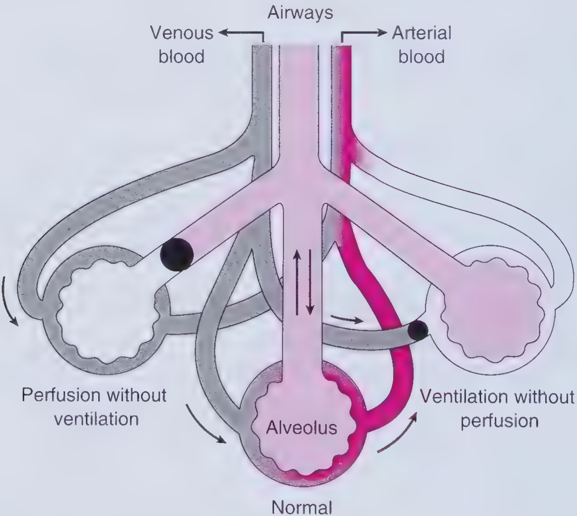
A V/Q of 0 indicates perfusion without ventilation, equivalent to a shunt. A very high V/Q, approaching infinity, indicates ventilation without perfusion, equivalent to alveolar dead space. Frameworks for Internal Medicine, p. 636.
Regional V/Q variation in an upright lung
Both ventilation and perfusion are greater at the lung bases than at the apices. However, perfusion increases much more than ventilation toward the bases. Therefore, the V/Q ratio rises from base to apex.
                 APEX
       Ventilation: low
       Perfusion: very low
       V/Q: high, about 3
       PAO2: higher | PACO2: lower
                  ↑
                  │ V/Q increases
                  │
       V/Q: low, about 0.6
       PAO2: lower | PACO2: higher
       Perfusion: very high
       Ventilation: high
                 BASE
Thus, the quoted overall V/Q ratio of 0.8 is an average, not the value in every lung region. Ganong's Review of Medical Physiology, 26th ed., p. 635.
Factors affecting the V/Q ratio
1. Gravity and body position
- In the upright position, gravity increases pulmonary blood flow markedly at dependent lung regions.
- Ventilation also rises toward the base because basal alveoli are more compliant, but this rise is less than the rise in perfusion.
- Hence: apex has high V/Q and base has low V/Q.
- In supine or lateral positions, the dependent lung regions receive relatively more perfusion.
2. Alveolar ventilation
Anything reducing airflow to an alveolus lowers V/Q:
- Airway obstruction: asthma, COPD, mucus plug, foreign body
- Alveolar filling/collapse: pneumonia, pulmonary edema, atelectasis
- Hypoventilation
Effect: low V/Q, low alveolar O₂, high alveolar CO₂.
3. Pulmonary perfusion
Anything reducing blood flow to an alveolus raises V/Q:
- Pulmonary embolism
- Low cardiac output or shock
- Compression or obstruction of pulmonary vessels
- Pulmonary vascular disease
Effect: high V/Q, dead-space-like ventilation.
4. Pulmonary vascular pressures and West zones
Regional blood flow depends on the relation between:
- pulmonary arterial pressure,
- pulmonary venous pressure, and
- alveolar pressure.
At the apex, lower vascular pressure may allow alveolar pressure to compress capillaries, reducing perfusion and increasing V/Q. At the base, vascular pressures are higher and perfusion is greater.
5. Lung volume and alveolar pressure
Very high or very low lung volumes can compress pulmonary vessels:
- Overdistended alveoli, such as during positive-pressure ventilation, may compress alveolar capillaries and reduce perfusion.
- This can increase V/Q and physiologic dead space.
6. Hypoxic pulmonary vasoconstriction
Low alveolar PO₂ causes local pulmonary arteriolar constriction. This diverts blood away from poorly ventilated alveoli toward better-ventilated areas, tending to improve V/Q matching. If widespread, such as in severe chronic lung disease or high altitude, it can cause pulmonary hypertension.
7. Disease-related unevenness
Regional V/Q inequality occurs commonly in:
- COPD and emphysema
- asthma
- pneumonia
- pulmonary edema
- interstitial lung disease
- pulmonary embolism
- atelectasis
V/Q mismatch is a common cause of hypoxemia and usually produces an increased alveolar-arterial oxygen gradient, as summarized in this NCBI physiology review.
This is a shared conversation. Sign in to Orris to start your own chat.