
Create detailed mbbs final year notes on leiomyoma of uterus
"Leiomyoma"[MeSH Terms] AND "uterine neoplasms"[MeSH Terms]
uterine leiomyoma fibroid gross specimen whorled cut surface
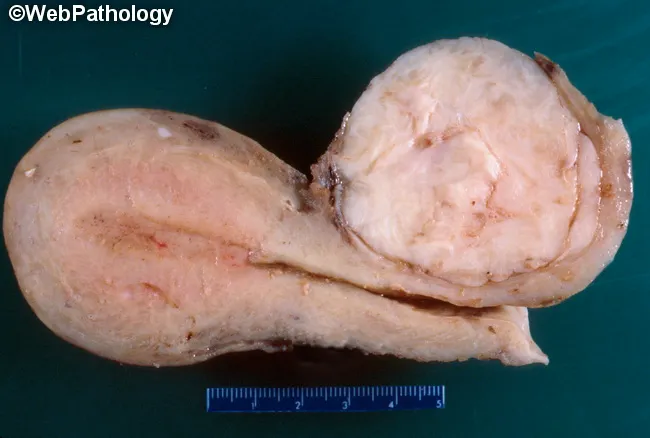
This is a gross pathology photograph of a uterine specimen demonstrating a leiomyoma (fibroid) arising within the uterine corpus, with the cervix oriented to indicate the uncommon cervical location. The image shows a pale, ivory-white to tan, firm nodular mass with a smooth, whorled cut surface embedded in myometrium and extending toward the left uterine body. The lesion appears well circumscribed, encapsulated by native myometrium, and measures on the order of several centimeters in greatest dimension as suggested by the included metric scale. The gross morphology is characteristic of benign smooth muscle neoplasia: firm consistency, whorled fascicles, and uniform pale coloration, with minimal surface necrosis or hemorrhage. The cervical portion remains recognizable; the corpus is to the left, consistent with the note that the corpus is the primary site in this specimen. The image lacks invasion into surrounding structures on gross inspection. This specimen supports a diagnosis of uterine leiomyoma (fibroid), a common benign tumor in women. Cervical involvement or atypical presentation must be considered. Clinically, fibroids cause menorrhagia, pelvic pressure, and reproductive dysfunction; management ranges from observation to myomectomy or hysterectomy depending on symptoms and fertility considerations. Correlation with histology confirms benign smooth muscle tumor.
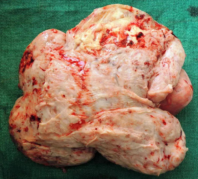
Gross macroscopic image of a giant subserosal uterine leiomyoma resected from a 42-year-old female. Modality: Gross pathology photography; specimen oriented with cut surface exposed for architectural assessment. The mass is large, firm, pale white to tan, with a lobulated contour and a lightly nodular surface. On cut, the lesion shows a homogeneous, whorled pattern of intersecting smooth muscle bundles with intervening fibrous stroma; margins are well defined, consistent with a benign neoplasm. No obvious hemorrhage or necrosis is evident on the exposed surface. Subserosal location implies growth toward the serosal surface with outward protrusion from the uterine wall. The uterus shows a single, predominant fibroid; the appearance is typical for leiomyoma, though giant size may cause bulk symptoms. Histology (to be confirmed) would demonstrate spindle-shaped myocytes in sweeping fascicles, with minimal atypia and low mitotic activity; hyaline degeneration may be present in larger lesions. Clinically, fibroids are estrogen-responsive and common in women of reproductive age; differential diagnoses include cellular leiomyoma and leiomyosarcoma, with clinical correlation and imaging guiding management. This specimen supports a benign diagnosis and is valuable for education, pathology atlases, and gynecologic oncology teaching.
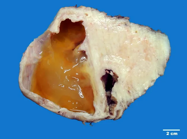
Imaging modality: gross pathology photograph of a hysterectomy specimen, sagittal cross-section through the uterus, highlighting a large cervical fibroid with degenerative changes. The cervix is enlarged by a well-circumscribed, firm mass measuring approximately 14 cm in maximum dimension. On the cut surface, cystic degeneration is evident, with multiple large, rounded cystic spaces filled by seromucinous fluid, imparting a yellow-orange gelatinous appearance. Intervening solid areas show dense, whorled fascicles of smooth muscle arranged in a classic 'whorled' or 'raw silk' pattern. The capsule is a fibrous envelope; the tumor coexists with adjacent myometrium and cervix, with no gross invasion into surrounding structures described. The external surface is pale white with occasional surface friability. The presence of cystic degeneration reflects long-standing growth and secondary ischemic changes commonly seen in large fibroids. These features help distinguish benign leiomyoma from malignant muscle tumors when evaluated grossly, though confirmation requires histology demonstrating benign smooth muscle cells with minimal mitotic activity. Clinically, a large cervical leiomyoma can cause mass effect and pelvic pain; surgical treatment—hysterectomy or myomectomy—may be indicated based on symptoms and patient fertility considerations. This specimen emphasizes classic gross hallmarks: mature smooth muscle whorling, degenerative changes, and seromucinous content within cystic spaces consistent with uterine fibroid pathology.
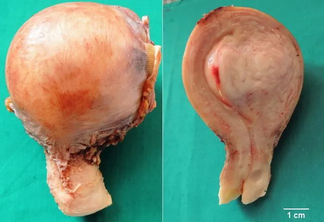
This gross pathology image shows a hysterectomy specimen from a 42-year-old female with a large intramural leiomyoma arising within the myometrium, the most frequent fibroid location. The tumor is centered in the uterine wall with a well-circumscribed, rounded appearance and an organized gray-pink to pink-tan cut surface. The surface exhibits a classic whorled pattern consistent with smooth muscle cell bundles and fibrous stroma, while the surrounding myometrium appears otherwise unremarkable macroscopically. External contour of the uterus is mildly enlarged by the intramural mass, with no obvious hemorrhage or necrosis identified on this gross examination. Ultrasound had detected a mass within the uterine wall correlating with the pathologic finding. Clinically, the patient presented with menorrhagia and back pain—typical symptoms of uterine fibroids. Histologically, intramural leiomyomas are benign neoplasms composed of uniform spindle-shaped smooth muscle cells arranged in interlacing fascicles; however a microscopic diagnosis is not shown here. The significance lies in distinguishing benign leiomyoma from malignant sarcoma, and in guiding treatment choices such as hysterectomy or myomectomy based on reproductive goals. This image provides educational demonstration of gross morphology, localization within the myometrium, and the characteristic cut-surface texture of intramural fibroids.
FIGO classification submucosal intramural subserosal fibroids diagram
leiomyoma histology interlacing fascicles smooth muscle cells
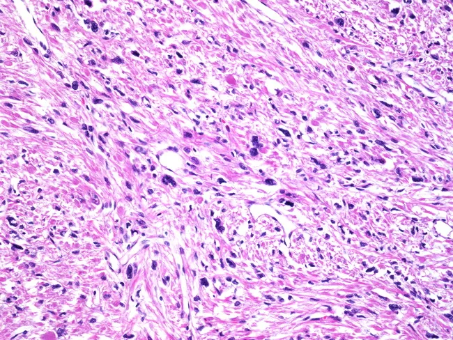
Imaging modality: Light microscopy of a hematoxylin and eosin stained uterine tissue section. Anatomical context: female reproductive system; uterus (myometrium); tissue type smooth muscle; region myometrial wall. Visual features: densely packed spindle to polygonal cells arranged in interlacing fascicles; marked cellular pleomorphism with bizarre, hyperchromatic nuclei; multinucleated smooth muscle cells; abundant eosinophilic cytoplasm; low to absent mitotic activity; no coagulative necrosis; scattered fibrous stroma and thick wavy collagen bands; occasional hyalinization. Interpretation: Symplastic (bizarre) leiomyoma, a histologic variant of benign uterine leiomyoma; despite alarming cellular atypia, lack of mitotic activity and necrosis supports benign behavior. Clinical significance: differentiate from leiomyosarcoma; crucial to avoid overtreatment; in premenopausal patients, often associated with progestin exposure; diagnosis guides conservative surgical management if fertility preservation is desired. Related terms: pleomorphic, symplastic, bizarre leiomyoma; also called unusual cell-variants of fibroid. Potential clinical use cases: educational histology demonstration; diagnostic training; differential diagnosis teaching; pathology review conferences; research on hormonal associations in leiomyoma variants. This image is helpful for learners to recognize that marked cellular atypia alone does not imply malignancy in smooth muscle tumors, provided mitotic activity and necrosis are absent.
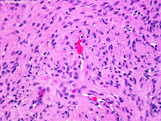
This histopathology image depicts a cutaneous smooth muscle tumor (leiomyoma) on light microscopy. The tissue shows interlacing fascicles of spindle-shaped smooth muscle cells with eosinophilic cytoplasm and elongated, blunt-ended nuclei. Cellular atypia is absent or minimal; occasional degenerative nuclear atypia may be present, but mitotic activity is not evident. The tumor is well circumscribed within dermal/subcutaneous tissue, and nerves are evident within or adjacent to the lesion, accounting for pain and sensitivity to manipulation described clinically. There is no overt necrosis or high-grade pleomorphism, supporting a benign behavior. The morphological features—uniform spindle cells, organized architecture, and lack of mitotic figures—are characteristic of leiomyoma and help distinguish it from malignant smooth muscle neoplasms such as leiomyosarcoma. Clinically, cutaneous leiomyomas present as firm, painful nodules and are typically cured by simple surgical excision with excellent prognosis; recurrence is uncommon when completely excised. Immunohistochemistry (not shown) would typically reveal smooth muscle actin (SMA) positivity. This image provides educational value for medical students and clinicians in recognizing benign smooth muscle tumors, differentiating from neural or fibrous lesions, and correlating histology with pain syndromes that drive therapeutic management. This correlation supports clinical decision-making and reinforces the benign, curable nature of cutaneous leiomyoma through complete excision.
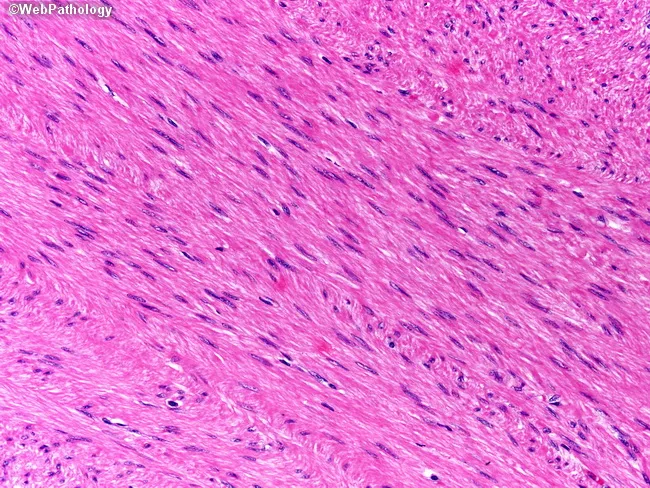
Histopathology: A formalin-fixed, paraffin-embedded tissue section stained with Hematoxylin and Eosin (H&E) shows interlacing fascicles of spindle-shaped smooth muscle cells with elongated, blunt-ended or cigar-shaped nuclei and abundant eosinophilic cytoplasm. The cells are uniform in size, with minimal nuclear atypia and rare mitotic figures; there is no evident necrosis. The architectural pattern is consistent with a benign leiomyoma arising from the smooth muscle layer, such as the muscularis propria of the gastrointestinal tract or uterine myometrium, depending on anatomic origin. Immunohistochemistry demonstrates tumor cells that are strongly positive for desmin and smooth muscle actin (SMA), confirming smooth muscle differentiation. The neoplastic cells are negative for gastrointestinal stromal tumor markers CD34, CD117 (c-KIT), and DOG1, effectively excluding GIST. Diagnostic significance: the combination of characteristic spindle cell morphology and desmin/SMA positivity with CD34/CD117/DOG1 negativity supports a diagnosis of leiomyoma over GIST or leiomyosarcoma. Clinical correlation: these lesions are typically benign with favorable prognosis following complete excision; differential diagnoses include leiomyosarcoma (increased mitotic activity, atypia, necrosis) and GIST (c-KIT or DOG1 positivity). This image exemplifies classical smooth muscle tumor histology and IHC profile, and is useful for educational purposes in surgical pathology, GI/hepatic pathology, and mesenchymal tumor differential diagnosis. Educational diagnostic utility.
uterine fibroids medical management relugolix elagolix linzagolix ulipristal withdrawn tranexamic acid guideline
Leiomyoma (Fibroid) of the Uterus: MBBS Final Year Notes
1. Definition
- A leiomyoma (fibroid, myoma) is a benign, monoclonal tumour of myometrial smooth muscle. It contains abundant extracellular matrix (collagen, elastin, fibronectin, proteoglycan) and is surrounded by a vascular pseudocapsule. - Berek & Novak's Gynecology, p. 497-499
- It is the commonest benign tumour of the female genital tract and the commonest pelvic tumour in women.
- Fewer than 1% undergo malignant change to leiomyosarcoma. - Bailey & Love, p. 1608
2. Epidemiology
- Fine serial sectioning of uteri from 100 consecutive hysterectomies found fibroids in 77%, some only 2 mm. - Berek & Novak's, p. 497
- Cumulative incidence in African American women is about 60% by age 35 and over 80% by age 50. In white women it is about 40% at 35 and almost 70% by 50. - Berek & Novak's, p. 497
- Fibroids are present in up to 40% of women during the reproductive years. - Goldman-Cecil, Uterine Fibroids
- Fibroids are rare before puberty, most common in the reproductive years, and regress after menopause.
3. Aetiology and Pathogenesis
- Fibroids are monoclonal. About 40-50% show tumour-specific chromosomal rearrangements involving chromosomes 6, 7, 12 and 14 (for example t(12;14)). - Berek & Novak's, p. 499
- MED12 mutations (X chromosome) occur in up to 70% of leiomyomas. - Robbins, p. 692
- A germline fumarate hydratase (FH) mutation causes Hereditary Leiomyomatosis and Renal Cell Carcinoma (HLRCC): multiple cutaneous and uterine leiomyomas plus renal cell cancer. - Robbins; Goldman-Cecil
- Fibroids are 2.5 times more common in first-degree relatives of affected women. - Berek & Novak's, p. 501
- Fibroids are oestrogen and progesterone dependent. They grow in the reproductive years, grow rapidly in early pregnancy, and shrink after menopause. - Robbins, p. 692
- Fibroids have higher aromatase activity, so they produce more oestradiol locally. They also have more progesterone receptors A and B than normal myometrium. - Berek & Novak's, p. 500
- Mitotic counts peak when progesterone is highest.
- GnRH agonists shrink fibroids, but adding a progestin blocks the shrinkage. Antiprogestins (RU486) block growth. - Berek & Novak's, p. 500
| Increase risk | Protective |
|---|---|
| Age (peak 40s), early menarche (<10 yr), obesity | Increased parity |
| Black race, family history | Smoking |
| Nulliparity | Exercise |
| Prolonged oestrogen exposure | Late menarche |
- Source: Berek & Novak's, p. 500-501
4. Pathology
Gross
- Sharply circumscribed, firm, grey-white masses with a whorled cut surface ("raw silk" or "trabeculated" look).
- They are often multiple and range from small nodules to huge masses.
- The pseudocapsule is compressed myometrium, so the tumour can be shelled out (enucleated) at myomectomy.
Microscopy
- Interlacing bundles and fascicles of uniform spindle-shaped smooth muscle cells with cigar-shaped nuclei and eosinophilic cytoplasm, resembling normal myometrium.
- There is minimal atypia and few or no mitoses, and no coagulative necrosis.
- Foci of fibrosis, calcification and degenerative softening may be present. - Robbins, p. 692
Location (classification)
- Intramural (commonest): within the myometrium.
- Subserosal: beneath the peritoneum. They may be pedunculated, can torse, or become "parasitic" by acquiring a blood supply from omentum or bowel.
- Submucosal: beneath the endometrium. They cause bleeding and infertility, and may be pedunculated and prolapse through the cervix (a "fibroid polyp").
- Rare sites: cervical and broad ligament. - Robbins, p. 692; Bailey & Love, p. 1608
| Type | Description |
|---|---|
| 0 | Pedunculated intracavitary |
| 1 | Submucosal, <50% intramural |
| 2 | Submucosal, ≥50% intramural |
| 3 | Intramural, contacts endometrium, 100% intramural |
| 4 | Intramural |
| 5 | Subserosal, ≥50% intramural |
| 6 | Subserosal, <50% intramural |
| 7 | Subserosal pedunculated |
| 8 | Other (cervical, parasitic) |
- Berek & Novak's uses types 0-2 to plan hysteroscopic myomectomy. - Berek & Novak's, p. 1363
Secondary changes (degenerations)
| Degeneration | Features |
|---|---|
| Hyaline (commonest) | Pale, glassy areas replace the whorled pattern |
| Cystic | Fluid-filled spaces after liquefaction of hyaline areas |
| Calcific | After menopause, in subserosal or pedunculated fibroids. Seen on X-ray as a "womb stone" |
| Red (carneous) | Haemorrhagic infarction, typically in pregnancy. Fish-like odour, beefy-red cut surface. Presents with acute pain, low fever and leucocytosis |
| Fatty | Rare |
| Infective / suppurative | After abortion or puerperium, or with submucous fibroid polyp |
| Sarcomatous | Very rare (<1%). Suspect if there is rapid growth after menopause |
Variants (standard teaching)
- Cellular leiomyoma
- Symplastic (bizarre) leiomyoma
- Mitotically active leiomyoma
- Lipoleiomyoma
- Hydropic leiomyoma
- Disseminated peritoneal leiomyomatosis
- Intravenous leiomyomatosis
- Benign metastasising leiomyoma, commonly to lung (usually in women with a prior myomectomy or hysterectomy)
5. Clinical Features
| Symptom | Detail |
|---|---|
| Abnormal uterine bleeding (most frequent) | Menorrhagia ± metrorrhagia. Mainly with submucosal and intramural fibroids that distort the cavity. The link to HMB is not fully proven, so exclude other causes (for example von Willebrand disease). Type 0 and 1 fibroids correlate most strongly with anaemia. - Berek & Novak's, p. 503 |
| Anaemia | Iron deficiency from chronic blood loss |
| Pressure / bulk symptoms | Pelvic heaviness, urinary frequency, urgency or retention, constipation, backache, varicosities. Ureteric compression can cause hydronephrosis |
| Pain | Not typical. Dysmenorrhoea is secondary to menorrhagia. Acute pain suggests red degeneration or torsion of a pedunculated fibroid. - Bailey & Love, p. 1609 |
| Infertility / recurrent pregnancy loss | Mainly submucosal and cavity-distorting fibroids |
| Abdominal lump | Large fibroids |
| Leucorrhoea | Submucous fibroid with infection |
- A firm, nodular, non-tender enlarged uterus that moves with the cervix.
- Palpable lump from the pelvis. The uterus cannot be separated from the mass.
- Pallor if anaemic.
6. Differential Diagnosis
- Adenomyosis (diffusely enlarged, globular, tender uterus)
- Ovarian tumour. This is the principal differential: the uterus is felt separately from an ovarian mass. - Bailey & Love, p. 1609
- Pregnancy
- Endometrial polyp or carcinoma
- Leiomyosarcoma
- Tubo-ovarian mass
7. Investigations
- Haemoglobin and iron studies; consider a bleeding profile.
- Pelvic ultrasound (transvaginal or transabdominal) is the first-line investigation. It shows well-defined hypoechoic whorled masses with shadowing and a pseudocapsule. - Bailey & Love, p. 1609
- MRI is used if ultrasound is inconclusive. It helps with mapping before surgery and with distinguishing adenomyosis and sarcoma. Fibroids appear as well-defined low T2 signal lesions. - Bailey & Love, p. 1609
- Hysteroscopy, saline infusion sonography or hysterosalpingography for submucosal fibroids and infertility work-up.
- Endometrial sampling in women over 40 or with persistent AUB.
- Pap smear and cervical assessment.
- Urine tests, serum creatinine, or IVU / CT urogram if ureteric obstruction is suspected.
8. Fibroids and Pregnancy
- Fibroids may grow in early pregnancy (oestrogen and hCG effect), and many remain stable or shrink later.
- Red degeneration is the commonest complication. It is managed conservatively with rest, analgesia (avoid NSAIDs late in pregnancy) and hydration.
- Complications (standard teaching):
- Miscarriage, preterm labour, abruption
- Malpresentation, obstructed labour (cervical or low-lying fibroid), caesarean section
- Retained placenta, PPH from uterine atony
- Puerperal infection and sepsis
- Myomectomy during caesarean section is generally avoided because of haemorrhage risk.
9. Management
A. Conservative
- Asymptomatic fibroids need observation with periodic review, usually every 6-12 months.
- Fibroids are not premalignant. - Goldman-Cecil
B. Medical (symptom control, not cure)
| Drug | Role |
|---|---|
| Tranexamic acid, NSAIDs | Reduce menstrual loss. Tranexamic acid cuts it by about 26-54% (non-fibroid-specific data) |
| Combined oral contraceptives, LNG-IUS, oral progestins | Reduce bleeding. LNG-IUS is poorly suited to cavity-distorting fibroids |
| GnRH agonists (leuprolide) | Amenorrhoea and 35-55% shrinkage. Short-term use only (3-6 months) because of hypo-oestrogenic effects and bone loss. Regrowth follows stopping. Used pre-operatively. Berek cites 8 weeks of leuprolide before surgery, which allowed vaginal rather than abdominal hysterectomy and raised haematocrit. - Berek & Novak's, p. 1409 |
| Selective progesterone receptor modulators (ulipristal, mifepristone) | Shrink fibroids. Ulipristal use was restricted after reports of serious liver injury (standard teaching; check current local regulation) |
| GnRH antagonists (oral elagolix, relugolix, linzagolix, usually with add-back therapy) | Reduce heavy bleeding in fibroids. Cecil cites the trials of elagolix and relugolix combination therapy. - Goldman-Cecil. NICE has issued guidance on linzagolix |
| Iron supplementation | Treat anaemia |
C. Surgical and interventional
- Myomectomy (abdominal, laparoscopic or robotic, hysteroscopic)
- Indicated for symptomatic women who desire fertility, and for infertility thought secondary to fibroids. Berek considers it a safe alternative to hysterectomy even for large fibroids. - Berek & Novak's, p. 495; Schwartz's
- Hysteroscopic resection is for submucous fibroids with HMB, infertility or recurrent miscarriage. It is limited by size, number and location. Type 0 fibroids of 3 cm or less are removed easily. Type 2 fibroids may need more than one procedure and careful fluid management to avoid overload. - Berek & Novak's, p. 1363-1364
- Risks: haemorrhage, adhesions, uterine rupture in later pregnancy, and recurrence of fibroids.
- Power morcellation carries a risk of spreading an occult sarcoma. Use contained morcellation or avoid it when sarcoma is suspected.
- Uterine artery embolisation (UAE)
- A radiological option for women who do not want surgery. It is contraindicated in those planning pregnancy, and it may cause acute fibroid degeneration needing admission for pain control. - Schwartz's, Uterine Corpus
- Cecil states myomectomy gives better outcomes than UAE. In a Berek-cited trial, 31% of UAE patients ultimately underwent hysterectomy. - Goldman-Cecil; Berek & Novak's, p. 1410
- Post-embolisation syndrome (pain, fever, nausea) is common.
- MR-guided focused ultrasound, radiofrequency ablation: other uterus-sparing options.
- Endometrial ablation: only for small or no cavity distortion with HMB.
- Hysterectomy for symptomatic women who do not want fertility, or after failed conservative treatment. Indications include AUB, pelvic pain or pressure, ureteric compression, and uterine growth after menopause. - Berek & Novak's, p. 1409
- Size greater than 12 weeks without symptoms, or inability to palpate the ovaries, is no longer an accepted indication. Abdominal surgical morbidity does not differ between a 12-week and a 20-week uterus. - Berek & Novak's, p. 1409-1410
- The route is vaginal, laparoscopic or abdominal, depending on size and mobility.
10. Complications
- Chronic blood loss anaemia
- Degeneration (red, hyaline, cystic, calcific)
- Torsion of a pedunculated fibroid
- Infection or abscess formation
- Sarcomatous change (<1%)
- Urinary retention or hydronephrosis, venous stasis
- Rare: polycythaemia (erythropoietin production), pseudo-Meigs syndrome (ascites with pleural effusion), uterine inversion from a submucous polyp
- Obstetric: see section 8
11. Leiomyoma vs Leiomyosarcoma
| Feature | Leiomyoma | Leiomyosarcoma |
|---|---|---|
| Number | Often multiple | Usually solitary |
| Age | Reproductive years | Mostly postmenopausal |
| Gross | Whorled, well circumscribed | Soft, necrotic, haemorrhagic, infiltrative |
| Microscopy | Bland spindle cells, no necrosis | Marked atypia, increased mitoses, coagulative necrosis |
| Genetics | MED12 in up to 70%, simple karyotype | Complex karyotype, TP53/RB/PTEN, MED12 in about 30% |
| Behaviour | Benign, shrinks after menopause | Aggressive, haematogenous spread to lung |
- Source: Robbins, p. 692-693
12. High-Yield Exam Points
- Commonest benign uterine tumour. Commonest site: intramural.
- Commonest symptom: menorrhagia. Commonest secondary change: hyaline degeneration.
- Red degeneration is typical of pregnancy. It is managed conservatively.
- Fibroids shrink after menopause. Growth after menopause suggests sarcoma.
- Sarcomatous change occurs in under 1% of cases.
- Submucous fibroids give the most bleeding and infertility. Subserosal fibroids give pressure symptoms.
- Parasitic fibroid is a subserosal fibroid that has acquired a blood supply from another organ.
- FIGO type 0: pedunculated intracavitary.
- Genetics: MED12, chromosomes 6/12/14, and FH (HLRCC).
- GnRH agonists give temporary shrinkage and are used before surgery. UAE is contraindicated when pregnancy is desired.