
General examination of body and its reason behind it
general physical examination body inspection
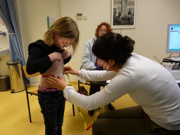
This clinical photograph depicts a general practitioner (GP) trainee performing an anthropometric measurement on a pediatric patient in a primary care setting. The professional is shown kneeling to maintain eye level with the standing child, using a flexible, non-stretch measuring tape to assess waist circumference. The child has appropriately lifted their shirt to expose the abdominal region, allowing for accurate tape placement midway between the lowest rib and the iliac crest. The procedure is conducted in a clinical environment, evidenced by the presence of an examination room sink, waste receptacle, medical privacy curtain, and a computer workstation. This image serves as an educational demonstration of standard physical examination techniques used in pediatrics to monitor nutritional status, assess metabolic risk, and calculate body mass index (BMI) correlates. The scene illustrates clinical best practices, including patient positioning and the use of standardized tools for longitudinal health tracking in general practice.
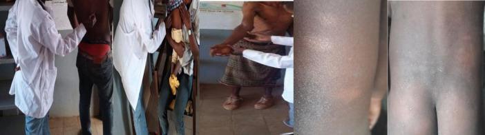
This composite of clinical photographs illustrates the physical examination process for leprosy (Hansen’s disease) in a community health setting. The images show healthcare professionals in white coats performing dermatological and neurological assessments on patients of varying ages. Key clinical features depicted include several hypopigmented, ill-defined anesthetic or hypoesthetic skin patches on the trunk and extremities, which are cardinal signs of the disease. The examination process shown includes visual inspection of the skin, palpation for thickened peripheral nerves, and sensory testing to identify loss of sensation within the lesions. Close-up panels highlight the morphological characteristics of the macules, showing a distinct contrast against the surrounding unaffected skin. This material is used to demonstrate active case-finding techniques and the clinical manifestations of leprosy, such as paucibacillary or multibacillary lesions, in endemic regions.
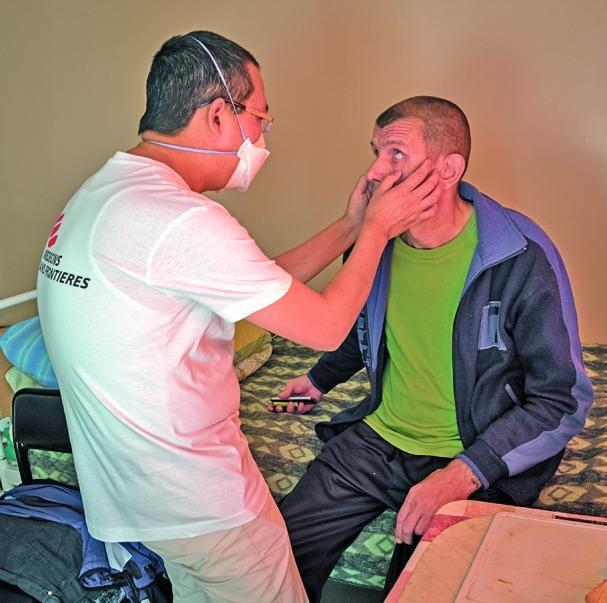
This clinical photograph depicts a physical examination during a patient consultation at a pulmonology and tuberculosis center. A healthcare professional, identified by 'Médecins Sans Frontières' branding on a white t-shirt, is performing a manual assessment of a seated patient's face. The clinician is wearing corrective glasses and a white surgical mask for infection control, which is essential in a respiratory health setting. The examination involves the clinician using both hands to palpate the patient's submandibular or malar region, while the patient looks upward, potentially allowing for the inspection of the conjunctivae for pallor or checking for lymphadenopathy. The setting is a clinical ward or residential treatment room, indicated by the bed and personal items. This image illustrates humanitarian medical aid, infectious disease screening (specifically tuberculosis monitoring), and the importance of personal protective equipment (PPE) during direct patient contact in respiratory specialty clinics.
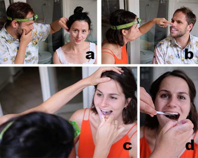
This composite of four clinical photographs (labeled a-d) illustrates fundamental Otorhinolaryngology (ENT) physical examination techniques performed by medical students using an examination kit and a headlamp for illumination. Image (a) depicts otoscopy, showing the examiner stabilizing the patient's head while inspecting the external auditory canal. Image (b) demonstrates anterior rhinoscopy, utilizing a nasal speculum to dilate the nares for visualization of the nasal mucosa and septum. Image (c) shows an oral cavity inspection, where a tongue depressor is used to visualize the oropharynx while the examiner's other hand stabilizes the patient's forehead. Image (d) provides a close-up of indirect laryngoscopy or posterior rhinoscopy, utilizing a laryngeal mirror and a tongue depressor to reflect light and visualize deeper structures. The series highlights the importance of proper ergonomic positioning, the use of external light sources (headlamps), and specific instrumentation for a comprehensive head and neck assessment. The clinical focus is on teaching propaedeutic skills for primary care and specialty ENT diagnostics.
clubbing fingers cyanosis jaundice pallor clinical signs
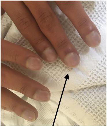
This clinical photograph shows a close-up of a patient's fingers demonstrating classic signs of digital clubbing and peripheral cyanosis. The distal phalanges exhibit a bulbous, 'drumstick' appearance with a noticeable loss of the normal Lovibond angle (the angle between the nail plate and the proximal nail fold). The nail beds appear convex and rounded. A distinct bluish-purple discoloration (cyanosis) is visible across the nail beds and distal fingertips, indicating poor blood oxygenation or circulation. A black arrow points to the most prominent area of clubbing on the index finger. These findings are clinically significant as they often indicate chronic hypoxemia secondary to congenital heart disease (such as Ebstein anomaly), pulmonary disease, or chronic inflammatory conditions. This image serves as a teaching tool for physical examination findings in cardiovascular and respiratory medicine.
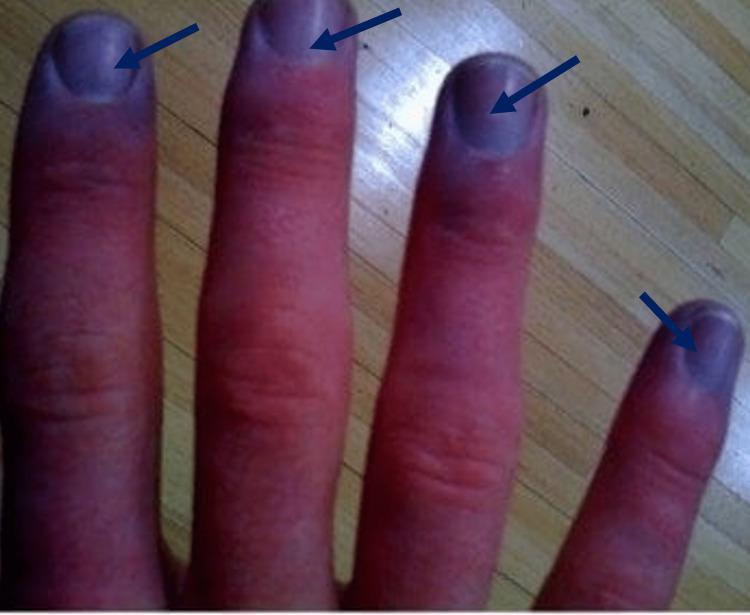
This clinical photograph displays the dorsal aspect of four human fingers, demonstrating classic signs of peripheral cyanosis. There is a distinct, localized bluish-purple discoloration (cyanosis) concentrated at the distal fingertips and involving the subungual regions (nail beds) of all visible digits, highlighted by dark blue arrows. The proximal and middle segments of the fingers exhibit a contrasting erythematous (reddish) hue. The skin appears intact without visible ulcerations, lesions, or significant edema. The nail plates maintain a normal morphology without evidence of digital clubbing. This visual finding is a hallmark of reduced peripheral oxygen saturation or impaired local circulation and is essential for medical students and clinicians in identifying hypoxia or vascular compromise. Target audience: medical students, nursing professionals, and emergency medicine practitioners.
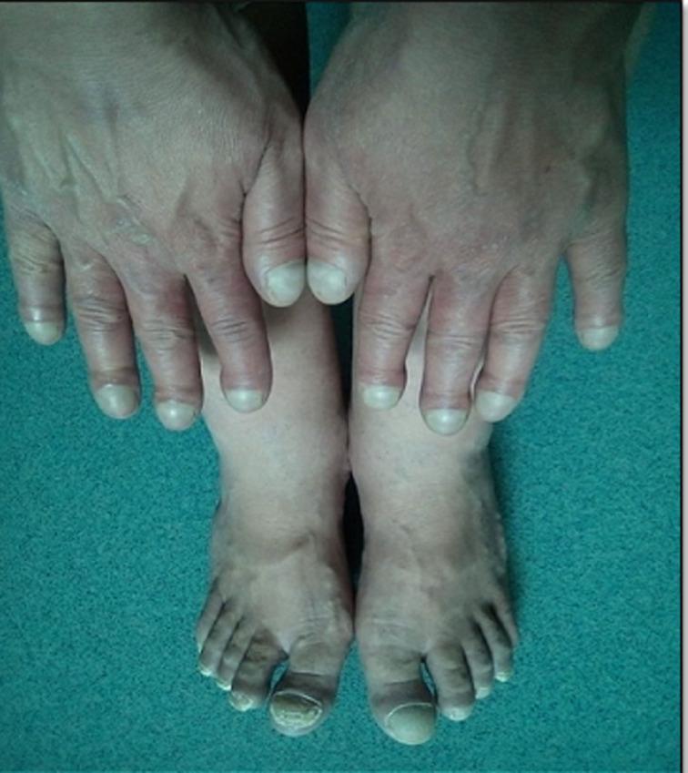
This clinical photograph displays the hands and feet of a patient, demonstrating classic physical signs of chronic hypoxemia. Both the fingers and toes exhibit significant digital clubbing (Hippocratic fingers), characterized by a bulbous, drumstick-like enlargement of the distal phalanges and increased longitudinal and transverse curvature of the nail plates. There is a noticeable loss of the normal Lovibond angle at the nail bed. Additionally, central cyanosis is evident as a dusky, bluish-purple discoloration of the nail beds and surrounding distal integument. These findings are clinically significant as classic markers for congenital cyanotic heart disease, such as Tetralogy of Fallot, or advanced pulmonary disease. The image serves as an educational reference for identifying cardinal signs of systemic oxygen desaturation during a physical examination.
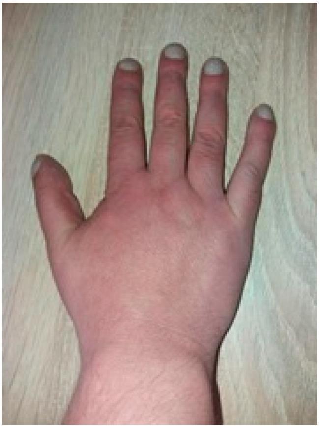
This clinical photograph displays a dorsal view of a human hand demonstrating classic digital clubbing (hippocratic fingers). The distal phalanges of all visible fingers appear bulbous and enlarged, with an increased longitudinal and transverse curvature of the nail plates, giving them a 'watch-glass' or convex appearance. There is a visible loss of the Lovibond angle (the normal 160-degree angle between the nail base and the proximal nail fold). The skin of the hand shows a generalized reddish-to-dusky hue, suggestive of polycythemia or chronic cyanosis. These physical findings are hallmark signs of chronic hypoxemia, frequently associated with underlying conditions such as congenital cyanotic heart disease (e.g., Tetralogy of Fallot) or chronic pulmonary disease. The image serves as an educational tool for identifying secondary physical manifestations of cardiovascular and respiratory pathology.
jaundice scleral icterus eyes yellow skin
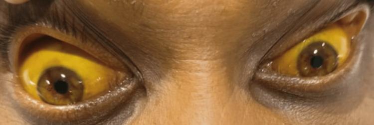
This clinical photograph displays a close-up view of a patient's eyes and upper face, illustrating severe scleral icterus. Both eyes exhibit deep, uniform yellow pigmentation of the sclera and bulbar conjunctiva, a hallmark sign of hyperbilirubinemia. The surrounding facial skin, particularly on the forehead and periorbital regions, also shows a subtle yellowish hue consistent with jaundice, though less pronounced than the ocular findings. The image serves as a clear educational example of the physical manifestations of liver dysfunction, biliary obstruction, or systemic toxicity—in this context, following rodenticide (zinc phosphide) ingestion. The visibility of the distinct yellowing against the patient's natural skin tone provides a significant diagnostic reference for internal medicine and gastroenterology students and practitioners.
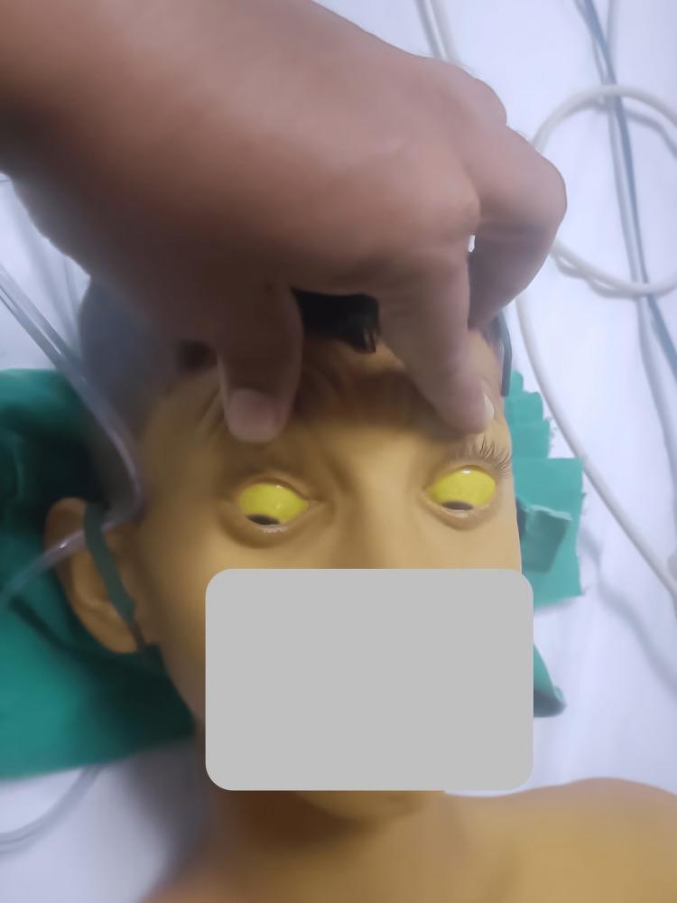
This clinical photograph displays a pediatric patient demonstrating severe systemic jaundice. The primary diagnostic feature is profound scleral icterus, characterized by a bright yellow discoloration of the entire visible sclera in both eyes. The upper eyelids are manually elevated to fully reveal the extent of the icterus. Additionally, the patient's facial skin, forehead, and upper chest exhibit a marked yellowish hue (jaundice), consistent with hyperbilirubinemia. The patient appears to be in a clinical setting, as evidenced by medical tubing and linens visible in the background. This image is an educational example of the physical manifestations of liver dysfunction or hemolytic processes, such as hepatitis, Wilsonian crisis, or acute toxicity, which can lead to significant accumulation of bilirubin in mucosal surfaces and skin.
pedal edema pitting lymph node examination
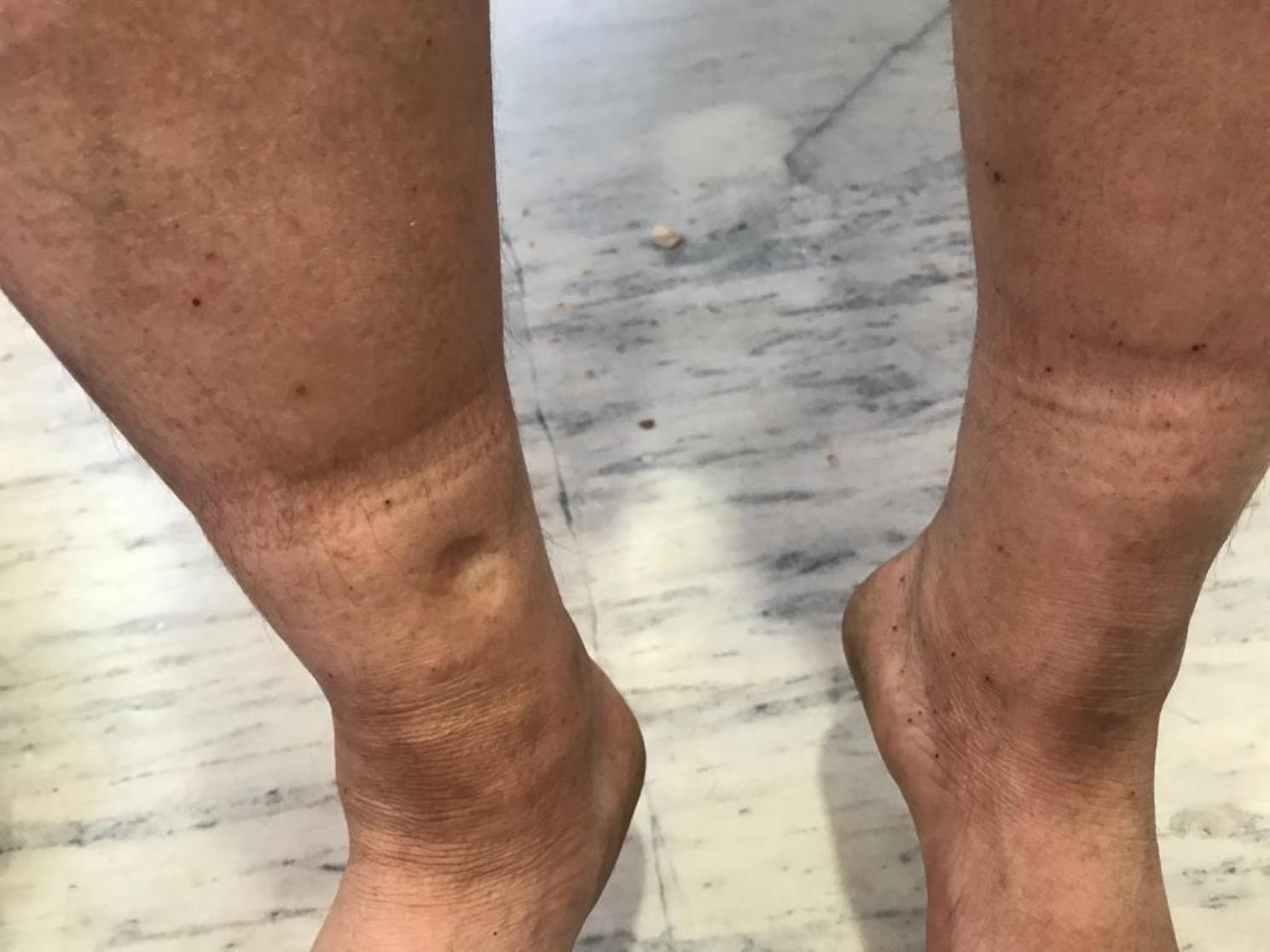
This clinical photograph displays bilateral pedal edema in the lower extremities. The image focuses on the distal legs and ankles, demonstrating significant swelling and tautness of the skin. A key diagnostic feature is visible on the anterior aspect of the lower leg, where a localized indentation or 'pit' remains after pressure was applied, characteristic of pitting edema. The skin shows slight discoloration with scattered hyperpigmented macules and a horizontal band of lighter pigmentation above the malleoli, possibly indicating chronic pressure from footwear or hosiery. The overall presentation is a classic clinical representation used in internal medicine and cardiology to illustrate fluid retention and systemic volume overload. The educational focus is on the assessment of peripheral edema and the visual identification of the 'pitting' sign in a clinical examination context.
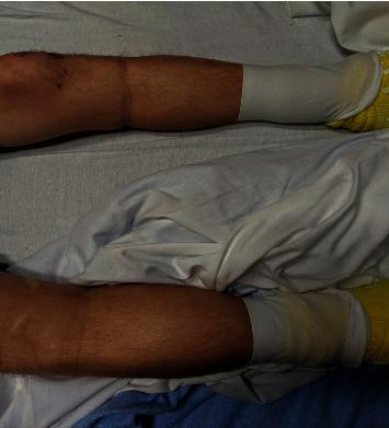
Clinical photograph of a patient's lower extremities in a supine position, demonstrating significant bilateral pedal edema. The swelling is symmetrical, extending from the feet proximally to the level of the knees. The skin over the shins and calves appears taut, shiny, and distended, characteristic of high-grade pitting edema (clinically noted as 4+). Both feet are covered with white medical compression wraps or dressings, with yellow non-slip hospital socks visible. This image serves as a clinical example of generalized peripheral edema, which in this educational context is associated with insulin-induced edema. Key landmarks include the visible distension of the pretibial area and the ankle region, contrasting with the proximal thigh area above the knee where the swelling appears to terminate. This visual is relevant for training in physical examination, fluid volume assessment, and recognizing secondary side effects of endocrine therapies.
General Examination of the Body
1. General Appearance / First Impression
- A patient who appears acutely ill (pale, diaphoretic, in pain) needs immediate attention.
- Facial characteristics can immediately suggest syndromes: Cushing's ("moon face"), Parkinson's (expressionless "mask face"), myxedema (puffy, coarse features), hyperthyroidism (anxious, wide-eyed), acromegaly (prominent jaw/brow), Down syndrome, or Marfanoid habitus.
- Apparent age greater than actual age suggests chronic illness, malnutrition, or prolonged stress.
Harrison's Principles of Internal Medicine 22E lists dozens of recognizable "faces" - Parkinsonian, Cushingoid, myxedematous, myasthenic, and more - as critical initial diagnostic cues.
2. Built, Nourishment, and Body Habitus
- Emaciation / cachexia - suggests malignancy, tuberculosis, chronic infection, malabsorption, or hyperthyroidism.
- Obesity - associated with diabetes, hypertension, sleep apnea, PCOS, hypothyroidism.
- Short stature - suggests growth hormone deficiency, chromosomal abnormalities, rickets.
3. Pallor (Anaemia)
- Blood loss (acute/chronic)
- Haemolysis (e.g., sickle cell, malaria)
- Bone marrow failure (aplastic anaemia, leukaemia)
- Nutritional deficiency (iron, B12, folate)
- Chronic kidney disease
Bailey and Love's Short Practice of Surgery, 28th Ed. emphasizes that "general inspection looking for evidence of... anaemia" is one of the first steps in surgical examination.
4. Jaundice (Icterus)
| Type | Cause | Clue |
|---|---|---|
| Pre-hepatic | Haemolysis | Mild jaundice, anaemia |
| Hepatic | Hepatitis, cirrhosis | Dark urine, tender liver |
| Post-hepatic | Bile duct obstruction (gallstones, cancer) | Pale stools, itching |
5. Cyanosis
| Type | Site | Cause |
|---|---|---|
| Central | Lips, tongue, oral mucosa | Lung disease, congenital heart disease (R-to-L shunt) |
| Peripheral | Fingertips, toes, ears | Poor circulation, cold, heart failure |
Barash's Clinical Anesthesia, 9th Ed.: "The presence of cyanosis and clubbing, the breathing pattern, and the type of breath sounds should be noted."
6. Clubbing of Fingers and Toes
- Pulmonary: lung cancer, chronic suppurative lung disease (bronchiectasis, abscess, empyema), fibrosing alveolitis
- Cardiac: cyanotic congenital heart disease, infective endocarditis
- GI: Crohn's disease, cirrhosis, hepatopulmonary syndrome
- Idiopathic
Robbins Pathologic Basis of Disease: Severe longstanding hypoxia leads to "peculiar distal blunting and enlargement ('clubbing') of the tips of the fingers."

7. Oedema (Edema)
| Cause | Clue |
|---|---|
| Cardiac failure | Bilateral, dependent, ascends with severity |
| Hypoalbuminaemia (nephrotic, cirrhosis, malnutrition) | Soft, bilateral, may be generalized (anasarca) |
| Renal failure | Periorbital edema (early morning), then peripheral |
| Lymphoedema | Non-pitting, usually unilateral |
| DVT | Unilateral, painful |
8. Dehydration
9. Lymphadenopathy
| Node characteristic | Likely cause |
|---|---|
| Tender, soft | Infection (reactive) |
| Hard, fixed | Metastatic carcinoma |
| Rubbery, non-tender | Lymphoma |
| Generalized | HIV, EBV, leukaemia, sarcoidosis |
Bailey and Love: Left supraclavicular lymphadenopathy (Virchow's node / Troisier's sign) is specifically noted as suggestive of intra-abdominal malignancy (e.g., stomach cancer).
10. Vital Signs
- Fever - infection, malignancy, autoimmune disease, drug reaction
- Tachycardia - pain, fever, anaemia, shock, hyperthyroidism
- Hypertension - essential or secondary (renal, endocrine)
- Tachypnoea - respiratory failure, acidosis, pain
- Low SpO2 - hypoxia, lung disease, cardiac failure
11. Skin, Hair, and Nails
- Hyperpigmentation - Addison's disease, haemochromatosis, chronic liver disease
- Spider naevi + palmar erythema - chronic liver disease
- Nicotine staining of nails - smoking-related lung/vascular disease
- Koilonychia (spoon nails) - iron deficiency anaemia
- Leukonychia (white nails) - hypoalbuminaemia
12. Gait and Posture
- Shuffling gait - Parkinson's disease
- High-stepping gait - foot drop (common peroneal nerve palsy)
- Trendelenburg gait - hip abductor weakness
- Hemiplegic gait - upper motor neuron lesion (stroke)
- Ataxic gait - cerebellar disease
13. Speech and Mental State
Summary Table
| Component | What You Look For | Key Diagnoses Suggested |
|---|---|---|
| General appearance | Distress, facies, habitus | Syndromes, systemic illness |
| Pallor | Conjunctivae, palms | Anaemia |
| Jaundice | Sclera, skin | Liver/haemolytic disease |
| Cyanosis | Lips, tongue, nails | Respiratory/cardiac failure |
| Clubbing | Fingertips | Lung cancer, congenital heart disease |
| Oedema | Ankles, sacrum | Heart failure, renal/liver disease |
| Dehydration | Skin turgor, tongue | Fluid deficit |
| Lymphadenopathy | Neck, axilla, groin | Infection, lymphoma, cancer |
| Vital signs | Pulse, BP, RR, Temp | Infection, shock, cardiac disease |
| Skin/nails | Pigmentation, koilonychia | Nutritional, liver, endocrine |
| Gait | Shuffle, limp, ataxia | Neuro/musculoskeletal |
Clinical examination of thyrod