
Explain invasion and metastasis refer robbins pathology
I'll start by reading the medical library skill, then search for invasion and metastasis content from Robbins Pathology.
Reading File
Now let me search the library for invasion and metastasis content from Robbins Pathology in parallel:
Finding Sources
Finding Sources
I have both Robbins books. Let me read the invasion and metastasis sections from both in parallel:
Reading File
Reading File
Good content from both books. Let me now read more from both - especially vascular dissemination and colonization sections, plus fetch the key diagrams:
Reading File
Reading File
Loading Image
Now let me read the detailed vascular dissemination section from Robbins Pathologic Basis of Disease (the larger textbook), which has more depth:
Reading File
Loading Image
Excellent - I now have very rich content and both key diagrams. I have all I need to construct a comprehensive, well-structured answer. Let me now compile the full explanation.
Invasion and Metastasis - Robbins Pathology
Invasion and metastasis are the major causes of cancer-related morbidity and mortality and represent the defining hallmarks of malignant tumors. They involve complex interactions between cancer cells, stromal cells, and the extracellular matrix (ECM). The entire process - called the metastatic cascade - is broken into two broad phases:
- Invasion of the ECM
- Vascular dissemination, tissue homing, and colonization
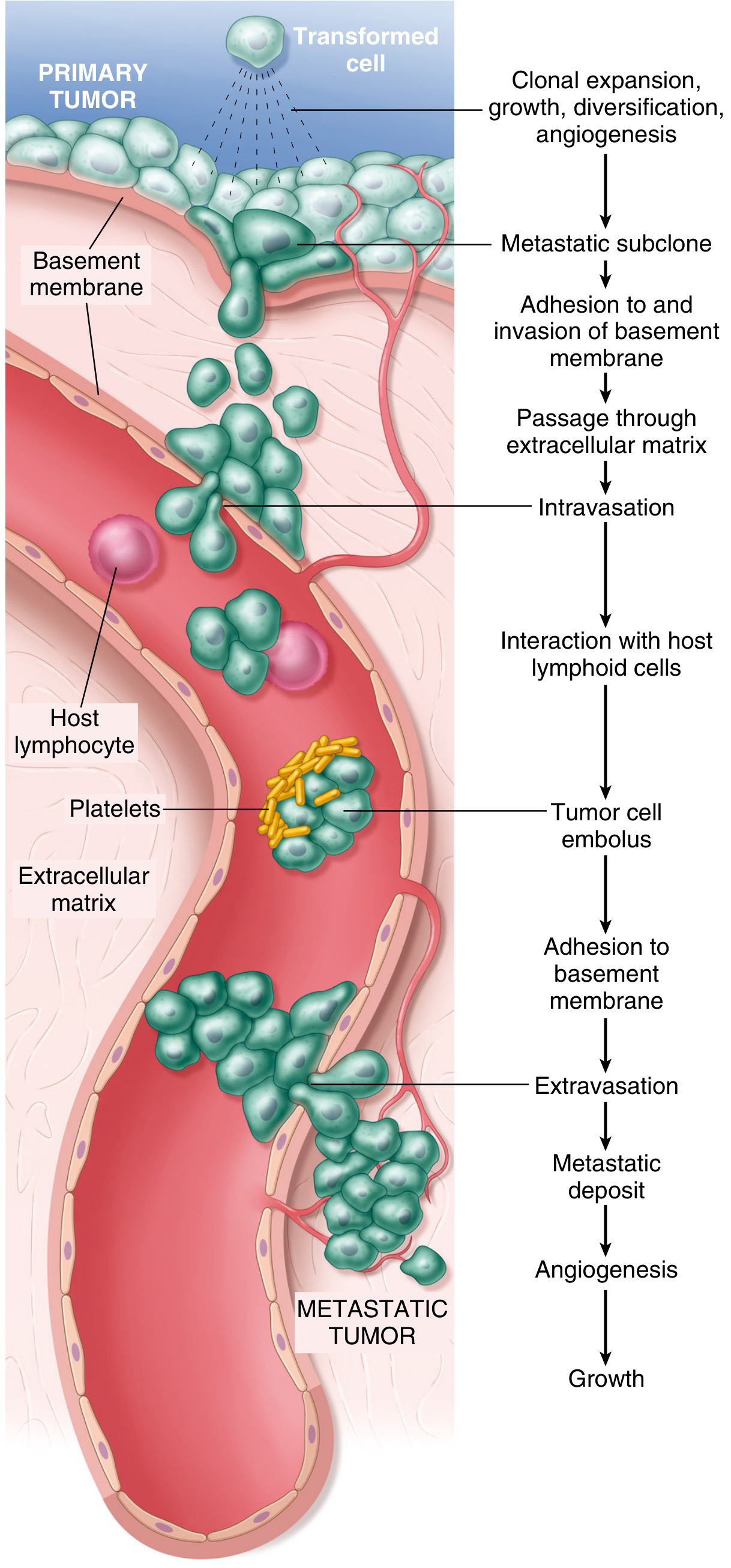
Fig. 7.36 - The metastatic cascade (Robbins, Cotran & Kumar Pathologic Basis of Disease)
Phase 1: Invasion of the Extracellular Matrix
Normal tissues are organized into compartments separated by two types of ECM: basement membranes and interstitial connective tissue, both composed of collagens, glycoproteins, and proteoglycans.
For a carcinoma to metastasize, it must:
- Breach the underlying basement membrane
- Traverse the interstitial connective tissue
- Penetrate the vascular basement membrane to enter circulation
This entire process reverses at the distant site during extravasation.
Invasion of the ECM proceeds through four sequential steps:
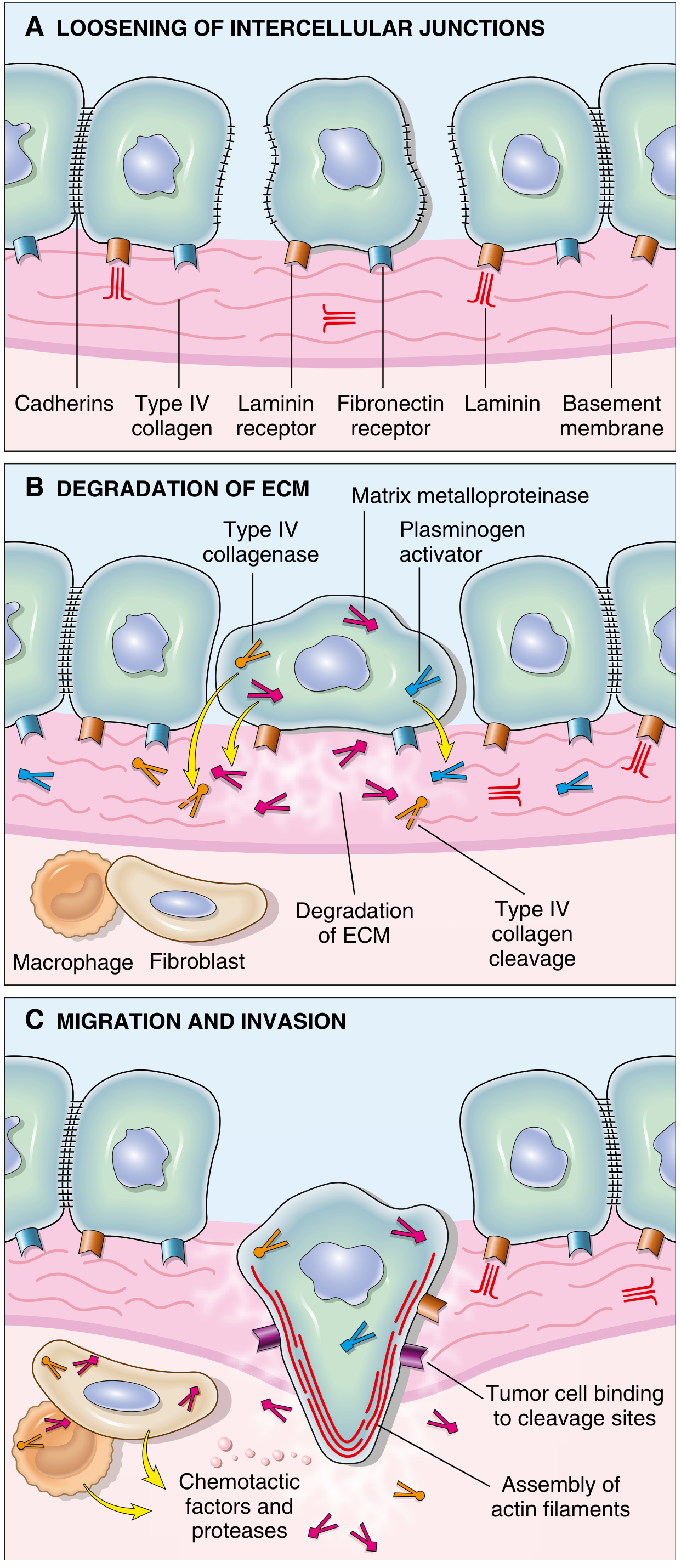
Fig. 7.37 - Invasion of epithelial basement membranes by tumor cells (Robbins, Cotran & Kumar)
Step 1 - Loosening of Tumor Cell-Tumor Cell Interactions
Normal epithelial cells are glued together by E-cadherin, a transmembrane glycoprotein that mediates homotypic adhesion. E-cadherin also relays antigrowth signals by sequestering beta-catenin.
In cancer, E-cadherin function is lost by:
- Mutational inactivation of E-cadherin genes
- Activation of beta-catenin genes
- Epithelial-Mesenchymal Transition (EMT) - controlled by transcription factors SNAIL and TWIST, which suppress E-cadherin expression
EMT is defined not only by downregulation of epithelial markers (E-cadherin) but also by upregulation of mesenchymal markers (vimentin, smooth muscle actin), promoting a pro-migratory phenotype. EMT is especially implicated in breast and prostate cancer metastasis.
Step 2 - Degradation of the Basement Membrane and Interstitial Matrix
Tumor cells accomplish ECM degradation either by secreting proteolytic enzymes themselves, or by inducing stromal cells (fibroblasts, inflammatory cells) to do so.
Key proteases involved:
- Matrix Metalloproteinases (MMPs) - especially MMP-9 (gelatinase), which cleaves Type IV collagen of the basement membrane
- Cathepsin D
- Urokinase plasminogen activator
MMPs do more than just degrade - they also:
- Release VEGF sequestered within the ECM (promoting angiogenesis)
- Generate chemotactic fragments from ECM glycoproteins
- Create angiogenic and growth-promoting cleavage products
Benign tumors have little MMP-9 activity, while their malignant counterparts overexpress it. Simultaneously, concentrations of metalloproteinase inhibitors (TIMPs) are reduced in many cancers, further tilting the balance toward tissue degradation.
Step 3 - Attachment to Novel ECM Components
After degradation, new ECM components are exposed. Tumor cells show complex changes in integrin expression - shifting from integrins that maintain normal tissue architecture to integrins that favor migration.
Key attachment interactions:
- Laminin receptors on tumor cells bind to laminin in the basement membrane - these are overexpressed in many aggressive carcinomas
- Fibronectin receptors facilitate attachment to interstitial matrix
The newly cleaved ECM fragments serve as binding tracks that guide tumor cell movement.
Step 4 - Migration and Invasion of Tumor Cells
Tumor cell locomotion is propelled by:
- Autocrine motility factors secreted by tumor cells themselves
- Paracrine factors from stromal/inflammatory cells (chemotactic gradients)
- Cleavage products of ECM components (collagen, laminin fragments) which are chemotactic
- Growth factors like IGF-I and IGF-II released from degraded ECM
Movement requires assembly of actin filaments at the leading edge of the migrating cell. Cells track along binding sites created by ECM cleavage.
Phase 2: Vascular Dissemination, Tissue Homing, and Colonization
Once tumor cells invade through the ECM, they enter the vasculature and face a new set of challenges.
Intravasation and Circulation
- Tumor cells enter blood vessels or lymphatics
- In the circulation, they are vulnerable to immune destruction (NK cells, T cells)
- They may form tumor cell emboli protected by aggregates of platelets and fibrin - this shields them from immune attack
- Most circulating tumor cells are destroyed; only rare cells produce metastases, explaining why the metastatic process is highly inefficient
Arrest and Extravasation
- Tumor cells arrest in the first capillary bed encountered - most commonly lungs and liver
- Liver is the first capillary bed for portal venous drainage
- Lungs are the first capillary bed for systemic venous drainage
- Once arrested, cells adhere to the vascular endothelium (via adhesion molecules) and then extravasate using the same mechanisms as intravasation
Organ Tropism
Some tumors show non-anatomic organ tropism not explained by circulatory patterns alone. This is explained by:
- Expression of chemokine receptors on tumor cells whose ligands are expressed by endothelial cells at the target site
- Example: Breast cancer cells express CXCR4, whose ligand CXCL12 (SDF-1) is abundantly expressed in lungs, liver, and bone marrow
- Expression of specific adhesion molecules on target organ endothelium
Classic examples of organ tropism:
- Prostate cancer - bone
- Breast cancer - lung, liver, bone, brain
- Colon cancer - liver
- Neuroblastoma - adrenal/liver
Colonization - Formation of Macroscopic Metastases
This is arguably the most difficult step and explains why many circulating tumor cells fail to produce overt metastases:
- Tumor cells first form micrometastases
- To grow into macroscopic tumors, they must adapt to a foreign microenvironment
- This requires:
- Evasion of local immune responses
- Induction of angiogenesis at the new site
- Modulation of stromal cells in the new niche
- Possible dormancy - micrometastases may remain dormant for years before reactivating
The reason individual "metastasis genes" have not been found is that the metastatic phenotype likely requires accumulation of multiple genetic and epigenetic changes. Evidence also suggests that metastases may arise from cells that migrate as cohesive groups (collective migration), with each subclone providing complementary functions.
Summary Table
| Phase | Step | Key Molecules |
|---|---|---|
| ECM Invasion | Loosening of cell-cell contacts | E-cadherin loss, SNAIL, TWIST, EMT |
| ECM Invasion | ECM degradation | MMPs (MMP-9), cathepsin D, urokinase |
| ECM Invasion | Attachment to remodeled ECM | Integrins, laminin receptors, fibronectin receptors |
| ECM Invasion | Migration | Autocrine motility factors, chemotactic ECM fragments, actin assembly |
| Vascular Spread | Intravasation | Platelet-fibrin emboli protection |
| Vascular Spread | Organ tropism | CXCR4/CXCL12, tissue-specific adhesion molecules |
| Vascular Spread | Colonization | Angiogenesis (VEGF), microenvironment adaptation |
Why is Metastasis so Inefficient?
Despite millions of tumor cells entering the bloodstream daily, very few produce metastases. The barriers include:
- Mechanical destruction in the circulation
- Immune attack by NK cells and CTLs
- Failure to adhere and extravasate successfully
- Inability to adapt to a foreign microenvironment
- Failure to induce angiogenesis at the new site
Each step in the cascade must be successfully completed - and most breakaway cells fail at one or more of these checkpoints.
Sources:
- Robbins & Kumar Basic Pathology (Robbins Pathology), Chapter 6 - Neoplasia
- Robbins, Cotran & Kumar Pathologic Basis of Disease, Chapter 7 - Neoplasia
Acute inflammation refers to robbins and cotran pathology
Finding Sources
Good. Now let me search for more specific headings and pull the main content sections in parallel:
Finding Sources
Reading File
Reading File
I have the table of contents. Now let me read the detailed content sections covering vascular permeability, leukocyte recruitment, mediators, morphologic patterns, and outcomes - all in parallel:
Reading File
Reading File
Good, rich content. Now let me read mediators, morphologic patterns, outcomes and the summary sections:
Reading File
Reading File
Reading File
Now let me get the morphologic patterns, outcomes and summary, plus fetch key images:
Reading File
Finding Sources
Loading Image
Now let me get the leukocyte recruitment details (rolling, adhesion, transmigration) and the mediators of inflammation (arachidonic acid, complement) sections:
Reading File
Reading File
I now have comprehensive, detailed content from Robbins, Cotran & Kumar Pathologic Basis of Disease covering all aspects of acute inflammation. Let me compile the full, well-structured answer.
Acute Inflammation - Robbins, Cotran & Kumar Pathologic Basis of Disease
Definition and Overview
Acute inflammation is the rapid host response to injurious agents - microbes, dead cells, foreign substances - that tries to eliminate them before they cause extensive damage. It has three major components:
- Dilation of small vessels leading to increased blood flow
- Increased permeability of the microvasculature - allowing plasma proteins and leukocytes to leave the circulation
- Emigration of leukocytes from the microcirculation, their accumulation at the focus of injury, and activation to eliminate the offending agent
Cardinal Signs of Inflammation
| Sign | Latin | Mechanism |
|---|---|---|
| Redness | Rubor | Vasodilation - increased blood flow |
| Heat | Calor | Increased blood flow |
| Swelling | Tumor | Edema from increased vascular permeability |
| Pain | Dolor | Prostaglandins, neuropeptides, cytokines |
| Loss of function | Functio laesa | Pain + tissue injury |
Phase 1: Vascular Reactions
A. Changes in Vascular Flow and Caliber
The sequence of events after injury:
- Transient vasoconstriction (seconds) - initial neurogenic reflex
- Vasodilation - induced by histamine and other mediators acting on vascular smooth muscle; involves mainly postcapillary venules; causes heat and redness
- Increased permeability - protein-rich fluid pours into the extravascular tissues
- Stasis - loss of fluid + increased vessel diameter = slower blood flow and red cell concentration in small vessels (vascular congestion)
- Margination - as stasis develops, neutrophils accumulate along the endothelium
B. Exudate vs. Transudate
| Exudate | Transudate | |
|---|---|---|
| Protein content | High | Low (mostly albumin) |
| Mechanism | Increased vascular permeability | Osmotic/hydrostatic imbalance |
| Inflammatory? | Yes | No |
| Cells | May contain leukocytes/debris | Little/none |
Edema = excess fluid in interstitial tissue or serous cavities (can be exudate or transudate).
Pus = purulent exudate rich in neutrophils, dead cell debris, and microbes.
C. Increased Vascular Permeability (Vascular Leakage)
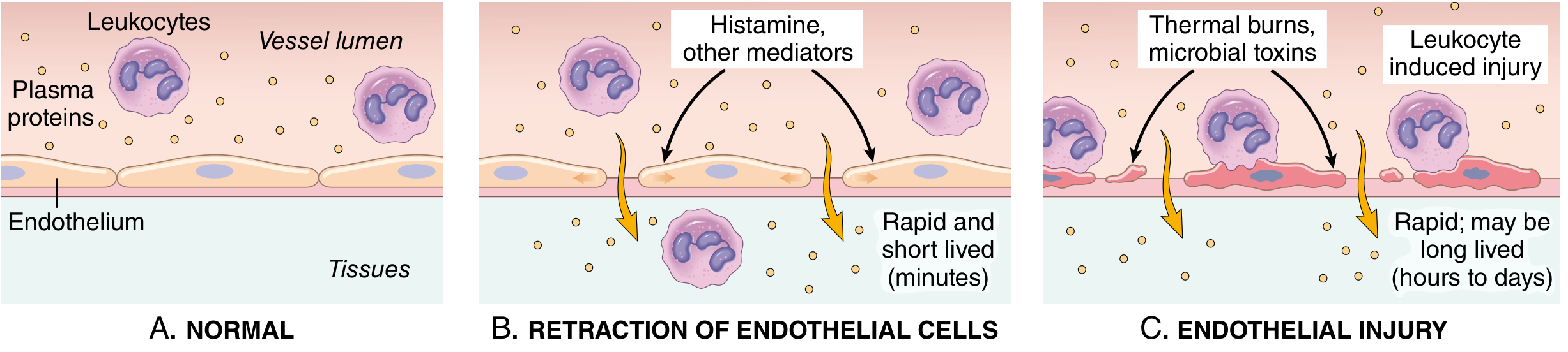
Fig. 3.3 - Principal mechanisms of increased vascular permeability (Robbins, Cotran & Kumar)
Two main mechanisms:
- Contraction of endothelial cells - opening of interendothelial gaps; elicited by histamine, bradykinin, leukotrienes; called the immediate transient response (15-30 minutes); the most common mechanism
- A delayed prolonged form (2-12 hours, lasting days) may occur with mild injury (e.g., sunburn) due to continued endothelial contraction or mild endothelial damage
- Endothelial injury - necrosis and detachment from severe physical injuries, thermal burns, microbial toxins, or neutrophil-induced damage; leakage starts immediately and is sustained until vessels are repaired
D. Lymphatic Response
- Lymph flow increases to drain excess edema fluid
- Leukocytes, cell debris, and microbes may enter lymphatics
- Lymphangitis (inflamed lymphatics) and lymphadenitis (enlarged, inflamed draining lymph nodes) may follow
- Red streaks near a skin wound = inflamed lymphatic channels = diagnostic of lymphangitis
Phase 2: Cellular Events - Leukocyte Recruitment
The journey of leukocytes from vessel lumen to tissue is a multistep, adhesion molecule-mediated process:
Steps of Leukocyte Recruitment
Step 1 - Margination and Rolling
- As blood flow slows (stasis), leukocytes (especially neutrophils) accumulate along the vessel wall (margination)
- They then roll along the endothelium via selectins
- P-selectin (stored in Weibel-Palade bodies of endothelium and platelet alpha granules; rapidly mobilized by histamine/thrombin)
- E-selectin (expressed by activated endothelium; induced by TNF, IL-1)
- L-selectin on leukocytes binds to ligands on endothelium
Step 2 - Firm Adhesion
- Rolling leukocytes are activated by chemokines on the endothelial surface
- Chemokines trigger conformational change in integrins on leukocytes - increasing their affinity for endothelial ligands
- Integrins (LFA-1, Mac-1 on leukocytes) bind to ICAM-1 on endothelium (induced by TNF and IL-1)
- This results in firm, stable adhesion
Step 3 - Transmigration (Diapedesis)
- Leukocytes squeeze between endothelial cells
- PECAM-1 (CD31) on leukocytes and endothelial cells mediates this migration
- After crossing the endothelium, leukocytes penetrate the basement membrane using collagenases
Step 4 - Chemotaxis
- Leukocytes migrate through tissues toward the site of injury guided by a chemotactic gradient
- Exogenous chemotactic agents: bacterial products (especially formyl-methionine peptides - f-Met-Leu-Phe)
- Endogenous chemotactic agents:
- Complement fragment C5a
- Leukotriene B4 (LTB4)
- IL-8 (CXCL8) / other chemokines
Sequence of Leukocytes at the Site
- 0-6 hours: Neutrophils dominate (first responders; short-lived in tissues - die by apoptosis within hours to a few days)
- 24-48 hours: Monocytes arrive and differentiate into macrophages (more long-lived, produce cytokines and growth factors for repair)
Phase 3: Phagocytosis and Destruction
Phagocytosis - Three Steps
-
Recognition and attachment - enhanced by opsonins:
- IgG antibodies (Fc receptors on phagocytes)
- C3b complement fragment (complement receptors)
-
Engulfment - pseudopods enclose the particle, forming a phagosome, which fuses with lysosomes to form a phagolysosome
-
Intracellular killing:Oxygen-dependent (most important):
- Respiratory burst - activation of NADPH oxidase converts O₂ to superoxide (O₂⁻), then to H₂O₂, then HOCl (hypochlorous acid via myeloperoxidase) - the most potent bactericidal agent
- Nitric oxide (NO) - produced by macrophages via iNOS
Oxygen-independent:- Lysozyme - degrades bacterial cell walls
- Lactoferrin - chelates iron
- Major basic protein (eosinophils) - against parasites
- Defensins - antimicrobial peptides
Neutrophil Extracellular Traps (NETs)
- Neutrophils can extrude their nuclear chromatin decorated with antimicrobial proteins (elastase, histones, MPO)
- These NETs trap and kill extracellular microbes
- Also seen in sepsis where they can cause vascular damage
Mediators of Inflammation
| Mediator | Source | Key Actions |
|---|---|---|
| Histamine | Mast cells, basophils, platelets | Vasodilation, increased vascular permeability (immediate) |
| Serotonin | Platelets | Similar to histamine |
| Prostaglandins (PGE2, PGI2) | Mast cells, leukocytes (via COX) | Vasodilation, pain, fever; PGI2 = prostacyclin |
| Thromboxane A2 | Platelets | Vasoconstriction, platelet aggregation |
| Leukotrienes (LTC4, D4, E4) | Mast cells, leukocytes (via 5-LOX) | Increased vascular permeability, bronchospasm |
| LTB4 | Leukocytes | Powerful chemotaxis for neutrophils |
| TNF and IL-1 | Macrophages, dendritic cells, mast cells | Endothelial activation, adhesion molecule expression; systemic fever, acute-phase response |
| IL-6 | Macrophages | Acute-phase response |
| Chemokines (IL-8/CXCL8) | Multiple cell types | Chemotaxis, leukocyte activation |
| Complement (C3a, C5a) | Plasma (liver) | Mast cell degranulation (C3a, C5a), chemotaxis (C5a), opsonization (C3b), MAC killing |
| Bradykinin | Plasma (via kinin system) | Vascular permeability, smooth muscle contraction, pain |
| PAF | Leukocytes, mast cells | Vasodilation, increased permeability, chemotaxis, oxidative burst |
Arachidonic Acid Metabolism
- Cyclooxygenase (COX) pathway → Prostaglandins + Thromboxane
- Aspirin/NSAIDs block COX (both COX-1 and COX-2)
- Selective COX-2 inhibitors (celecoxib) spare gastric mucosa
- 5-Lipoxygenase pathway → Leukotrienes (LTB4, LTC4, LTD4, LTE4)
- Leukotriene receptor antagonists (montelukast) used in asthma
- Lipoxins - produced via the 5-LOX pathway; anti-inflammatory - they suppress neutrophil recruitment (switch from LT to lipoxin production is one active termination mechanism)
Termination of Acute Inflammation
Active termination is tightly regulated to prevent excess tissue damage:
- Mediators have short half-lives and are degraded after release
- Neutrophils die by apoptosis within hours to days
- Switch from leukotrienes → lipoxins (antiinflammatory)
- Liberation of TGF-beta and IL-10 from macrophages
- Cholinergic neural impulses inhibit TNF production in macrophages
Morphologic Patterns of Acute Inflammation
1. Serous Inflammation
- Accumulation of protein-rich but cell-poor fluid (watery exudate) in body cavities or into tissue
- Example: blister formation in skin burns; pleural effusion in early pleuritis
- The fluid originates from serum or mesothelial cell secretion
2. Fibrinous Inflammation
- More severe vascular leakage allows large proteins (fibrinogen) to pass out → deposited as fibrin
- Occurs on organ surfaces, especially pericardium ("bread-and-butter pericarditis") and pleura
- May be resolved by fibrinolysis (resolution) or organized into scar (organization)
3. Purulent (Suppurative) Inflammation
- Production of large amounts of pus (neutrophils + dead cells + microbes)
- Caused by pyogenic bacteria (e.g., staphylococci)
- Abscess = localized collection of pus with central necrosis surrounded by neutrophils, then granulation tissue
4. Ulcers
- A local defect (excavation) of the surface of an organ or tissue, caused by sloughing of inflamed necrotic tissue
- Typical sites: stomach, duodenum, lower legs (vascular insufficiency)
- Characterized by acute and chronic inflammation at the base and margins
Outcomes of Acute Inflammation
Three possible outcomes:
- Resolution - complete restoration of normal structure and function; occurs when injury is limited and tissue is capable of regeneration (e.g., lobar pneumonia resolving)
- Healing by fibrosis (scarring) - when substantial tissue destruction occurs or in non-regenerating tissues; inflammation resolves but is replaced by connective tissue scar
- Progression to chronic inflammation - when the injurious agent cannot be eliminated or there is interference with normal healing; leads to persistent inflammation with ongoing tissue damage and repair
Summary
"When a host encounters an injurious agent, resident phagocytes try to eliminate it. Sentinel cells react by liberating cytokines, lipid messengers, and other mediators. These act on small blood vessels, promoting efflux of plasma proteins and recruitment of circulating leukocytes to the site. Recruited leukocytes are activated and try to remove the offending agent by phagocytosis. As the injurious agent is eliminated and antiinflammatory mechanisms become active, the process subsides and the host returns to a normal state of health."
- Robbins, Cotran & Kumar Pathologic Basis of Disease, Chapter 3
Source: Robbins, Cotran & Kumar Pathologic Basis of Disease, Chapter 3 - Inflammation and Repair (p. 67-86)
This is a shared conversation. Sign in to Orris to start your own chat.