
Closed heart surgery detailed note for clinical cardio 15 marks bpt mgr university exam
Reading File
This is a broad clinical topic. Let me search the medical library for closed heart surgery content.
Finding Sources
Finding Sources
Finding Sources
Finding Sources
Finding Sources
Good - there's content in Miller's Anesthesia and Braunwald's Heart Disease. Let me pull the relevant sections.
Reading File
Finding Sources
Finding Sources
Excellent! Found good references. Let me now read the key sections from Sabiston (history + closed mitral commissurotomy), Harrison's, and the Miller's Anesthesia closed-heart operations section.
Reading File
Reading File
Reading File
Excellent material gathered. Let me also get the closed-heart section image and read more of the Sabiston history section, plus fetch the CMC image for closed mitral commissurotomy.
Reading File
Finding Sources
Finding Sources
I now have comprehensive material. Let me fetch the key image of closed mitral commissurotomy and compile the full detailed answer.
Loading Image
I now have all the material needed for a comprehensive 15-mark exam answer. Let me compile the full detailed note.
Closed Heart Surgery - Detailed Clinical Note
(15 Marks | BPT Clinical Cardiology | MGR University)
Definition
Closed heart surgery refers to cardiac operations performed without cardiopulmonary bypass (CPB) and without opening (incising) the cardiac chambers. The heart continues to beat throughout the procedure, and the operation is carried out through thoracotomy incisions. These procedures contrast with "open heart surgery," in which CPB is used and the heart is arrested and opened for direct visualization.
Historical Background
The history of closed heart surgery is fundamental to understanding modern cardiac surgery:
| Year | Surgeon | Procedure |
|---|---|---|
| 1902 | Sir Thomas Lauder Brunton | Proposed closed repair of rheumatic mitral stenosis (never attempted clinically) |
| 1923 | Elliot Cutler & Samuel Levine | First closed transventricular MV commissurotomy (tenotomy knife) - Peter Bent Brigham Hospital, Boston |
| 1925 | Sir Henry Souttar | First successful closed digital commissurotomy via left atrial appendage |
| 1948 | Charles Bailey (Philadelphia) & Dwight Harken (Boston) | First widely adopted closed digital mitral commissurotomy |
| 1944 | Alfred Blalock & Helen Taussig | Blalock-Taussig shunt (subclavian artery to pulmonary artery) |
| 1940 | Horace Smithy | First successful aortic valvotomy |
(Sabiston Textbook of Surgery, p. 2527)
Classification of Closed Heart Procedures
Closed heart surgeries are classified as:
A. Corrective (Definitive) Closed-Heart Procedures
These operations achieve complete anatomical correction without CPB:
- PDA (Patent Ductus Arteriosus) Ligation
- Repair of Coarctation of the Aorta
- Closed Mitral Valvotomy / Commissurotomy
B. Non-Corrective (Palliative) Closed-Heart Procedures
These operations improve hemodynamics but do not correct the underlying defect:
- Pulmonary Artery Banding - for large VSD / excessive pulmonary blood flow
- Systemic-to-Pulmonary Artery Shunts (Blalock-Taussig shunt, Potts shunt, Waterston shunt)
(Miller's Anesthesia, 10e)
1. Closed Mitral Valvotomy (CMV) / Closed Mitral Commissurotomy (CMC)
Indication
- Symptomatic rheumatic mitral stenosis (NYHA Class II-IV)
- Mitral valve area < 1.5 cm² in a normal-sized adult (or < 1 cm²/m² BSA)
- Pliable, non-calcified leaflets with minimal subvalvular fusion
- No left atrial thrombus; MR ≤ 2+/4+
Surgical Technique
The classic Harken procedure is illustrated below:
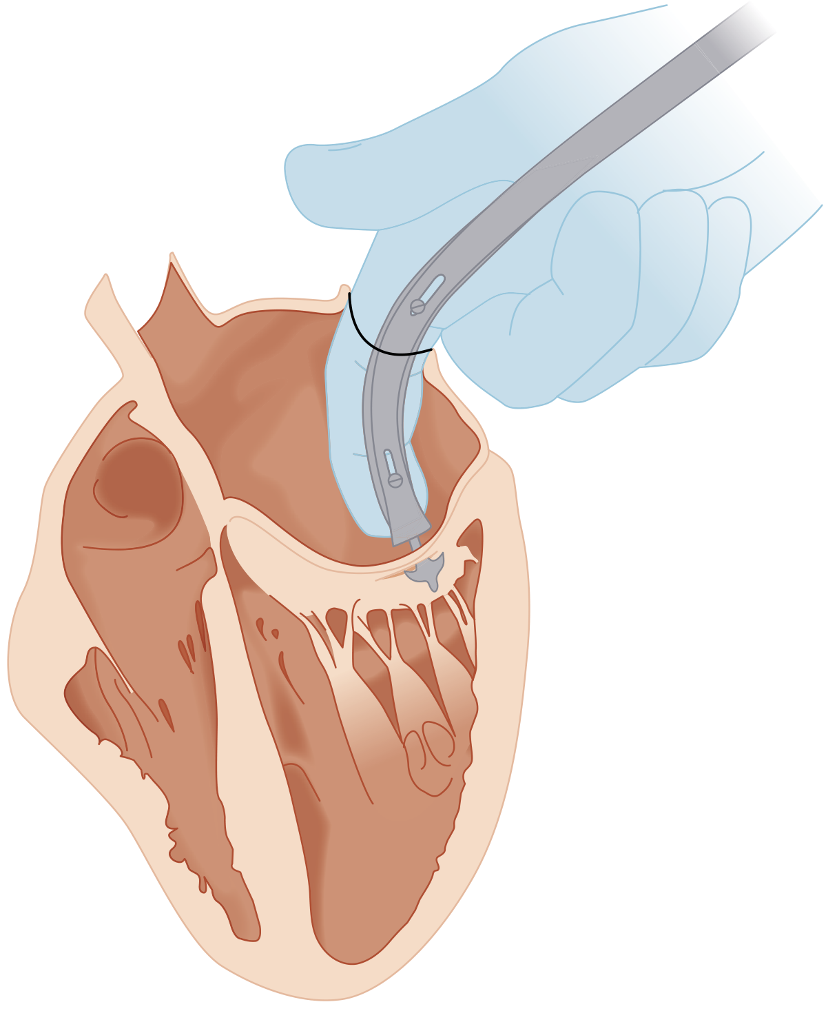
FIGURE: Closed Mitral Commissurotomy - Dwight Harken's technique using a transatrial dilator. (From Sabiston Textbook of Surgery, 7e)
Steps:
- Approach: Left posterolateral thoracotomy (4th or 5th intercostal space)
- Pericardiotomy: Pericardium is incised to expose the left atrium and left ventricle
- Purse-string suture: Placed on the left atrial appendage and on the left ventricular apex
- Digital exploration: Surgeon's index finger introduced through the left atrial appendage to assess the valve (palpate commissural fusion, subvalvular thickening, presence of thrombus)
- Commissurotomy: A mechanical dilator (Tubbs dilator or Brock's dilator) is introduced through the left ventricular apex and advanced under digital guidance to the mitral orifice; the dilator is opened to split the fused commissures
- Assessment: The surgeon assesses the adequacy of commissurotomy by finger palpation; auscultation for diastolic murmur reduction
- Closure: Purse-string sutures tightened; routine chest closure
Advantages of CMV
- No cardiopulmonary bypass required
- Lower morbidity and shorter operating time
- Suitable for resource-limited settings (historically important in India)
Disadvantages / Limitations
- "Blind" procedure - no direct visualization
- Cannot address subvalvular disease adequately
- Cannot remove atrial thrombus
- Risk of inducing mitral regurgitation
- Largely replaced by Percutaneous Balloon Mitral Commissurotomy (PBMC) and open surgical commissurotomy in modern practice
Contraindications to CMV
- Significant mitral regurgitation (> Grade 2)
- Left atrial thrombus
- Heavily calcified valve
- Combined aortic + mitral disease
- Previous commissurotomy with restenosis
Results
- Event-free survival in younger patients (< 45 years) with pliable valves: 80-90% over 3-7 years (Harrison's Principles, 22e)
- Operative mortality: approximately 2% for surgical commissurotomy
2. Patent Ductus Arteriosus (PDA) Ligation
Pathophysiology
The ductus arteriosus normally connects the pulmonary artery to the descending aorta in fetal life. Failure to close postnatally creates a left-to-right shunt, causing volume overload of the left heart and excessive pulmonary blood flow.
Indication for Surgery
- Symptomatic PDA not responding to medical treatment (indomethacin + fluid restriction in premature infants)
- Large PDA causing congestive heart failure
- Prevention of Eisenmenger syndrome
Surgical Technique
- Approach: Left thoracotomy (3rd intercostal space) OR video-assisted thoracoscopic surgery (VATS) - increasingly common
- Exposure: Pleura is opened; aortic arch and descending aorta identified; left recurrent laryngeal nerve carefully protected
- Ligation: The ductus is isolated between ligatures and divided, OR clipped with surgical clips; in premature infants, simple ligation without division is performed
- Verification: Pulse oximeter on lower extremity detects any inadvertent ligation of the descending aorta
Complications
- Inadvertent ligation of the left pulmonary artery or descending aorta
- Recurrent laryngeal nerve damage (left-sided hoarseness)
- Excessive bleeding from PDA disruption
- Post-ligation: worsening pulmonary compliance in premature infants; acute increase in left ventricular afterload
(Miller's Anesthesia, 10e, p. 10852-10853)
3. Repair of Coarctation of the Aorta
Definition
Coarctation of the aorta is a narrowing of the descending aorta near the insertion of the ductus arteriosus, causing obstruction to aortic flow. Results in hypertension proximal to the coarctation and reduced pressure distally.
Surgical Techniques (all without CPB)
| Technique | Description |
|---|---|
| Resection + End-to-End Anastomosis | Segment of coarctation excised; direct anastomosis of aortic ends |
| Subclavian Flap Angioplasty (Waldhausen procedure) | Left subclavian artery divided and turned down as a patch to widen the coarctation |
| Patch Aortoplasty | Dacron or pericardial patch sewn to enlarge the narrowed segment |
| Interposition Graft | Segment resected; replaced with a synthetic graft - used for longer coarctations |
Intraoperative Monitoring
- Bilateral radial arterial lines (right arm = pre-ductal; left arm shows subclavian sacrifice effect)
- Femoral arterial line = post-ductal pressure
- NIRS (near-infrared spectroscopy) monitors spinal cord perfusion
- Cross-clamp time should be minimized to prevent paraplegia from spinal cord ischemia
Complications
- Paraplegia (from spinal cord ischemia during aortic cross-clamping)
- Paradoxical hypertension post-repair (mesenteric arteritis)
- Recoarctation (especially after subclavian flap in neonates as they grow)
- Bleeding, chylothorax
4. Systemic-to-Pulmonary Artery Shunts (Palliative)
These are performed for cyanotic congenital heart disease (e.g., Tetralogy of Fallot, pulmonary atresia) to increase pulmonary blood flow without CPB.
| Shunt | Connection | Notes |
|---|---|---|
| Classic Blalock-Taussig (BT) Shunt | Subclavian artery to ipsilateral pulmonary artery (end-to-side) | Original 1944 procedure |
| Modified BT Shunt | Gore-Tex tube graft - subclavian artery to pulmonary artery | More common currently; preserves subclavian artery |
| Waterston Shunt | Ascending aorta to right pulmonary artery | Now abandoned (too difficult to control flow) |
| Potts Shunt | Descending aorta to left pulmonary artery | Used as palliative shunt for end-stage pulmonary hypertension (Reverse Potts) |
| Glenn Shunt | Superior vena cava to right pulmonary artery | For single ventricle physiology |
Purpose
- Increases pulmonary blood flow in duct-dependent circulation
- Allows the child to grow before definitive repair (e.g., total correction of TOF)
- Reduces hypoxic spells (Tet spells)
5. Pulmonary Artery Banding (PAB)
Indication
- Large VSD or other causes of excessive pulmonary blood flow (Qp:Qs >> 1) when complete repair cannot be performed (premature neonate, complex anatomy)
- Preparation for biventricular repair or univentricular palliation
Procedure
- Left thoracotomy or median sternotomy
- A tape or band placed around the main pulmonary artery and tightened until:
- Distal PA pressure = 25-50% of systemic pressure
- Qp/Qs approaches 1:1
- Acceptable oxygen saturation maintained
- The band is secured with sutures to prevent migration
Complications
- Over-tightening: cyanosis, right ventricular hypertrophy
- Under-tightening: persistent pulmonary overcirculation
- Band migration
- Pulmonary artery distortion (makes future corrective surgery difficult)
Key Differences: Closed vs. Open Heart Surgery
| Feature | Closed Heart Surgery | Open Heart Surgery |
|---|---|---|
| Cardiopulmonary bypass | NOT used | Required |
| Heart | Beating | Arrested (cardioplegia) |
| Visualization | Indirect / tactile | Direct |
| Incision | Thoracotomy | Median sternotomy (usually) |
| Complexity | Simpler procedures | Complex repairs |
| Examples | PDA ligation, CMV, coarctation repair | VSD repair, valve replacement, CABG |
| Morbidity | Lower (no bypass complications) | Higher |
| Indications | Select congenital lesions, mitral stenosis | Most cardiac surgery |
Anesthetic Considerations in Closed Heart Surgery
- No CPB - arterial and central venous access still essential
- Intra-arterial monitoring mandatory (site depends on procedure - avoid right radial for BT shunt, avoid left radial for subclavian flap coarctation repair)
- NIRS monitoring for cerebral and somatic (spinal cord) perfusion
- Pulse oximetry on both upper and lower extremities
- One-lung ventilation may be required for thoracoscopic PDA ligation
- Anticipate hemodynamic shifts from aortic cross-clamping (coarctation) or shunt manipulation
(Miller's Anesthesia, 10e)
Summary Points for Exam
- Closed heart surgery = no CPB, beating heart, through thoracotomy
- CMV was the first widespread cardiac operation; replaced by PBMC and open surgery
- PDA ligation is corrective; commonest closed heart procedure in neonates
- Coarctation repair uses aortic cross-clamping; paraplegia is a feared complication
- BT shunt is palliative for cyanotic CHD to increase pulmonary blood flow
- PA banding is palliative to reduce pulmonary overcirculation
- All procedures avoid CPB, thus avoiding pump-related complications (hemolysis, coagulopathy, systemic inflammatory response)
- Early corrective repair in infancy has significantly reduced the need for palliative closed-heart operations (Miller's Anesthesia, 10e)
Sources:
- Sabiston Textbook of Surgery, 7e - History of Heart Valve Surgery, pp. 2527-2530
- Harrison's Principles of Internal Medicine, 22e - Mitral Commissurotomy, Chapter 274
- Miller's Anesthesia, 10e - Anesthesia for Closed-Heart Operations, pp. 10851-10857
- Fuster & Hurst's The Heart, 15e - Mitral Valve Disease
This is a shared conversation. Sign in to Orris to start your own chat.