
dudenum and its relation at every part and i aslo want picture.
Reading File
The duodenum is the first and shortest part of the small intestine, divided into 4 parts (D1-D4), with key anatomical relations at each part - the pylorus proximally, head of pancreas medially, and it ends at the duodenojejunal flexure.
Searching sources...
Finding Sources
Searching the Web
duodenum anatomy parts relations diagram
Reading File
Loading Image
Loading Image
Now let me fetch the textbook diagram and get the Bailey & Love surgical relations content:
Loading Image
Reading File
I now have excellent content and images. Let me compile the full answer.
The Duodenum - Anatomy and Relations at Every Part
The duodenum is the first and shortest part of the small intestine, approximately 20-25 cm (about 10 inches) long. Its name comes from "duodeni" (Latin: twelve finger-breadths). It is C-shaped, mostly retroperitoneal, and wraps around the head of the pancreas. It is divided into 4 parts.
Overview Diagram (Gray's Anatomy for Students)
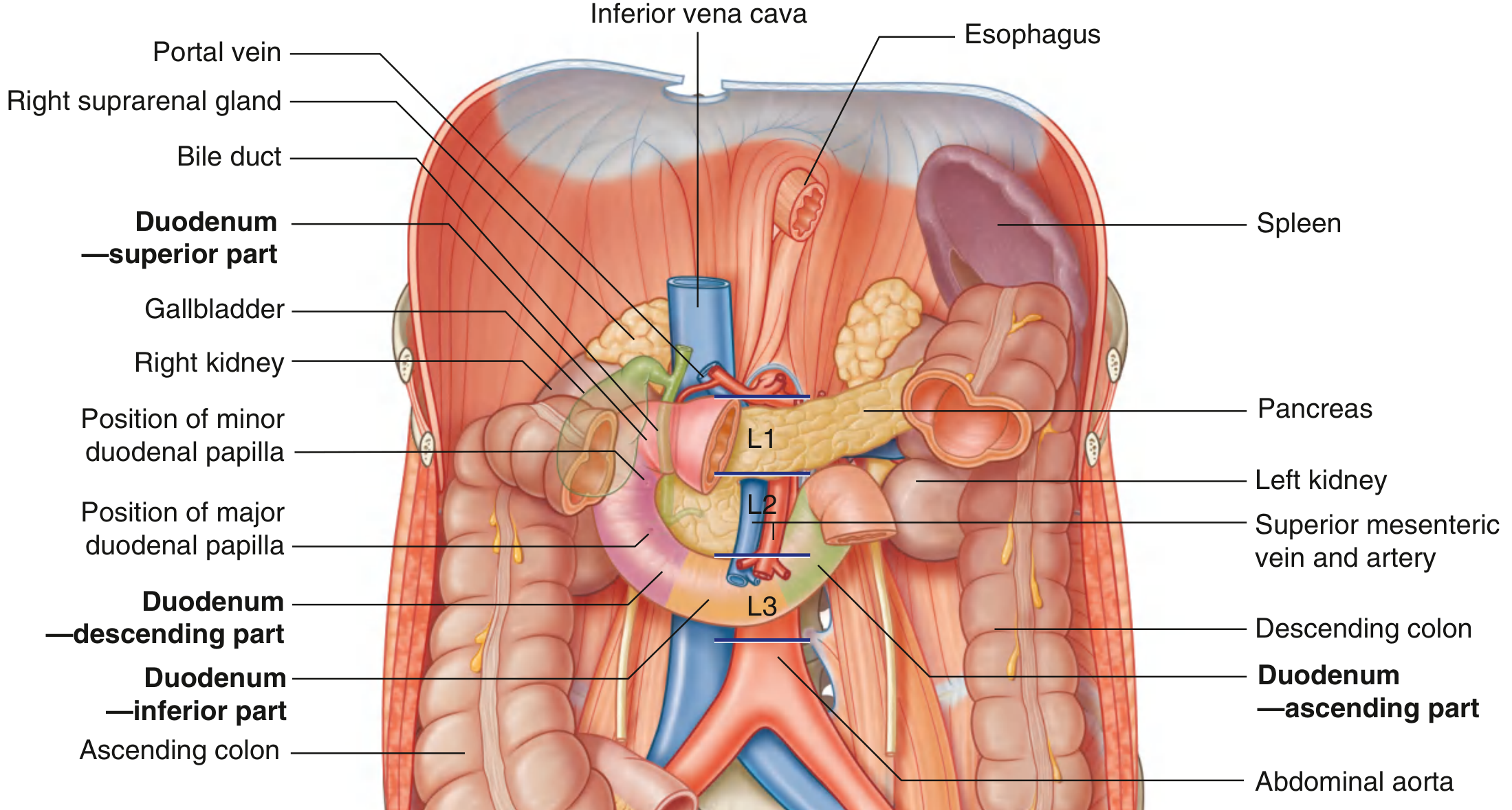
Relations Diagram

3D Anatomical View
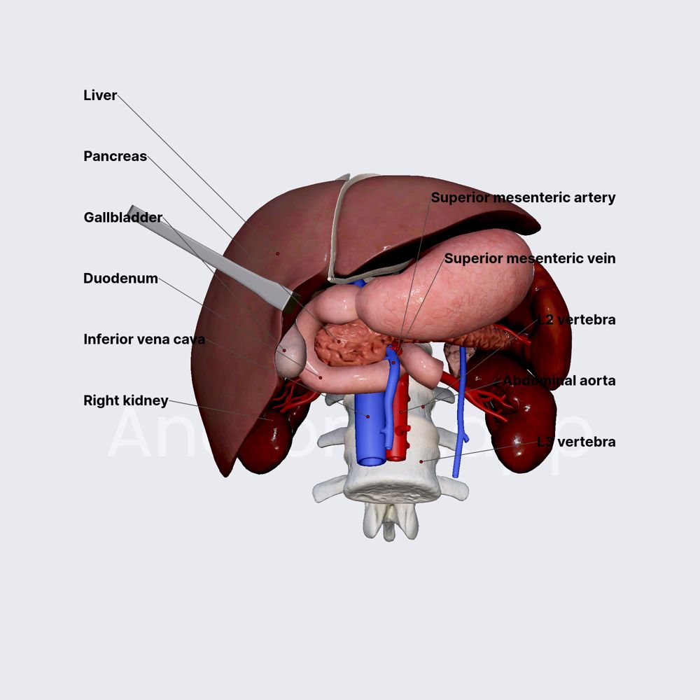
Part 1 - Superior Part (D1)
- Level: Vertebra L1
- Length: ~5 cm
- Peritoneum: Intraperitoneal at its start (the duodenal cap / ampulla), then retroperitoneal
- Connected to the liver via the hepatoduodenal ligament (part of the lesser omentum)
- Clinically important: most duodenal ulcers occur here
| Direction | Relation |
|---|---|
| Anterior | Liver (quadrate lobe), gallbladder |
| Posterior | Bile duct (CBD), gastroduodenal artery, portal vein, inferior vena cava (IVC) |
| Superior | Hepatoduodenal ligament, epiploic foramen (of Winslow) |
| Inferior | Neck of pancreas |
Part 2 - Descending Part (D2)
- Level: L1 down to L3 (right of midline)
- Length: ~7-8 cm
- Contains the major duodenal papilla (of Vater) - the opening of the common bile duct + main pancreatic duct
- Contains the minor duodenal papilla - opening of the accessory pancreatic duct
- The foregut/midgut junction occurs just below the major papilla
| Direction | Relation |
|---|---|
| Anterior | Transverse colon, liver (right lobe), coils of jejunum |
| Posterior | Right kidney (hilum), right ureter, right psoas major |
| Medial | Head of pancreas, bile duct, main pancreatic duct |
| Lateral | Right colic flexure, ascending colon, right lobe of liver |
Part 3 - Horizontal (Inferior) Part (D3)
- Level: L3 (crosses the vertebral column horizontally)
- Length: ~10 cm (the longest part)
- Passes in front of major vessels
| Direction | Relation |
|---|---|
| Anterior | Superior mesenteric artery (SMA) and vein (SMV), mesentery of small intestine, coils of jejunum |
| Posterior | Inferior vena cava (IVC), abdominal aorta, vertebral column (L3), right ureter, right psoas major |
| Superior | Head of pancreas |
| Inferior | Coils of jejunum |
Clinical note: The SMA can compress D3 against the aorta, causing Superior Mesenteric Artery Syndrome (SMA syndrome / Wilkie's disease) - a rare cause of high intestinal obstruction.
Part 4 - Ascending Part (D4)
- Level: Ascends from L3 to L2 (upper border)
- Length: ~2-3 cm
- Ends at the duodenojejunal (DJ) flexure
- The DJ flexure is held in place by the ligament of Treitz (suspensory muscle of duodenum) - a fibromuscular band anchored to the right crus of the diaphragm
| Direction | Relation |
|---|---|
| Anterior | Beginning of mesentery, coils of jejunum |
| Posterior | Left psoas major, left sympathetic trunk, left renal vessels, aorta |
| Right | Aorta (close), body of pancreas |
| Left | Left kidney, left ureter |
Summary Table
| Part | Level | Key Anterior Relation | Key Posterior Relation | Key Medial Relation |
|---|---|---|---|---|
| D1 - Superior | L1 | Liver, gallbladder | Bile duct, portal vein, IVC | Epiploic foramen |
| D2 - Descending | L1-L3 | Transverse colon | Right kidney, right ureter | Head of pancreas |
| D3 - Horizontal | L3 | SMA + SMV | IVC, aorta | Head of pancreas |
| D4 - Ascending | L3-L2 | Mesentery, jejunum | Left psoas, aorta | Body of pancreas |
Blood Supply
- Foregut supply (above the major papilla): Anterior and posterior superior pancreaticoduodenal arteries - from the gastroduodenal artery (branch of hepatic artery)
- Midgut supply (below the major papilla): Anterior and posterior inferior pancreaticoduodenal arteries - from the superior mesenteric artery (SMA)
These vessels form arcades (anastomotic loops) along the head of the pancreas, which is why pancreatic head pathology (e.g., cancer) can affect duodenal blood supply.
Venous and Lymphatic Drainage
- Veins drain into the portal vein (via superior mesenteric and splenic veins)
- Lymphatics drain to pancreaticoduodenal nodes → coeliac and superior mesenteric nodes
Key Clinical Points
- Duodenal ulcers most commonly occur in D1 (the duodenal cap), due to acid exposure and H. pylori
- A posterior D1 ulcer can erode the gastroduodenal artery - causing life-threatening haemorrhage
- The ampulla of Vater in D2 is where bile and pancreatic juice enter - obstruction here (e.g., stone, tumour) causes obstructive jaundice + pancreatitis
- The ligament of Treitz marks the duodenojejunal junction - surgically important as it defines the upper GI tract; it is the landmark for identifying the start of the jejunum
- The duodenum is fixed and retroperitoneal, making it vulnerable to crush injuries (e.g., seatbelt injury) since it is pressed against the vertebral column
- Gray's Anatomy for Students, p. 372
- Bailey and Love's Short Practice of Surgery 28th Ed.
- Sleisenger and Fordtran's Gastrointestinal and Liver Disease
This is a shared conversation. Sign in to Orris to start your own chat.