
Pituitary hormons
pituitary gland hormones diagram anterior posterior
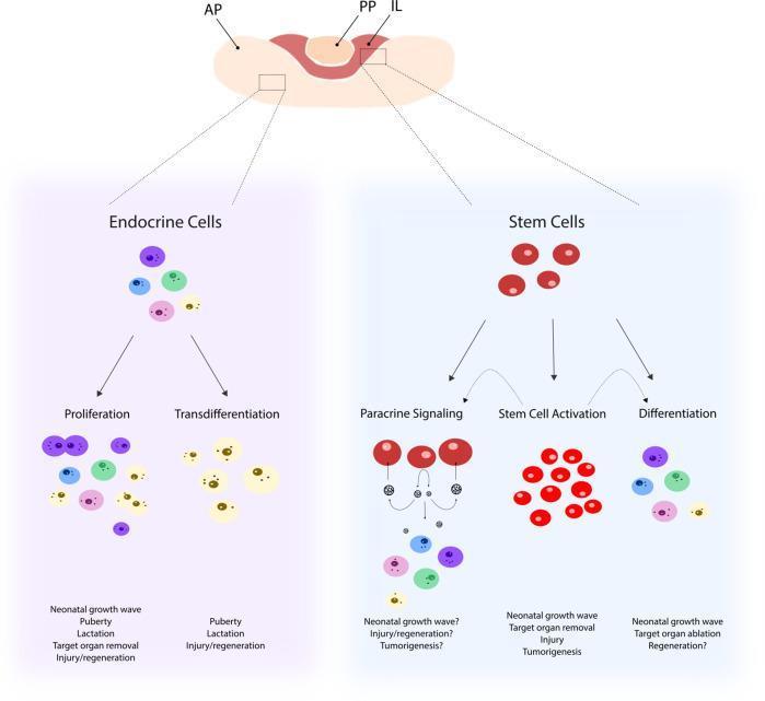
This medical pathophysiology diagram illustrates the cellular mechanisms underlying the remodeling of the pituitary gland landscape. At the top, a cross-section of the pituitary gland is shown with labels for the anterior pituitary (AP), posterior pituitary (PP), and intermediate lobe (IL). The diagram branches into two main pathways: Endocrine Cells and Stem Cells. The Endocrine Cells section demonstrates two modulation pathways: Proliferation (increase in cell number) and Transdifferentiation (transformation into different endocrine cell types), influenced by triggers like puberty, lactation, and injury. The Stem Cells section depicts three potential fates: Paracrine Signaling (releasing signals to surrounding cells), Stem Cell Activation (multiplication and upregulation of stemness), and Differentiation (maturation into specialized endocrine cells). These processes are linked to physiological and pathological states including the neonatal growth wave, target organ removal, tumorigenesis, and tissue regeneration. The illustration uses color-coded circles to represent various differentiated hormone-producing cells and red circles for undifferentiated stem cells.
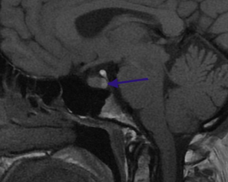
This diagnostic image is a T1-weighted sagittal Magnetic Resonance Imaging (MRI) scan of the human brain, specifically centered on the sella turcica and the midbrain structures. A blue arrow points to a distinct, focal hyperintense signal within the posterior aspect of the pituitary gland, known as the 'posterior pituitary bright spot.' This finding represents a normal radiological landmark indicating the storage of neurohypophyseal hormones (vasopressin and oxytocin) within the neurohypophysis. Anatomical landmarks clearly visible include the anterior pituitary (adenohypophysis), the pituitary stalk (infundibulum), the optic chiasm located superiorly, the sphenoid sinus inferiorly, and the pons and cerebellum posteriorly. The image serves as a clinical reference for normal pituitary anatomy in neuroimaging, distinguishing physiological hyperintensity from pathological lesions such as microadenomas or cysts.
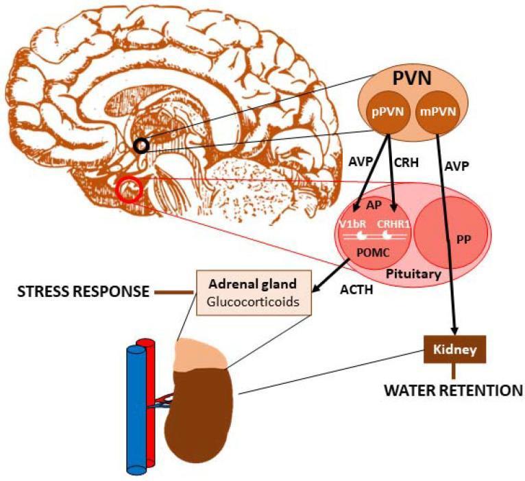
This pathophysiology diagram illustrates the role of vasopressin (AVP) in the regulation of the Hypothalamic-Pituitary-Adrenal (HPA) axis and renal water retention. A sagittal brain illustration highlights the Paraventricular Nucleus (PVN), which is subdivided into parvocellular (pPVN) and magnocellular (mPVN) regions. The pPVN releases both AVP and Corticotropin-Releasing Hormone (CRH) into the anterior pituitary (AP). Within the AP, these hormones bind to V1bR and CRHR1 receptors, respectively, on proopiomelanocortin (POMC) cells to stimulate the release of Adrenocorticotropic Hormone (ACTH). ACTH subsequently acts on the adrenal gland to stimulate glucocorticoid production, mediating the systemic stress response. Simultaneously, the mPVN produces AVP, which is transported through the posterior pituitary (PP) into the bloodstream to act on the kidney, promoting water retention. The diagram effectively maps the dual neuroendocrine pathways of AVP, linking hypothalamic activity to endocrine stress responses and physiological osmoregulation. Key labels include ACTH, pPVN, mPVN, V1bR, CRHR1, and POMC.
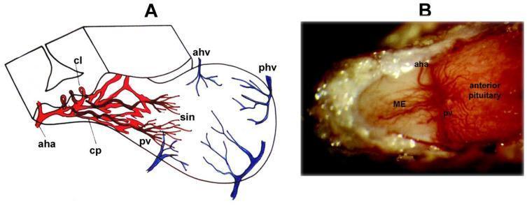
This composite image illustrates the anatomy and physiology of the hypophyseal portal system, which facilitates neurohumoral regulation of the anterior pituitary. Panel A is a schematic three-dimensional diagram showing the blood supply to the median eminence (ME) and anterior pituitary. It delineates the anterior hypophyseal artery (aha) branching into a primary capillary plexus (cp) with capillary loops (cl). These transition into portal veins (pv) that drain into the sinusoids (sin) of the anterior pituitary, with venous drainage via the anterior (ahv) and posterior (phv) hypophyseal veins. Panel B is a high-power clinical photograph taken through a dissecting microscope of an anesthetized rat model. It provides a real-world visualization of the hypophyseal portal vessels on the pituitary stalk surface. The portal vessels (pv) can be seen fanning out from the ME toward the anterior pituitary gland. The tuberoinfundibular artery (aha) is visible arching across the stalk-pituitary junction. This material serves as an educational resource for understanding the hypothalamic-hypophyseal portal system and the vascular pathways for neurosecretory transport.
Pituitary Hormones - Complete Overview
The Hypothalamic-Pituitary System
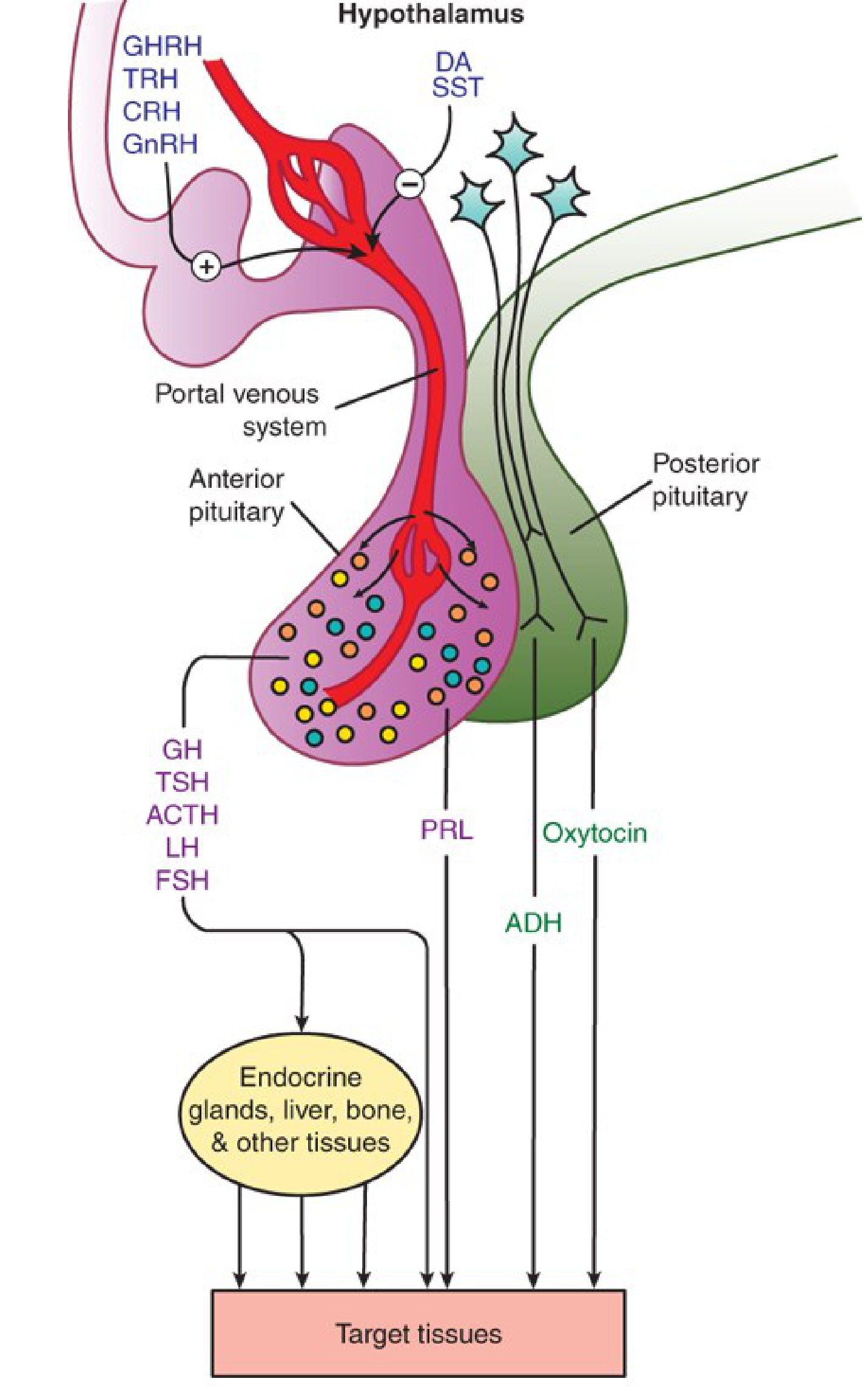
ANTERIOR PITUITARY (Adenohypophysis) Hormones
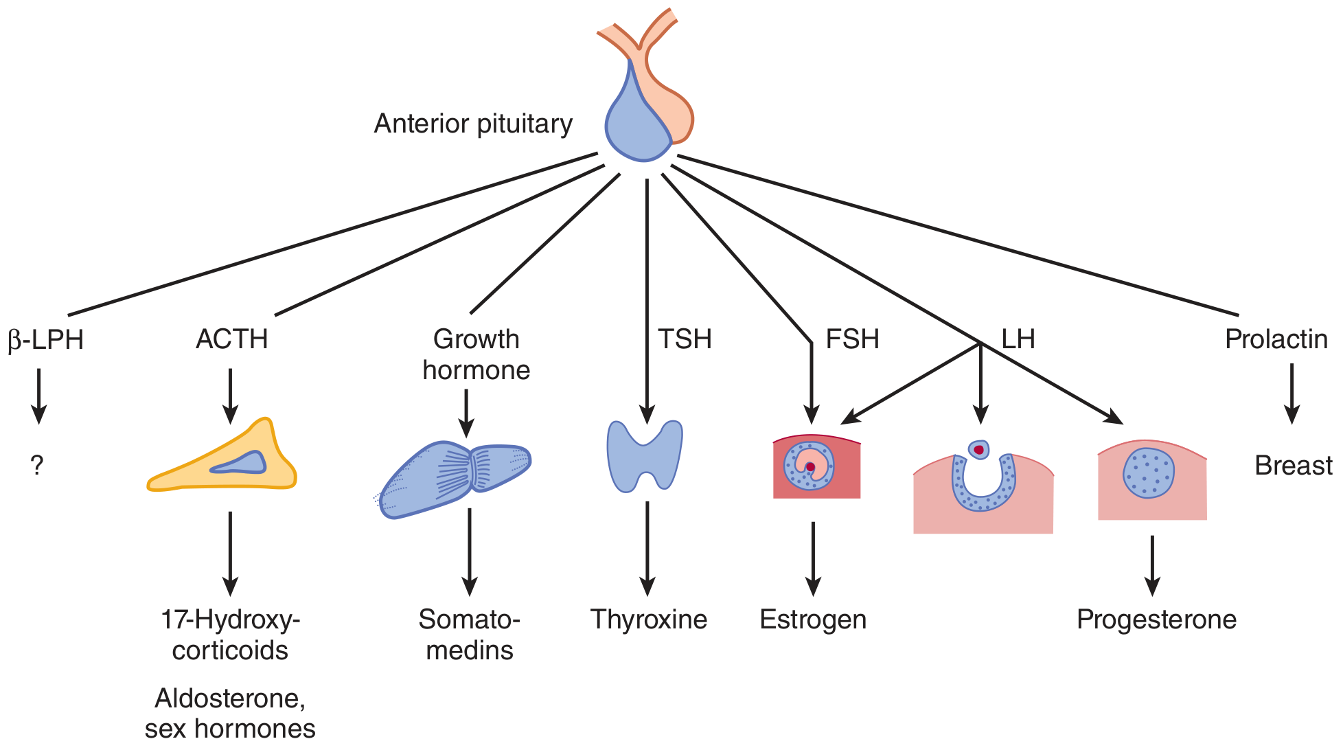
Group 1: GH/PRL Family (activate JAK/STAT receptors)
| Hormone | Hypothalamic Control | Target | Action |
|---|---|---|---|
| Growth Hormone (GH / Somatotropin) | GHRH (+) / Somatostatin (-) | Liver, bone, muscle, kidney | Stimulates IGF-I (somatomedins) production; promotes growth, protein anabolism, lipolysis, anti-insulin effect |
| Prolactin (PRL) | Dopamine (-) predominantly | Breast | Stimulates lactation; dopamine is its primary inhibitor - loss of hypothalamic input causes hyperprolactinemia |
Group 2: Glycoprotein Hormones - TSH, FSH, LH (activate G-protein coupled receptors)
| Hormone | Hypothalamic Control | Target | Action |
|---|---|---|---|
| TSH (Thyrotropin) | TRH (+) | Thyroid gland | Stimulates synthesis and release of T3/T4 (thyroxine, triiodothyronine) |
| FSH (Follicle-Stimulating Hormone) | GnRH (+) | Gonads | Females: follicle development, estrogen production; Males: spermatogenesis (via Sertoli cells) |
| LH (Luteinizing Hormone) | GnRH (+) | Gonads | Females: ovulation trigger, corpus luteum maintenance, progesterone; Males: testosterone production (via Leydig cells) |
Key note on GnRH: Pulsatile endogenous GnRH stimulates LH/FSH release. When given continuously (as a drug), GnRH and its analogs inhibit LH/FSH via receptor downregulation - used clinically in prostate cancer, endometriosis.
Group 3: POMC-Derived Peptides (activate G-protein coupled receptors)
| Hormone | Hypothalamic Control | Target | Action |
|---|---|---|---|
| ACTH (Adrenocorticotropin / Corticotropin) | CRH (+) | Adrenal cortex | Stimulates cortisol, aldosterone, and sex hormone synthesis |
| Beta-Lipotropin (β-LPH) | Co-secreted with ACTH | Unknown | Physiologic role unclear; precursor to endorphins |
Hypothalamic Releasing/Inhibiting Hormones Summary
| Hypothalamic Hormone | Effect on Anterior Pituitary |
|---|---|
| CRH (Corticotropin-Releasing Hormone) | Stimulates ACTH release |
| TRH (Thyrotropin-Releasing Hormone) | Stimulates TSH (and PRL) release |
| GHRH (Growth Hormone-Releasing Hormone) | Stimulates GH release |
| Somatostatin (GIH) | Inhibits GH (and TSH) release |
| GnRH (Gonadotropin-Releasing Hormone) | Stimulates LH and FSH release (pulsatile) |
| Dopamine (DA) | Inhibits Prolactin release |
POSTERIOR PITUITARY (Neurohypophysis) Hormones
| Hormone | Synthesized In | Target | Action |
|---|---|---|---|
| ADH / Vasopressin (AVP) | Supraoptic nucleus | Kidney collecting duct (V2 receptor) / vessels (V1 receptor) | Water reabsorption (anti-diuresis); vasoconstriction at high doses |
| Oxytocin | Paraventricular nucleus | Uterus, breast | Uterine contraction during labor; milk ejection (let-down reflex) |
- ADH deficiency → Central Diabetes Insipidus (polyuria, polydipsia)
- ADH excess (SIADH) → hyponatremia, water retention
Clinical Significance at a Glance
| Hormone | Excess | Deficiency |
|---|---|---|
| GH | Gigantism (children), Acromegaly (adults) | Growth failure, dwarfism |
| PRL | Galactorrhea, amenorrhea, infertility | Failure of lactation post-partum |
| TSH | Secondary hyperthyroidism | Secondary hypothyroidism |
| ACTH | Cushing's disease (bilateral adrenal hyperplasia) | Secondary adrenal insufficiency |
| FSH/LH | Precocious puberty | Hypogonadotropic hypogonadism, infertility |
| ADH | SIADH (hyponatremia) | Diabetes insipidus |
| Oxytocin | - | Failure to progress in labor |
Receptor Types
- JAK/STAT receptors: GH, PRL
- G-protein coupled receptors (GPCRs): TSH, ACTH, FSH, LH, ADH (V2: Gs; V1: Gq), Oxytocin
- Feedback regulation is primarily negative: target organ hormones (cortisol, T3/T4, sex steroids, IGF-I) feed back to suppress both the hypothalamus and the pituitary.
Explain thyroid gland fully ppt
thyroid gland anatomy hormones synthesis
Make with diagram and images available on google
thyroid gland anatomy hormones diagram
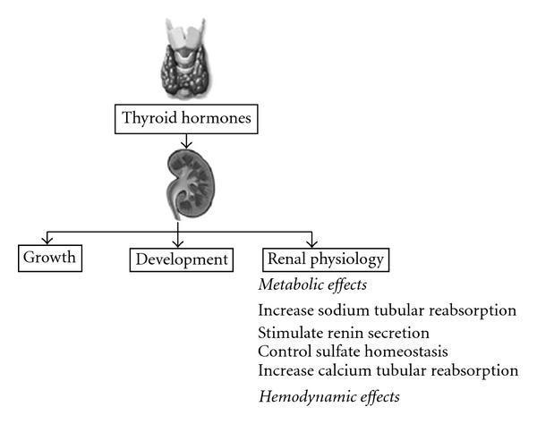
A pathophysiology diagram illustrating the regulatory effects of thyroid hormones on the human kidney. The visual layout follows a vertical top-down hierarchy: an illustration of the thyroid gland leads to a box labeled 'Thyroid hormones', which in turn points to an anatomical illustration of a kidney. From the kidney, the diagram branches into three primary educational concepts: 'Growth', 'Development', and 'Renal physiology'. Under 'Renal physiology', the diagram details metabolic and hemodynamic effects. Specific metabolic actions listed include increased sodium tubular reabsorption, stimulated renin secretion, control of sulfate homeostasis, and increased calcium tubular reabsorption. The diagram serves as a conceptual overview of endocrine-renal interactions, highlighting the influence of thyroid function on renal growth, maturation, and electrolyte/fluid homeostasis. It is designed for medical students and clinicians studying nephrology and endocrinology.
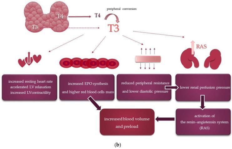
This medical flowchart illustrates the systemic and cardiovascular effects of thyroid hormones. The diagram begins with the thyroid gland releasing Thyroxine (T4) and Triiodothyronine (T3), highlighting the peripheral conversion of T4 to the more active T3. The downstream physiological impacts of T3 are categorized into four primary pathways: 1) Cardiac effects, including increased resting heart rate, accelerated left ventricular (LV) relaxation, and increased LV contractility. 2) Hematologic effects, specifically increased erythropoietin (EPO) synthesis leading to higher red blood cell mass. 3) Vascular effects, showing reduced peripheral resistance and lower diastolic pressure. 4) Renal effects, where reduced peripheral resistance leads to lower renal perfusion pressure. These combined pathways culminate in the activation of the renin-angiotensin system (RAS) and an overall increase in blood volume and preload. The illustration uses anatomical icons of the heart, blood cells, vessels, and kidneys to correlate physiological mechanisms with organ-level changes, demonstrating how hyperthyroidism or thyroid hormone signaling influences hemodynamic stability and cardiac output.
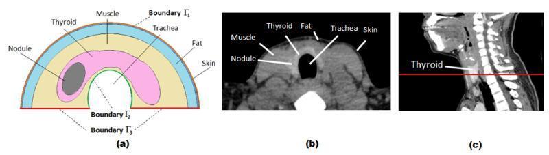
This composite educational graphic details the cross-sectional anatomy of the human neck for Finite Element (FE) thyroid simulation. (a) A 2D schematic diagram presents a simplified semicircular model of the neck. It labels the layers concentrically: skin (outermost), fat, and muscle. Inside the muscle layer, the thyroid gland is depicted in pink, containing an elliptical dark grey thyroid nodule. Boundaries are marked as Γ1 (skin surface), Γ2 (trachea boundary), and Γ3 (prescribed temperature base). (b) A corresponding axial CT scan image shows the actual anatomical cross-section, with labels identifying the skin, fat, muscle, thyroid gland, trachea (central air-filled void), and an internal nodule. The CT highlights varying tissue densities, from the dark fat layer to the intermediate density of muscle and thyroid tissue. (c) A sagittal CT view of the head and neck shows a red horizontal line indicating the anatomical plane of the axial slice in (b), positioned at the level of the thyroid gland. This visual serves as a reference for modeling heat transfer and infrared thermography in patients with thyroid nodules.
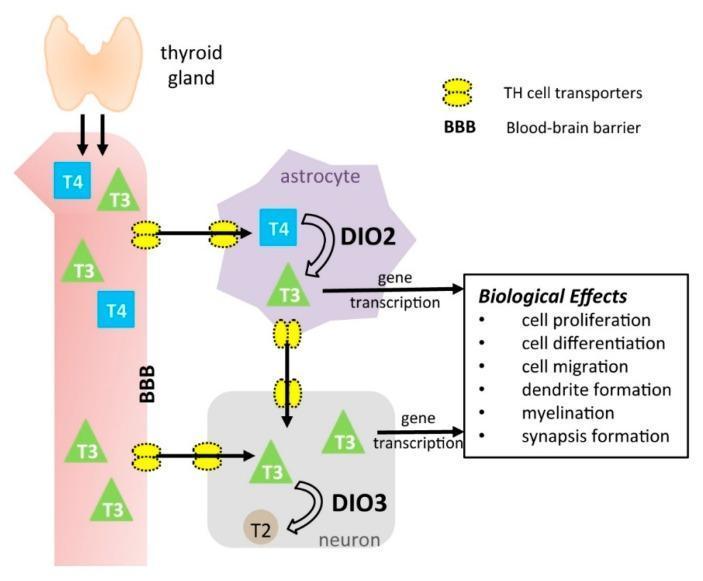
This pathophysiology diagram illustrates the transport and metabolic pathways of thyroid hormones (TH) within the central nervous system. The process begins with the thyroid gland releasing thyroxine (T4) and triiodothyronine (T3) into the systemic circulation. TH cell transporters (yellow structures) facilitate the passage of T4 and T3 across the blood-brain barrier (BBB) and into astrocytes and neurons. Within the astrocyte, T4 is converted to active T3 by the enzyme type 2 deiodinase (DIO2). T3 from the astrocyte can then be transported to neurons. In the neuron, type 3 deiodinase (DIO3) inactivates T3 by converting it to diiodothyronine (T2). In both cell types, T3 triggers gene transcription, leading to critical neurodevelopmental and biological effects: cell proliferation, cell differentiation, cell migration, dendrite formation, myelination, and synapse formation. The diagram highlights the localized regulation of TH action in the brain through specific transporters and deiodinase enzymes, emphasizing their roles in neural development and homeostasis.
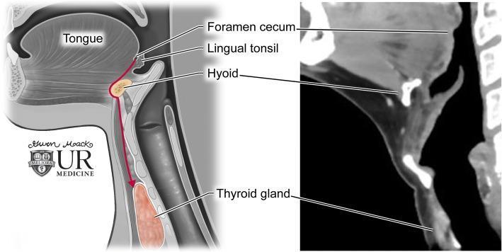
This dual-panel educational graphic illustrates thyroglossal duct anatomy and the embryological descent of the thyroid gland. On the left is a sagittal anatomical diagram showing the path of descent (indicated by a red arrow) from the foramen cecum at the base of the tongue, passing through the hyoid bone, to its final position at the thyroid gland anterior to the trachea. Key labeled structures include the tongue, lingual tonsil, foramen cecum, hyoid, and thyroid gland. The right panel displays a corresponding sagittal contrast-enhanced CT image of the neck. The CT scan highlights the radiologic appearance of these landmarks: the hyoid bone appears as a high-attenuation U-shaped structure, and the thyroid gland exhibits homogenous enhancement inferior to the thyroid cartilage. This comparison demonstrates the clinical relevance of embryological pathways in understanding the location of thyroglossal duct cysts, which can occur anywhere along this midline tract.
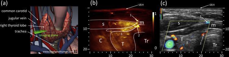
This composite educational graphic illustrates the cross-sectional anatomy of the anterior neck, focusing on the left thyroid lobe through three modalities: (a) a 3D anatomical diagram, (b) an optoacoustic image, and (c) a correlative ultrasound with Directional Power Doppler. The 3D diagram establishes the spatial orientation of the common carotid artery, jugular vein, trachea, and thyroid lobe. The optoacoustic cross-section (b) displays high-sensitivity vascular mapping, identifying the sternocleidomastoid (s) and infrahyoid (m) muscles, the carotid artery (C), and the trachea (Tr). Intraglandular vascularity (3) and superficial vessels (1, 2) are highlighted in a high-contrast heatmap. The corresponding ultrasound image (c) provides structural grayscale context with superimposed Doppler signals (red and blue) indicating blood flow direction. This multimodal comparison demonstrates how optoacoustic imaging provides superior visualization of microvascular networks within the thyroid gland (T) compared to standard Doppler ultrasound, while maintaining consistent anatomical landmarks such as the trachea's medial position and the lateral placement of the carotid artery.
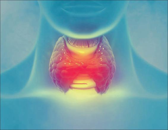
Anatomical diagram depicting the thyroid gland situated in the anterior neck, inferior to the larynx. The illustration utilizes a stylized, translucent blue silhouette of a human neck and torso as a background to provide anatomical context. The central focus is a bipedal, butterfly-shaped organ representing the thyroid gland, characterized by its right and left lobes connected by a central isthmus. The gland is rendered with a textured, lobulated surface and a vibrant color gradient ranging from deep red in the center to bright yellow at the superior and inferior margins, simulating metabolic activity or localized inflammation. Visible above the thyroid is the laryngeal prominence, and the tracheal rings are suggested beneath the isthmus. The image is designed for educational purposes to demonstrate the anatomical positioning of the thyroid gland within the endocrine system and its relationship to airway structures. It serves as a visual aid for discussions on thyroid disorders such as goiter, hyperthyroidism, or thyroiditis.
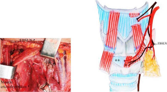
This composite image illustrates the surgical anatomy of the thyroid region, specifically focusing on the anatomical variations of the external branch of the superior laryngeal nerve (EBSLN). On the left, a clinical photograph shows an intraoperative surgical field during thyroidectomy, with labels identifying the EBSLN and the cricothyroid muscle. Metallic retractors are used to provide exposure. On the right, a corresponding anatomical diagram depicts the Type IV Kierner classification. The diagram shows the EBSLN (labeled) descending and passing directly between the trifurcation branches of the superior thyroid artery (STA) just above the upper pole of the thyroid gland. The cricothyroid muscle is shown as the destination for the nerve fibers. This visual illustrates a critical surgical risk zone where the nerve is susceptible to injury during ligation of the superior thyroid pedicle. The educational focus is on identifying variations in nerve-vessel relationships to prevent postoperative vocal cord dysfunction in endocrine surgery.
thyroid hormone synthesis steps iodine T3 T4 diagram site:wikimedia.org OR site:commons.wikimedia.org
thyroid hormone synthesis iodination thyroglobulin T3 T4
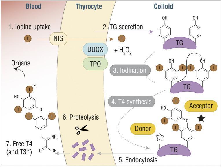
A pathophysiology diagram illustrating the seven-step process of thyroid hormone synthesis (T3 and T4) across three compartments: Blood, Thyrocyte, and Colloid. (1) Iodine uptake: Iodide (I-) enters the thyrocyte from the blood via the Sodium/Iodide Symporter (NIS). (2) TG secretion: Thyroglobulin (TG) is secreted into the colloid. (3) Iodination: Within the colloid, iodide is oxidized by Thyroid Peroxidase (TPO), dual oxidase (DUOX), and H2O2, leading to the attachment of iodine to tyrosine residues on TG. (4) T4 synthesis: A coupling reaction occurs between 'Donor' and 'Acceptor' iodinated tyrosine residues on the TG scaffold. (5) Endocytosis: The modified TG molecule is re-absorbed into the thyrocyte. (6) Proteolysis: Lysosomal enzymes break down TG, releasing individual hormones. (7) Release: Free T4 (thyroxine) and T3 (triiodothyronine) are secreted into the blood for systemic distribution to organs. The diagram includes detailed chemical structures of the phenolic rings and iodine substitutions characteristic of thyroid hormones.
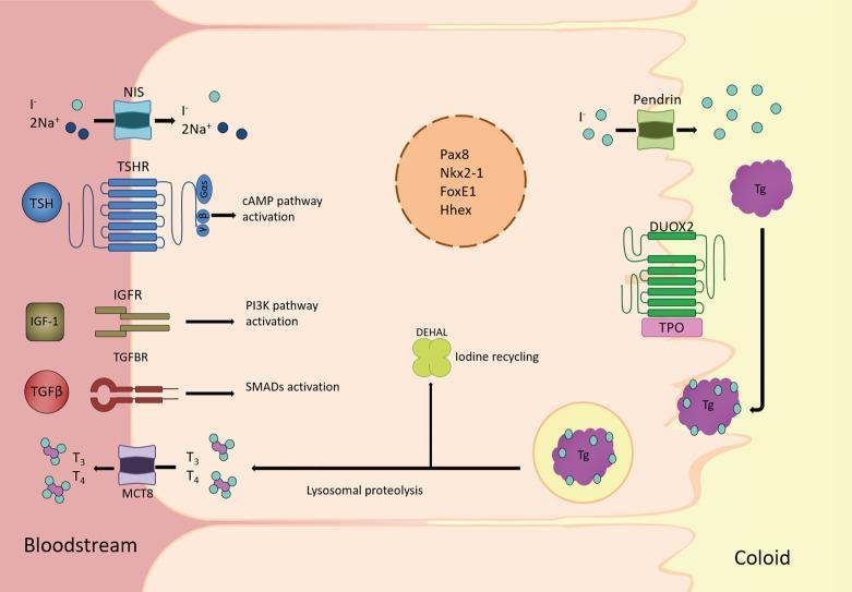
This medical schematic illustrates the pathophysiology and cellular mechanisms of a thyroid follicular cell, detailing the synthesis and secretion of thyroid hormones. The diagram is divided into three compartments: the bloodstream (basolateral side), the follicular cell cytoplasm, and the colloid lumen (apical side). Key transport proteins at the basolateral membrane include the Sodium-Iodide Symporter (NIS), which co-transports I- and Na+, and the Monocarboxylate Transporter 8 (MCT8), which facilitates the secretion of T3 and T4 into the blood. Signaling receptors TSHR, IGFR, and TGFBR are shown triggering cAMP, PI3K, and SMAD pathways respectively. At the apical membrane, Pendrin transports iodide into the colloid, where DUOX2 and Thyroid Peroxidase (TPO) facilitate the iodination of Thyroglobulin (Tg). The diagram further depicts the endocytosis of Tg, followed by lysosomal proteolysis to release T3 and T4. The enzyme DEHAL is shown recycling iodine from MIT and DIT. Centrally, the nucleus contains transcription factors Pax8, Nkx2-1, FoxE1, and Hhex, which regulate gene expression for thyroid differentiation.
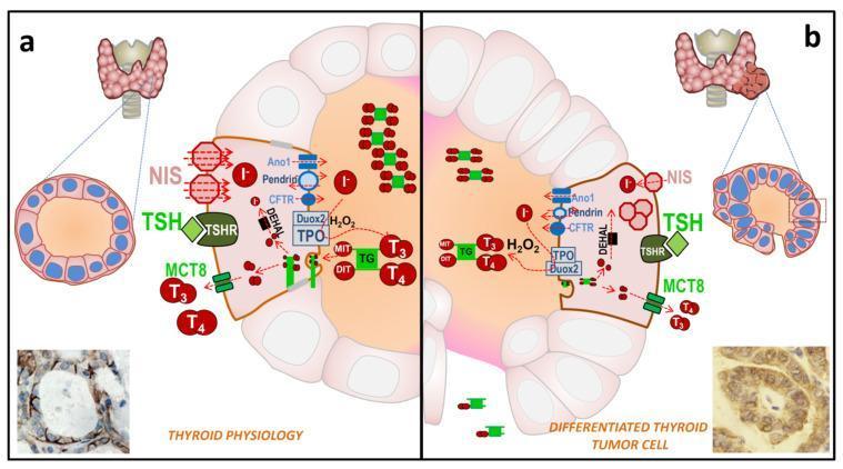
This comparative medical illustration depicts thyroid hormone (TH) biosynthesis in a normal follicle (Panel A) versus a differentiated thyroid cancer (DTC) cell (Panel B). Panel A illustrates normal physiology: a well-organized follicular structure with follicular epithelial cells surrounding a central colloid. Key proteins shown include the sodium/iodide symporter (NIS) localized strictly at the basolateral membrane, the TSH receptor (TSHR), and apical transporters like pendrin and Ano1. Iodide (I-) organification via thyroperoxidase (TPO) and Duox2-generated H2O2 is shown creating iodinated thyroglobulin (TG-I), leading to T3 and T4 synthesis. Panel B represents the pathophysiology of DTC, highlighting structural irregularities in the follicle and cellular alterations. Pathological changes include the delocalization of NIS from the basolateral membrane into the cytoplasm and a reduction in the expression of functional proteins like pendrin and TSHR. While H2O2 production persists, the synthesis and storage of TG-I and TH (T3/T4) in the colloid are visibly diminished. Bottom insets provide immunohistochemistry (IHC) correlations, confirming linear basolateral NIS expression in normal tissue versus diffuse, cytoplasmic NIS staining in tumor tissue.
This medical flowchart illustrates the systemic and cardiovascular effects of thyroid hormones. The diagram begins with the thyroid gland releasing Thyroxine (T4) and Triiodothyronine (T3), highlighting the peripheral conversion of T4 to the more active T3. The downstream physiological impacts of T3 are categorized into four primary pathways: 1) Cardiac effects, including increased resting heart rate, accelerated left ventricular (LV) relaxation, and increased LV contractility. 2) Hematologic effects, specifically increased erythropoietin (EPO) synthesis leading to higher red blood cell mass. 3) Vascular effects, showing reduced peripheral resistance and lower diastolic pressure. 4) Renal effects, where reduced peripheral resistance leads to lower renal perfusion pressure. These combined pathways culminate in the activation of the renin-angiotensin system (RAS) and an overall increase in blood volume and preload. The illustration uses anatomical icons of the heart, blood cells, vessels, and kidneys to correlate physiological mechanisms with organ-level changes, demonstrating how hyperthyroidism or thyroid hormone signaling influences hemodynamic stability and cardiac output.
ls -la /tmp/workspace/ 2>/dev/null || mkdir -p /tmp/workspace && echo "created"
mkdir -p /tmp/workspace/thyroid-ppt && echo "done"
python /tmp/skills/shared/scripts/fetch_images.py \ "https://cdn.orris.care/cdss_images/329aa70c4ec5b194046e2fe5959567caffac89e18971f56f6b28d804b9711e0d.jpg" \ "https://cdn.orris.care/cdss_images/9744bd054a9f867efd4f076ea214077a90eb333ea94604f81b0eaba8002bc3fa.png" \ "https://cdn.orris.care/cdss_images/pmc_clinical_VQA_218547f733b850129fb471806cfb5f2b9a21773d7af4d62542aa81d17c24b0ef.jpg" \ "https://cdn.orris.care/cdss_images/pmc_clinical_VQA_b5fd571082c8f0678dc4ec12a34fac02413cf46bf79d8855193f207d1a8b3103.jpg" \ "https://cdn.orris.care/cdss_images/pmc_clinical_VQA_6626509e52d3136ca2625bdf08aedfc5106f1629bcc33cc7543694c294e65a84.jpg" \ "https://upload.wikimedia.org/wikipedia/commons/8/82/Thyroid_hormone_synthesis.png" 2>&1 | head -5
An error occurred (ValidationException) when calling the ConverseStream operation: The model returned the following errors: prompt is too long: 210244 tokens > 200000 maximum
We encountered an error while processing your request. Please try again or reach out to us for help.
Make in 2 parts
- What the topic/subject is
- What format you want (e.g., document, presentation, notes, flashcards, essay, etc.)
Thyroid gland Image if thyroid gland with labelling Introduction part Synthesis Storage of T3 T4 Release of T3 T4 Actions Regulations of them Applied
thyroid gland anatomy labelled diagram with lobes isthmus trachea
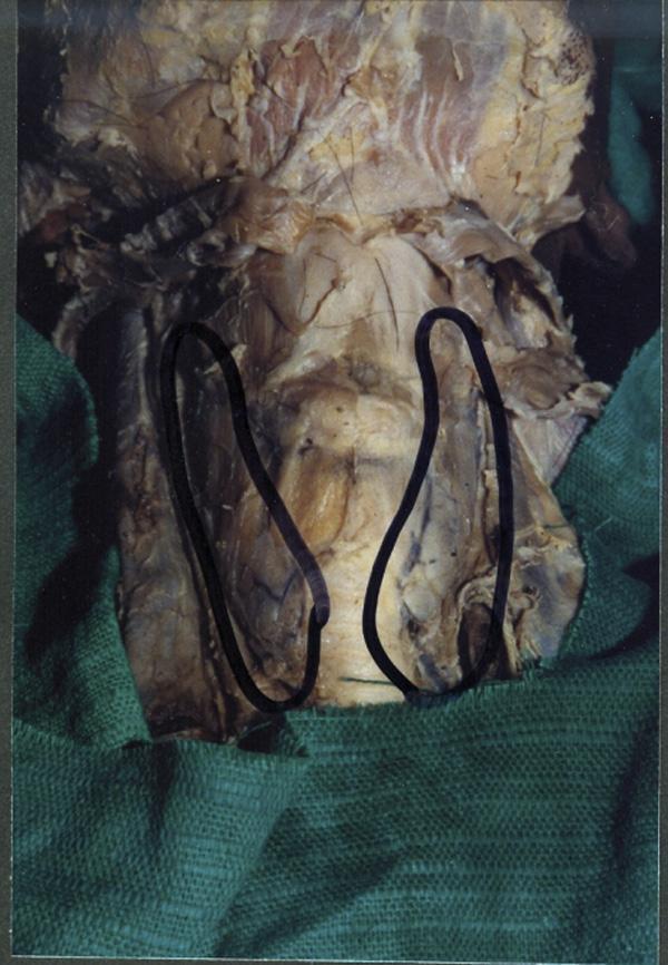
This clinical photograph of a gross anatomical specimen shows a midline dissection of the human neck, highlighting a congenital variation of the thyroid gland. The image depicts the thyroid gland consisting of two distinct, independent lateral lobes (outlined in black) positioned on either side of the trachea. Notably, there is a complete absence (agenesis) of the thyroid isthmus, which normally connects the two lobes across the second to fourth tracheal rings. In the area where the isthmus typically resides, only a thin, translucent layer of pre-tracheal fascia is visible covering the anterior surface of the trachea. The specimen demonstrates the anatomical relationship between the glandular lobes, the respiratory pathway, and the surrounding connective tissue structures. This visual is significant for medical education in endocrinology, anatomy, and surgery, as it illustrates a rare developmental anomaly where the median diverticulum of the thyroglossal duct failed to form the glandular bridge between the bifurcated lobes.
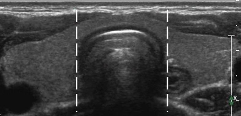
This diagnostic image is a transverse greyscale ultrasound of the anterior neck, specifically showing the thyroid gland. The image functions as an anatomical diagram to define the boundaries of the thyroid isthmus. The thyroid parenchyma appears homogeneous and moderately echogenic. Two vertical dashed white lines are superimposed on the image to delineate the lateral side-lines (lateral borders) of the trachea, which lies immediately posterior to the thyroid isthmus. The tissue situated between these two markers is identified as the isthmus, while the thyroid tissue lateral to these lines represents the left and right thyroid lobes. The trachea itself exhibits characteristic sonographic features, including an echogenic anterior border with posterior acoustic shadowing. This visual representation is used for clinical classification, particularly in determining the location of thyroid nodules or Papillary Thyroid Microcarcinomas (PTMC) for procedures like ultrasound-guided Radiofrequency Ablation (RFA).
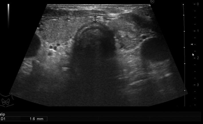
Diagnostic ultrasound image of the thyroid gland in a transverse plane, showing the right and left lobes connected by the isthmus overlying the trachea. The thyroid gland appears normal in size but exhibits a heterogeneous echotexture. Multiple small, predominantly hypoechoic nodules are visible within the parenchyma, particularly in the right lobe (left side of the image). These nodules vary in shape and possess relatively ill-defined to partially well-defined margins. Caliper measurements are visible at the isthmus, indicating a thickness of 1.6 mm. The surrounding structures, including the carotid arteries and internal jugular veins, appear as anechoic circular structures lateral to the thyroid lobes. This imaging demonstrates typical findings associated with a multinodular goiter or focal thyroiditis in the clinical context of hypothyroidism and positive thyroid antibodies. The educational focus is on identifying nodular pathology within the thyroid using ultrasonography.
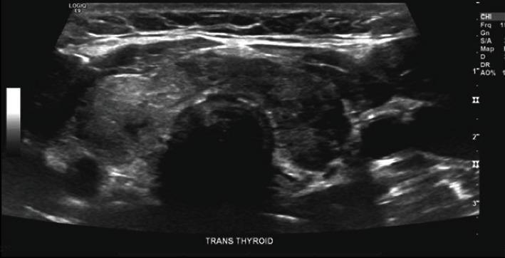
This diagnostic ultrasound image displays a transverse view of the thyroid gland. The gland exhibits thyromegaly, or general enlargement, with a notably heterogeneous echotexture across both the right and left lobes. Located within the central isthmus are two small, solid, hypoechoic nodules, approximately 4 mm in size, which appear darker relative to the surrounding thyroid parenchyma. The trachea is visible as a central shadowed area inferior to the isthmus. This imaging study is representative of subacute thyroiditis or inflammatory thyroid conditions where diffuse parenchymal changes and focal nodules are present. The image provides clinical evidence for evaluating anterior neck swelling and tenderness in an endocrine context.
thyroid hormone synthesis T3 T4 iodination thyroglobulin follicle diagram
A pathophysiology diagram illustrating the seven-step process of thyroid hormone synthesis (T3 and T4) across three compartments: Blood, Thyrocyte, and Colloid. (1) Iodine uptake: Iodide (I-) enters the thyrocyte from the blood via the Sodium/Iodide Symporter (NIS). (2) TG secretion: Thyroglobulin (TG) is secreted into the colloid. (3) Iodination: Within the colloid, iodide is oxidized by Thyroid Peroxidase (TPO), dual oxidase (DUOX), and H2O2, leading to the attachment of iodine to tyrosine residues on TG. (4) T4 synthesis: A coupling reaction occurs between 'Donor' and 'Acceptor' iodinated tyrosine residues on the TG scaffold. (5) Endocytosis: The modified TG molecule is re-absorbed into the thyrocyte. (6) Proteolysis: Lysosomal enzymes break down TG, releasing individual hormones. (7) Release: Free T4 (thyroxine) and T3 (triiodothyronine) are secreted into the blood for systemic distribution to organs. The diagram includes detailed chemical structures of the phenolic rings and iodine substitutions characteristic of thyroid hormones.
This medical schematic illustrates the pathophysiology and cellular mechanisms of a thyroid follicular cell, detailing the synthesis and secretion of thyroid hormones. The diagram is divided into three compartments: the bloodstream (basolateral side), the follicular cell cytoplasm, and the colloid lumen (apical side). Key transport proteins at the basolateral membrane include the Sodium-Iodide Symporter (NIS), which co-transports I- and Na+, and the Monocarboxylate Transporter 8 (MCT8), which facilitates the secretion of T3 and T4 into the blood. Signaling receptors TSHR, IGFR, and TGFBR are shown triggering cAMP, PI3K, and SMAD pathways respectively. At the apical membrane, Pendrin transports iodide into the colloid, where DUOX2 and Thyroid Peroxidase (TPO) facilitate the iodination of Thyroglobulin (Tg). The diagram further depicts the endocytosis of Tg, followed by lysosomal proteolysis to release T3 and T4. The enzyme DEHAL is shown recycling iodine from MIT and DIT. Centrally, the nucleus contains transcription factors Pax8, Nkx2-1, FoxE1, and Hhex, which regulate gene expression for thyroid differentiation.
This comparative medical illustration depicts thyroid hormone (TH) biosynthesis in a normal follicle (Panel A) versus a differentiated thyroid cancer (DTC) cell (Panel B). Panel A illustrates normal physiology: a well-organized follicular structure with follicular epithelial cells surrounding a central colloid. Key proteins shown include the sodium/iodide symporter (NIS) localized strictly at the basolateral membrane, the TSH receptor (TSHR), and apical transporters like pendrin and Ano1. Iodide (I-) organification via thyroperoxidase (TPO) and Duox2-generated H2O2 is shown creating iodinated thyroglobulin (TG-I), leading to T3 and T4 synthesis. Panel B represents the pathophysiology of DTC, highlighting structural irregularities in the follicle and cellular alterations. Pathological changes include the delocalization of NIS from the basolateral membrane into the cytoplasm and a reduction in the expression of functional proteins like pendrin and TSHR. While H2O2 production persists, the synthesis and storage of TG-I and TH (T3/T4) in the colloid are visibly diminished. Bottom insets provide immunohistochemistry (IHC) correlations, confirming linear basolateral NIS expression in normal tissue versus diffuse, cytoplasmic NIS staining in tumor tissue.
thyroid regulation HPT axis TSH TRH negative feedback diagram
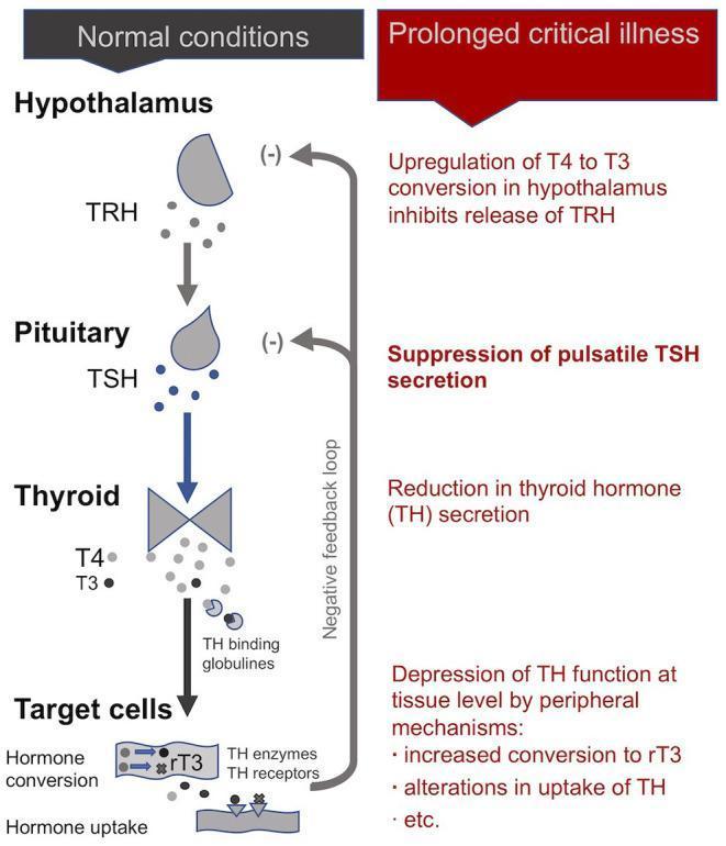
This pathophysiology diagram illustrates the Hypothalamic-Pituitary-Thyroid (HPT) axis, comparing normal physiological conditions with the alterations observed during prolonged critical illness. The 'Normal conditions' section shows the standard cascade: the Hypothalamus secretes TRH, which stimulates the Pituitary to release TSH, leading the Thyroid gland to produce T4 and T3. These hormones reach target cells via TH binding globulins for hormone conversion and uptake, with a negative feedback loop inhibiting TRH and TSH secretion. In contrast, the 'Prolonged critical illness' section details the central and peripheral suppression of the axis. Key pathological features include: upregulation of T4 to T3 conversion in the hypothalamus (inhibiting TRH release), suppression of pulsatile TSH secretion by the pituitary, reduced thyroid hormone secretion, and depression of thyroid function at the tissue level. Peripheral mechanisms shown include increased conversion to inactive rT3 and altered hormone uptake. This comparison illustrates the endocrine maladaptation typical of Non-Thyroidal Illness Syndrome (NTIS) in intensive care settings.
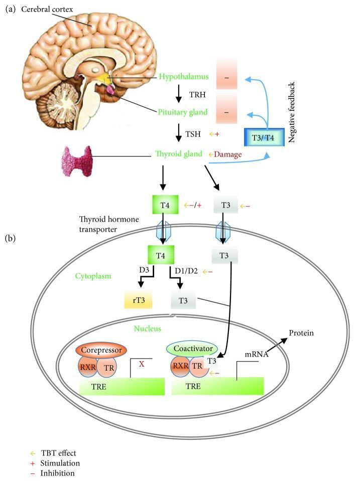
A two-part medical illustration depicting the pathophysiology of the Hypothalamic-Pituitary-Thyroid (HPT) axis and cellular thyroid hormone action. Section (a) shows the systemic HPT axis, beginning with the cerebral cortex and hypothalamus. The hypothalamus releases thyrotropin-releasing hormone (TRH), which stimulates the pituitary gland to secrete thyroid-stimulating hormone (TSH). TSH subsequently acts on the thyroid gland to stimulate the production of T4 and T3 hormones, which exert negative feedback on the hypothalamus and pituitary. Section (b) details the intracellular mechanism of action within a target cell. T4 and T3 enter the cell via thyroid hormone transporters. In the cytoplasm, deiodinases (D1/D2) convert T4 to T3, while D3 converts T4 to inactive reverse T3 (rT3). T3 enters the nucleus, binding to the thyroid hormone receptor (TR) which heterodimerizes with the retinoid X receptor (RXR). This complex binds to the thyroid hormone response element (TRE) on DNA, displacing corepressors and recruiting coactivators to initiate gene transcription (mRNA) and subsequent protein synthesis. The diagram also highlights potential sites of endocrine disruption (e.g., by Tributyltin/TBT), including receptor inhibition and deiodinase interference.
🦋 THYROID GLAND - PART 1
Image | Introduction | Synthesis | Storage of T3/T4
LABELLED DIAGRAM OF THE THYROID GLAND
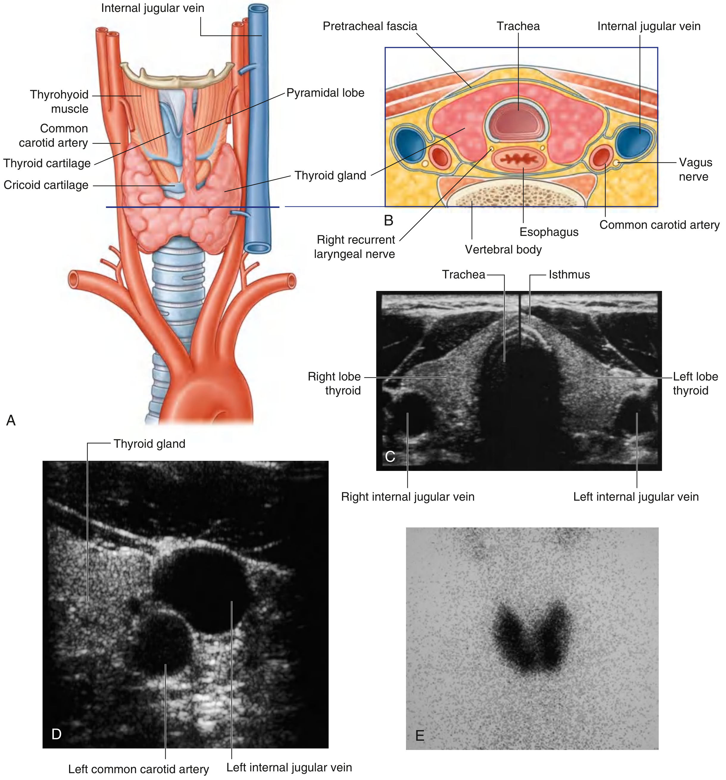
1. INTRODUCTION
Location and General Description
Gross Anatomy
- Two lateral lobes - cover the anterolateral surfaces of the trachea, the cricoid cartilage, and the lower part of the thyroid cartilage
- Isthmus - connects the lateral lobes and crosses the anterior surfaces of the 2nd and 3rd tracheal cartilages
- Pyramidal lobe - may extend upward from the isthmus along the path of the thyroglossal duct (present in ~50% of individuals)
- Weight - approximately 25-30 g in a normal adult
- Colour - brownish-red (highly vascular)
Blood Supply
- Superior thyroid artery - first branch of the external carotid artery; supplies the upper pole
- Inferior thyroid artery - branch of the thyrocervical trunk (from subclavian artery); supplies the lower pole
- Thyroidea ima artery - occasionally present, arises directly from the aorta or brachiocephalic artery
- Venous drainage - via superior, middle, and inferior thyroid veins draining into the internal jugular and brachiocephalic veins
Nerve Supply
- Sympathetic fibres from the superior and middle cervical ganglia (vasomotor)
- Parasympathetic fibres via the vagus nerve
Relations of Importance
| Relation | Structure |
|---|---|
| Anterior | Sternohyoid, sternothyroid, omohyoid muscles |
| Posterior | Parathyroid glands (4 in number), recurrent laryngeal nerve |
| Lateral | Common carotid artery, internal jugular vein, vagus nerve |
| Medial | Trachea, esophagus, larynx |
Development
Microscopic Structure (Histology)
- Follicles - spherical units, the functional unit of the gland
- Follicular cells (thyrocytes) - cuboidal epithelium; synthesize T3 and T4
- Colloid - proteinaceous material in the lumen; contains thyroglobulin (Tg)
- Parafollicular C cells - scattered between follicles; secrete calcitonin (regulates calcium)
2. SYNTHESIS OF T3 AND T4
Overview of the Process
- Collect and transport iodine
- Synthesize thyroglobulin and secrete it into the colloid
- Fix iodine to thyroglobulin to generate thyroid hormones
- Remove thyroid hormones from thyroglobulin and secrete them into circulation
- Ganong's Review of Medical Physiology, 26th Ed.
Thyroid Hormone Biosynthesis Diagram
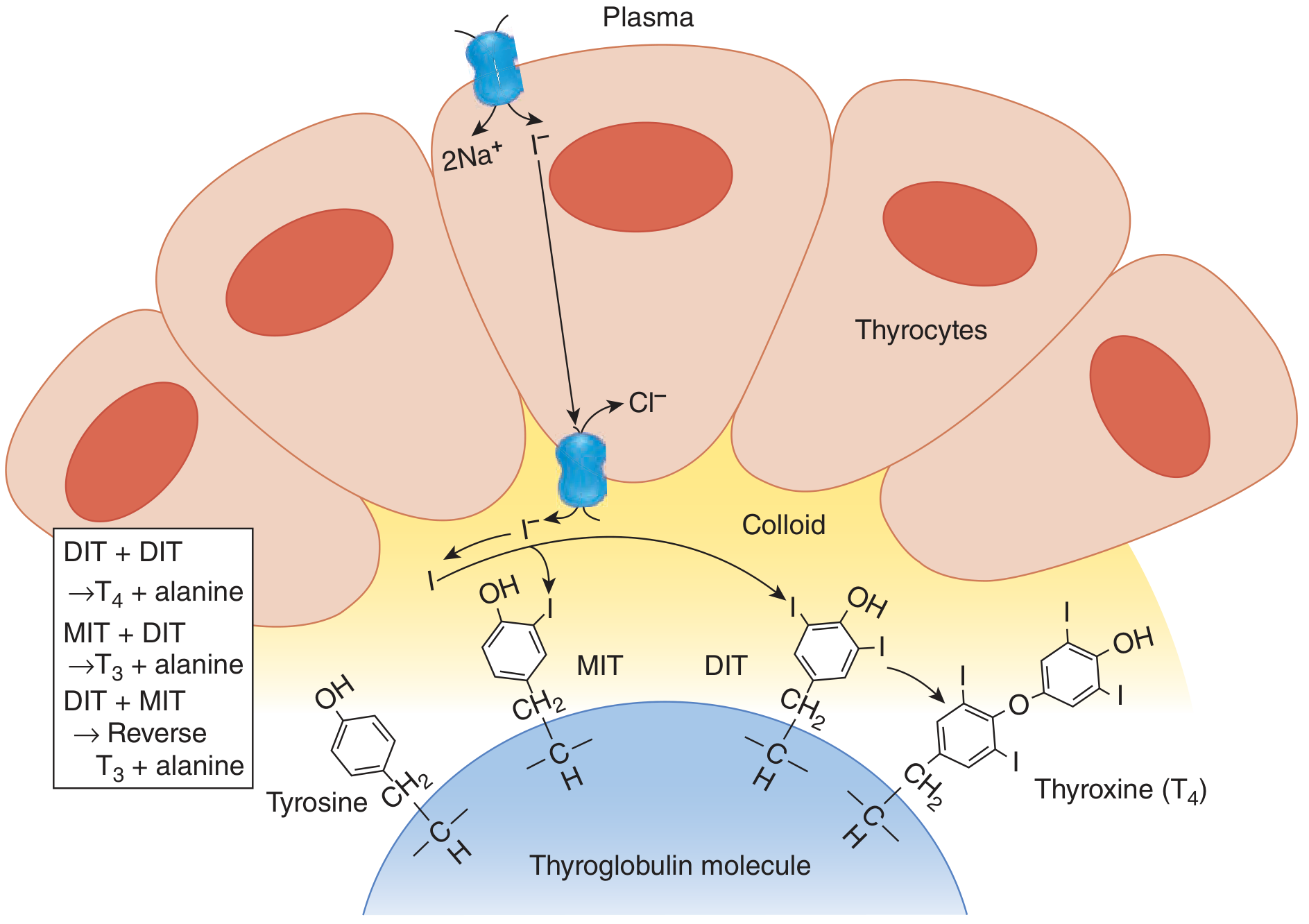
Step-by-Step Synthesis
-
Dietary iodine is absorbed as iodide (I-) in the gut
-
Iodide is actively pumped from plasma into the thyrocyte by the Sodium-Iodide Symporter (NIS) at the basolateral membrane
-
NIS co-transports 2 Na+ with 1 I- (secondary active transport; driven by Na+/K+-ATPase)
-
This creates a 25-fold concentration gradient of iodide inside the cell vs. plasma
-
NIS is stimulated by TSH and inhibited by high iodide (Wolff-Chaikoff effect)
-
Pendrin (on the apical surface) mediates iodide efflux into the colloid lumen
-
Harrison's Internal Medicine, 22nd Ed.
- Tg is a large glycoprotein (MW ~660 kDa) made up of two identical subunits
- It contains 123 tyrosine residues, of which only 4-8 are normally incorporated into thyroid hormones
- Tg is synthesized in the rough ER of thyrocytes and secreted into the colloid by exocytosis
- Iodide in the colloid is oxidized to iodine (I2) by Thyroid Peroxidase (TPO) using H2O2 generated by DUOX (dual oxidase)
- Iodine is then rapidly incorporated into carbon-3 position of tyrosine residues on Tg
- This takes place at the apical border of thyrocytes while tyrosine remains bound to Tg
- Results in formation of:
- MIT - Monoiodotyrosine (one iodine on tyrosine)
- DIT - Diiodotyrosine (two iodines on tyrosine)
-
Also catalyzed by TPO: iodinated tyrosine residues are coupled together:
- DIT + DIT → T4 (thyroxine, tetraiodothyronine) + alanine
- MIT + DIT → T3 (triiodothyronine) + alanine
- DIT + MIT → rT3 (reverse T3, biologically inactive) + alanine
-
Ratio of T4:T3 produced is approximately 20:1 (the thyroid secretes much more T4 than T3)
-
Ganong's Review of Medical Physiology, 26th Ed.
Key Enzymes in Synthesis
| Enzyme/Protein | Location | Function |
|---|---|---|
| NIS (Na+/I- Symporter) | Basolateral membrane | Active iodide uptake |
| Pendrin | Apical membrane | Iodide efflux into colloid |
| DUOX 1/2 | Apical membrane | Generates H2O2 |
| Thyroid Peroxidase (TPO) | Apical membrane | Oxidation, organification, coupling |
| Thyroglobulin (Tg) | Colloid | Scaffold/precursor for T3/T4 |
Iodine Requirements
- Daily requirement: 150 μg/day in adults; 200 μg/day in pregnancy
- Minimum required: if iodine intake falls below 50 μg/day, hormone synthesis becomes inadequate
- Iodine deficiency is a leading cause of preventable intellectual disability worldwide (WHO)
3. STORAGE OF T3 AND T4
Key Features of Storage
-
Thyroid hormones remain part of the thyroglobulin molecule until needed
-
Colloid represents a reservoir of thyroid hormones bound to Tg
-
A normal thyroid gland stores enough hormones for 2-3 months of metabolic needs
-
Humans can ingest a diet completely devoid of iodide for up to 2 months before a decline in circulating thyroid hormone levels is observed
-
This prolonged storage capacity explains the slow onset and offset of thyroid dysfunction
-
Ganong's Review of Medical Physiology, 26th Ed.
Factors Regulating Storage
- TSH stimulates all steps including synthesis and increases the storage pool
- High iodide (pharmacological doses) transiently inhibits organification = Wolff-Chaikoff effect; the thyroid "escapes" after a few days by reducing NIS expression
- Low iodide upregulates NIS expression to maximize trapping
🦋 THYROID GLAND - PART 2
Release | Actions | Regulation | Applied (Clinical)
4. RELEASE OF T3 AND T4
Mechanism of Release
- Colloid (containing Tg with bound T3/T4) is internalized by thyrocytes by endocytosis
- TSH stimulates this process via cAMP-dependent mechanisms
- Colloid droplets merge with lysosomes to form phagolysosomes
- Lysosomal proteases hydrolyze the peptide bonds of Tg
- This releases free T4, T3, MIT, and DIT into the cytosol
-
Free T4 and T3 are discharged into the cytosol and then into the capillaries
-
MIT and DIT are NOT secreted - they are deiodinated by the enzyme iodotyrosine dehalogenase (DEHAL), which recycles the iodide back into the synthesis pathway
-
T4 and T3 are secreted in a 20:1 ratio (predominantly T4)
-
Ganong's Review of Medical Physiology, 26th Ed.
Plasma Transport
| Binding Protein | Proportion Bound | Notes |
|---|---|---|
| Thyroxine-Binding Globulin (TBG) | ~70% | Primary carrier; increased by estrogen, pregnancy |
| Transthyretin (TTR/TBPA) | ~15% | Also carries retinol |
| Albumin | ~10% | Low affinity, high capacity |
| Free (unbound) | ~0.03% T4; ~0.3% T3 | Biologically active fraction |
- Only the free fraction is metabolically active and enters cells
- T3 is 3-5x more potent than T4 and has a shorter half-life (T3: ~1 day; T4: ~7 days)
- T4 is the major circulating form but is a prohormone - peripherally converted to the active T3 by deiodinases (D1, D2, D3):
- D1 and D2 - convert T4 → T3 (active) mainly in liver, kidney, muscle, pituitary
- D3 - converts T4 → reverse T3 (rT3, inactive) - inactivation pathway
Normal Serum Values
| Hormone | Normal Range |
|---|---|
| Total T4 | 5-12 μg/dL (64-154 nmol/L) |
| Free T4 | 0.9-1.7 ng/dL |
| Total T3 | 80-180 ng/dL |
| Free T3 | 2.3-4.2 pg/mL |
| TSH | 0.4-4.5 mIU/L |
5. ACTIONS OF THYROID HORMONES
Mechanism of Action
- Thyroid hormones enter cells by passive diffusion and via specific transporters (MCT8, MCT10)
- Inside the cell, T3 binds to nuclear Thyroid Hormone Receptors (TRs) - members of the superfamily of hormone-sensitive nuclear transcription factors (T4 can also bind, but with lower affinity)
- The hormone-receptor complex migrates to the nucleus, binds to Thyroid Hormone Response Elements (TREs) on DNA via zinc fingers
- This increases (or in some cases decreases) expression of genes regulating cell function
-
TRα gene (chromosome 17): forms TRα1 and TRα2 isoforms; widely distributed; dominant in heart, brain, bone, skeletal muscle
-
TRβ gene (chromosome 3): forms TRβ1 and TRβ2; TRβ2 is found only in brain/pituitary (key for feedback control); TRβ1 is high in liver and pituitary
-
Ganong's Review of Medical Physiology, 26th Ed. | Harrison's Internal Medicine 22e
Systemic Actions (by System)
- Increase basal metabolic rate (BMR) - stimulate O2 consumption in most tissues (except brain, testes, uterus, spleen, anterior pituitary)
- Stimulate Na+/K+-ATPase activity (major contributor to thermogenesis)
- Uncouple oxidative phosphorylation (increase heat production)
- Increase glucose absorption from the intestine
- Stimulate glycogenolysis and gluconeogenesis
- Chronotropic - increase heart rate (↑ β-adrenergic receptor number)
- Inotropic - increase contractility (↑ α-myosin heavy chain with higher ATPase activity)
- Increase cardiac output and blood volume
- Decrease peripheral vascular resistance
- Enhance responses to circulating catecholamines
- Catabolic - stimulate lipolysis in adipose tissue
- Lower circulating cholesterol levels - increase LDL receptor formation in the liver → increased hepatic removal of LDL-cholesterol from circulation (this effect precedes the metabolic rate rise)
- At physiological levels: promote protein synthesis (anabolic in growing tissues)
- At high levels: protein catabolism - increased muscle protein breakdown
- Increase GH secretion and potentiate GH effects on tissues
- Essential for normal brain development (critical period: fetal life to 3 years)
- Deficiency during fetal/neonatal period → cretinism (intellectual disability, deafness, motor deficits)
- Increase reflexes and neural excitability in adults
- Promote normal bone growth and skeletal maturation
- Required for normal ossification and epiphyseal closure
- In hypothyroid children: slowed bone growth, delayed epiphyseal closure, and delayed puberty
- Maintain normal hypoxic and hypercapnic drive in the respiratory center
- Increase 2,3-DPG in red blood cells → shifts O2-Hb dissociation curve to the right → enhanced O2 delivery to tissues
| Target Tissue | Effect | Mechanism |
|---|---|---|
| Heart | Chronotropic + inotropic | ↑ β-adrenergic receptors; ↑ α-MHC ATPase |
| Adipose | Catabolic | Stimulated lipolysis |
| Muscle | Catabolic (excess) | ↑ protein breakdown |
| Bone | Developmental | Normal growth, skeletal maturation |
| Nervous system | Developmental | Normal brain development |
| Gut | Metabolic | ↑ carbohydrate absorption |
| Liver/LDL | Metabolic | ↑ LDL receptor formation → ↓ cholesterol |
| Whole body | Calorigenic | ↑ O2 consumption, ↑ BMR |
- Ganong's Review of Medical Physiology, Table 20-3
6. REGULATION OF THYROID HORMONES
HPT (Hypothalamic-Pituitary-Thyroid) Axis
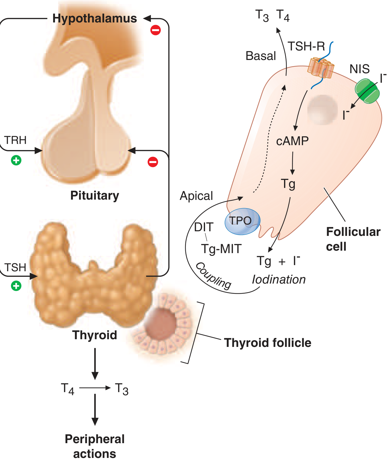
- The paraventricular nucleus of the hypothalamus secretes Thyrotropin-Releasing Hormone (TRH)
- TRH is a tripeptide (pyroGlu-His-ProNH2)
- TRH acts on the anterior pituitary to stimulate TSH release
- TRH stimulates thyrotroph cells in the anterior pituitary to release TSH (Thyroid-Stimulating Hormone / Thyrotropin)
- TSH is a glycoprotein with α and β subunits; shares the α subunit with LH, FSH, and hCG
- TSH binds to TSH-R (G-protein coupled receptor) on thyroid follicular cells
- TSH acts via cAMP to stimulate ALL steps: iodide trapping, Tg synthesis, organification, coupling, and hormone secretion
-
T3 and T4 exert negative feedback at BOTH the hypothalamus (inhibit TRH) and pituitary (inhibit TSH release and blunt TSH response to TRH)
-
The feedback is mediated primarily by T3, including T3 converted locally from T4 in pituitary by D2 deiodinase
-
TRβ2 isoform plays a specific role in pituitary feedback
-
Ganong's Review of Medical Physiology, 26th Ed.
Autoregulation by Iodide
- Low iodide → upregulates NIS, increases trapping efficiency
- High iodide (pharmacological doses) → transiently inhibits organification = Wolff-Chaikoff effect
- Thyroid normally "escapes" from Wolff-Chaikoff effect in 1-2 weeks by downregulating NIS
- Failure to escape → iodide-induced hypothyroidism (seen in some patients with underlying thyroid disease)
Other Regulatory Factors
| Factor | Effect on Thyroid |
|---|---|
| Cold exposure | ↑ TRH → ↑ TSH → ↑ T3/T4 (significant in neonates; minimal in adults) |
| Stress | ↓ TRH (inhibitory) |
| Glucocorticoids | ↓ TSH secretion |
| Dopamine | ↓ TSH secretion |
| Somatostatin | ↓ TSH secretion |
| Estrogen | ↑ TBG → ↑ total T4 (but free T4 unchanged) |
| Propylthiouracil (PTU) | Blocks organification + inhibits D2 deiodinase |
| Methimazole | Blocks organification |
7. APPLIED (CLINICAL ASPECTS)
A. Hypothyroidism
- Most common cause worldwide: iodine deficiency
- Most common cause in iodine-replete regions: Hashimoto thyroiditis (autoimmune - anti-TPO and anti-Tg antibodies)
- Other causes: thyroidectomy, radioiodine therapy, antithyroid drugs, congenital dysgenesis
- Causes: iodine deficiency, thyroid dysgenesis, inborn errors of thyroid hormone synthesis, maternal antithyroid antibodies
- Features: intellectual disability, growth retardation, coarse facial features, macroglossia, umbilical hernia, hypothermia
- Neonatal screening (TSH) allows early detection and treatment
- Cold intolerance, weight gain, bradycardia, constipation
- Dry skin, hair loss, periorbital puffiness
- Slow reflexes (delayed relaxation of deep tendon reflexes)
- Menstrual irregularities, infertility
- Raised cholesterol (↓ LDL receptor activity)
- In severe cases: myxedema coma (life-threatening)
B. Hyperthyroidism (Thyrotoxicosis)
- Graves' disease - most common; TSH-receptor stimulating antibodies (TSI/TRAb) mimic TSH action
- Toxic multinodular goiter
- Solitary toxic adenoma
- TSH-secreting pituitary tumor (secondary hyperthyroidism)
- Thyroiditis (transient - early Hashimoto, subacute, postpartum)
- Exogenous T3/T4 (factitious/iatrogenic)
- Heat intolerance, weight loss, increased appetite, diarrhea
- Palpitations, tachycardia, atrial fibrillation
- Tremor, anxiety, restlessness
- Exophthalmos (Graves' disease - due to retroorbital inflammation)
- Pretibial myxedema, thyroid acropachy (Graves' specific)
- Thyroid bruit (in Graves')
- Proximal muscle weakness
- Thioureylenes - Propylthiouracil (PTU) and Methimazole: compete with tyrosine for iodination; block organification and coupling. PTU also inhibits D2 deiodinase (blocks T4→T3 conversion peripherally - preferred in thyroid storm)
- Radioiodine (131I) - taken up by NIS; destroys follicular cells
- Surgery (thyroidectomy)
- Beta blockers (propranolol) - control adrenergic symptoms; also inhibits D2 deiodinase
- Potassium iodide (Lugol's solution) - pre-operative preparation; exploits Wolff-Chaikoff effect to temporarily reduce T3/T4 synthesis and decrease vascularity
C. Goiter
- Diffuse enlargement of the thyroid gland
- Endemic goiter - iodine deficiency; ↑ TSH drives hyperplasia
- Toxic goiter (Graves') - diffuse + hyperthyroid
- Non-toxic multinodular goiter - variable function, commonly euthyroid
- Complications: compressive symptoms (dysphagia, dyspnea, hoarseness from RLN involvement)
D. Pendred Syndrome
- Autosomal recessive mutation in Pendrin gene
- Results in defective iodide organification → goiter + hypothyroidism
- Also causes sensorineural deafness (pendrin expressed in cochlea)
E. Thyroid Hormone Resistance
- Mutations in TRβ gene → resistance to T3/T4 in peripheral tissues and pituitary
- Patients have high T3/T4 with inappropriately normal or raised TSH
- Usually clinically euthyroid (TRα unaffected compensates)
- Higher incidence of ADHD seen in this population - Ganong's Review
F. NIS and Radioiodine Therapy
- NIS expression in thyroid allows radioiodine (131I) uptake for:
- Imaging (thyroid scan with 123I or 99mTc pertechnetate)
- Treatment of hyperthyroidism and thyroid cancer
- Low-level NIS expression in salivary glands, lactating breast, and placenta - basis for some side effects of radioiodine
G. Key Drug Targets Summary
| Drug | Mechanism | Use |
|---|---|---|
| Methimazole | Blocks TPO (organification + coupling) | Hyperthyroidism |
| Propylthiouracil (PTU) | Blocks TPO + inhibits D2 (T4→T3) | Hyperthyroidism; thyroid storm; pregnancy 1st trimester |
| Radioiodine (131I) | Concentrated by NIS, destroys tissue | Hyperthyroidism, thyroid cancer |
| Levothyroxine (T4) | Replaces T4 | Hypothyroidism |
| Potassium iodide | Wolff-Chaikoff → ↓ synthesis; ↓ vascularity | Pre-op thyroid surgery; radiation emergencies |
| Propranolol | ↓ β-adrenergic effects; inhibits D2 | Symptomatic control in thyrotoxicosis |
Quick Reference - What's in Each Part
PART 1 covers:
- Labelled Diagram - Full anatomical diagram from Gray's Anatomy (anterior view, transverse section, ultrasound, nuclear scan) with all key structures labelled
- Introduction - Location, gross anatomy (lobes, isthmus, pyramidal lobe), blood supply, nerve supply, important relations, development (thyroglossal duct, foramen cecum), histology (follicles, thyrocytes, colloid, C cells)
- Synthesis of T3/T4 - Complete step-by-step: NIS iodide trapping → Tg synthesis → Organification (TPO, DUOX, H2O2) → Coupling (MIT+DIT, DIT+DIT), with enzyme table and iodine requirements
- Storage - Extracellular colloid storage (2-3 month reserve), Wolff-Chaikoff effect, TSH regulation
PART 2 covers:
- Release - Endocytosis of colloid → lysosomal proteolysis → MIT/DIT deiodination + recycling → T3/T4 secretion; plasma transport proteins (TBG, TTR, albumin); T4 → T3 conversion by deiodinases
- Actions - Nuclear TRα/TRβ mechanism; calorigenic, cardiovascular, lipid, protein, neural, growth, and hematopoietic effects - with full table
- Regulation - HPT axis diagram; TRH/TSH/T3/T4 negative feedback; autoregulation by iodide; modifying factors (cold, stress, glucocorticoids, dopamine)
- Applied - Hypothyroidism (cretinism, myxedema, Hashimoto's), Hyperthyroidism (Graves', toxic goiter, thyroid storm), Goiter, Pendred syndrome, Thyroid hormone resistance, Radioiodine therapy, Drug targets table
I want it in ppt form
ls -la /tmp/workspace/ 2>/dev/null || echo "workspace does not exist"
mkdir -p /tmp/workspace/thyroid-ppt && echo "done"
~/thyroid-ppt/build_thyroid_ppt.py
import json, subprocess, base64, sys
from io import BytesIO
from pptx import Presentation
from pptx.util import Inches, Pt, Emu
from pptx.dml.color import RGBColor
from pptx.enum.text import PP_ALIGN, MSO_ANCHOR
from pptx.oxml.ns import qn
from lxml import etree
# ── Colour palette (teal/navy medical theme) ──────────────────────────────────
C_NAVY = RGBColor(0x0D, 0x2B, 0x55) # slide header bg
C_TEAL = RGBColor(0x00, 0x7B, 0x8E) # accent / divider
C_LTBLUE = RGBColor(0xE8, 0xF4, 0xF8) # body bg tint
C_WHITE = RGBColor(0xFF, 0xFF, 0xFF)
C_YELLOW = RGBColor(0xFF, 0xD7, 0x00)
C_GREEN = RGBColor(0x21, 0x8C, 0x3A)
C_RED = RGBColor(0xC0, 0x39, 0x2B)
C_GRAY = RGBColor(0x55, 0x55, 0x55)
C_DARK = RGBColor(0x1A, 0x1A, 0x2E)
C_ORANGE = RGBColor(0xE6, 0x7E, 0x22)
W = Inches(13.333)
H = Inches(7.5)
def new_prs():
prs = Presentation()
prs.slide_width = W
prs.slide_height = H
return prs
def blank(prs):
return prs.slides.add_slide(prs.slide_layouts[6])
# ── Low-level helpers ─────────────────────────────────────────────────────────
def rect(slide, x, y, w, h, fill_rgb, line_rgb=None, line_w=None):
from pptx.enum.shapes import MSO_SHAPE_TYPE
shp = slide.shapes.add_shape(1, x, y, w, h) # 1 = RECTANGLE
shp.fill.solid()
shp.fill.fore_color.rgb = fill_rgb
shp.line.fill.background()
if line_rgb:
shp.line.color.rgb = line_rgb
shp.line.width = Pt(line_w or 1)
else:
shp.line.fill.background()
return shp
def txt(slide, text, x, y, w, h, size=18, bold=False, color=C_DARK,
align=PP_ALIGN.LEFT, italic=False, wrap=True, anchor=MSO_ANCHOR.TOP,
font_name="Calibri"):
tb = slide.shapes.add_textbox(x, y, w, h)
tf = tb.text_frame
tf.word_wrap = wrap
tf.vertical_anchor = anchor
tf.margin_left = Inches(0.05)
tf.margin_right = Inches(0.05)
tf.margin_top = 0
tf.margin_bottom= 0
p = tf.paragraphs[0]
p.alignment = align
run = p.add_run()
run.text = text
run.font.name = font_name
run.font.size = Pt(size)
run.font.bold = bold
run.font.italic= italic
run.font.color.rgb = color
return tb
def header_bar(slide, title, subtitle=None):
"""Navy top bar with white title."""
rect(slide, 0, 0, W, Inches(1.15), C_NAVY)
txt(slide, title, Inches(0.4), Inches(0.1), Inches(11), Inches(0.75),
size=30, bold=True, color=C_WHITE, align=PP_ALIGN.LEFT)
if subtitle:
txt(slide, subtitle, Inches(0.4), Inches(0.78), Inches(12), Inches(0.38),
size=14, bold=False, color=C_YELLOW, align=PP_ALIGN.LEFT)
# teal accent line
shp = slide.shapes.add_shape(1, 0, Inches(1.15), W, Inches(0.06))
shp.fill.solid(); shp.fill.fore_color.rgb = C_TEAL
shp.line.fill.background()
def bullet_box(slide, items, x, y, w, h, title=None,
title_bg=C_TEAL, body_bg=C_LTBLUE,
font_size=15, title_size=16, indent_char=" "):
"""Rounded card with optional title and bullet list."""
# background card
shp = slide.shapes.add_shape(1, x, y, w, h)
shp.fill.solid(); shp.fill.fore_color.rgb = body_bg
shp.line.color.rgb = C_TEAL; shp.line.width = Pt(1)
off = 0
if title:
th = Inches(0.38)
hdr = slide.shapes.add_shape(1, x, y, w, th)
hdr.fill.solid(); hdr.fill.fore_color.rgb = title_bg
hdr.line.fill.background()
txt(slide, title, x + Inches(0.1), y + Inches(0.03),
w - Inches(0.2), th - Inches(0.04),
size=title_size, bold=True, color=C_WHITE, align=PP_ALIGN.LEFT)
off = th
tb = slide.shapes.add_textbox(x + Inches(0.12), y + off + Inches(0.06),
w - Inches(0.25), h - off - Inches(0.1))
tf = tb.text_frame; tf.word_wrap = True
tf.margin_left = tf.margin_right = tf.margin_top = tf.margin_bottom = 0
first = True
for item in items:
if first:
p = tf.paragraphs[0]; first = False
else:
p = tf.add_paragraph()
level = 0
text = item
if item.startswith(" "):
level = 2; text = item.strip()
elif item.startswith(" "):
level = 1; text = item.strip()
p.level = level
run = p.add_run()
run.text = text
run.font.name = "Calibri"
run.font.size = Pt(font_size - level)
run.font.color.rgb = C_DARK
p.space_before = Pt(2)
def fetch_image(url):
"""Download image bytes via the skill helper."""
result = json.loads(subprocess.check_output(
["python", "/tmp/skills/shared/scripts/fetch_images.py", url],
timeout=30
))
item = result[0]
if item.get("base64"):
raw = base64.b64decode(item["base64"].split(",", 1)[1])
return BytesIO(raw)
return None
def add_img(slide, url, x, y, w=None, h=None):
buf = fetch_image(url)
if buf:
if w and h:
slide.shapes.add_picture(buf, x, y, width=w, height=h)
elif w:
slide.shapes.add_picture(buf, x, y, width=w)
elif h:
slide.shapes.add_picture(buf, x, y, height=h)
else:
slide.shapes.add_picture(buf, x, y)
return True
return False
# ═══════════════════════════════════════════════════════════════════════════════
# SLIDE BUILDERS
# ═══════════════════════════════════════════════════════════════════════════════
def slide_title(prs):
sl = blank(prs)
# full background
rect(sl, 0, 0, W, H, C_NAVY)
# decorative teal band
rect(sl, 0, Inches(4.5), W, Inches(0.08), C_TEAL)
# title
txt(sl, "THYROID GLAND", Inches(1), Inches(1.2), Inches(11.3), Inches(1.4),
size=54, bold=True, color=C_WHITE, align=PP_ALIGN.CENTER)
txt(sl, "Anatomy · Synthesis · Actions · Regulation · Applied",
Inches(1), Inches(2.8), Inches(11.3), Inches(0.6),
size=20, color=C_YELLOW, align=PP_ALIGN.CENTER)
txt(sl, "Sources: Gray's Anatomy | Ganong's Physiology | Harrison's Internal Medicine",
Inches(1), Inches(5.2), Inches(11.3), Inches(0.5),
size=13, color=RGBColor(0xAA,0xCC,0xDD), align=PP_ALIGN.CENTER)
# butterfly icon (text)
txt(sl, "🦋", Inches(6.1), Inches(3.5), Inches(1.2), Inches(0.8),
size=40, align=PP_ALIGN.CENTER)
def slide_anatomy_image(prs):
sl = blank(prs)
rect(sl, 0, 0, W, H, C_LTBLUE)
header_bar(sl, "Anatomy of the Thyroid Gland",
subtitle="Gray's Anatomy for Students – Anterior & Transverse Views")
# Main anatomy image (left side)
added = add_img(sl,
"https://cdn.orris.care/cdss_images/2ec58523e023bd33efeb9ba12b34b76158eb9fa16b3ce47605088bd30921c13e.png",
Inches(0.2), Inches(1.3), h=Inches(5.9))
# right column labels
labels = [
("GROSS ANATOMY", True, C_TEAL),
("Two lateral lobes", False, C_DARK),
("Isthmus – connects lobes at 2nd–3rd tracheal rings", False, C_DARK),
("Pyramidal lobe (present in ~50%)", False, C_DARK),
("Weight: 25–30 g", False, C_DARK),
("", False, C_DARK),
("BLOOD SUPPLY", True, C_TEAL),
("Superior thyroid a. (ext. carotid)", False, C_DARK),
("Inferior thyroid a. (thyrocervical trunk)", False, C_DARK),
("Thyroidea ima a. (occasionally)", False, C_DARK),
("", False, C_DARK),
("KEY RELATIONS", True, C_TEAL),
("Ant: strap muscles", False, C_DARK),
("Post: parathyroids + RLN", False, C_DARK),
("Lat: CCA, IJV, vagus nerve", False, C_DARK),
("Med: trachea, esophagus", False, C_DARK),
]
y_pos = Inches(1.28)
for lbl, bold, color in labels:
if lbl == "":
y_pos += Inches(0.12)
continue
txt(sl, lbl, Inches(8.2), y_pos, Inches(4.9), Inches(0.3),
size=13 if not bold else 14, bold=bold, color=color)
y_pos += Inches(0.33 if bold else 0.29)
def slide_development_histology(prs):
sl = blank(prs)
rect(sl, 0, 0, W, H, C_LTBLUE)
header_bar(sl, "Development & Histology", subtitle="Embryology and microscopic structure")
# Development card
bullet_box(sl, [
"Arises as median outgrowth from pharynx floor",
"Site of origin: Foramen cecum of tongue",
"Migrates via Thyroglossal duct to final position",
"Thyroglossal duct normally disappears",
" Remnant → Thyroglossal cyst or fistula",
" Ectopic thyroid: Lingual thyroid (at base of tongue)",
"Pyramidal lobe = upward extension along duct path",
], Inches(0.3), Inches(1.3), Inches(6.1), Inches(3.0),
title="DEVELOPMENT", font_size=14)
# Histology card
bullet_box(sl, [
"Functional unit: Thyroid follicle (spherical)",
"Follicular cells (Thyrocytes) – cuboidal epithelium",
" → Synthesize T3 and T4",
"Colloid – proteinaceous fluid in lumen",
" → Contains Thyroglobulin (Tg)",
"Parafollicular C cells – between follicles",
" → Secrete Calcitonin (calcium regulation)",
"Active gland: tall cells, little colloid",
"Resting gland: flat cells, abundant colloid",
], Inches(6.7), Inches(1.3), Inches(6.3), Inches(3.2),
title="HISTOLOGY (Microscopic Structure)", font_size=14)
# caption
txt(sl, "Nerve supply: Sympathetic (superior/middle cervical ganglia) – vasomotor | "
"Parasympathetic (vagus nerve)",
Inches(0.3), Inches(4.55), Inches(12.7), Inches(0.4),
size=12, italic=True, color=C_GRAY)
# RLN clinical note box
shp = slide.shapes if False else sl.shapes
bullet_box(sl, [
"Recurrent Laryngeal Nerve (RLN) lies in tracheoesophageal groove — closely related to inferior thyroid artery",
"At RISK during thyroid surgery → hoarseness if damaged",
"Superior laryngeal nerve (external branch) → cricothyroid muscle → pitch of voice",
], Inches(0.3), Inches(5.0), Inches(12.7), Inches(2.25),
title="CLINICAL: Nerve Relations in Surgery", title_bg=C_RED, font_size=13)
def slide_synthesis_overview(prs):
sl = blank(prs)
rect(sl, 0, 0, W, H, C_LTBLUE)
header_bar(sl, "Synthesis of T3 & T4 – Overview",
subtitle="Multi-step biosynthesis at thyrocyte–colloid interface | Ganong's Review of Medical Physiology")
steps = [
("1", "IODIDE TRAPPING", "NIS (Na⁺/I⁻ Symporter) at basolateral membrane\nActive transport – 2Na⁺ : 1I⁻\n25× concentration gradient created\nStimulated by TSH; inhibited by excess iodide"),
("2", "Tg SYNTHESIS", "Thyroglobulin (Tg) – large glycoprotein, 660 kDa\n123 tyrosine residues; only 4–8 used for hormones\nSynthesized in RER → secreted into colloid by exocytosis"),
("3", "ORGANIFICATION", "I⁻ oxidised to I₂ by Thyroid Peroxidase (TPO)\nH₂O₂ generated by DUOX1/2\nI₂ added to C-3 of tyrosine residues on Tg\nForms MIT (1 iodine) and DIT (2 iodines)"),
("4", "COUPLING", "TPO catalyses coupling:\nDIT + DIT → T4 + alanine\nMIT + DIT → T3 + alanine\nDIT + MIT → rT3 + alanine (inactive)\nRatio T4:T3 ≈ 20:1"),
]
x_positions = [Inches(0.2), Inches(3.55), Inches(6.9), Inches(10.25)]
colors = [C_NAVY, C_TEAL, RGBColor(0x16, 0x61, 0x7A), C_ORANGE]
for i, (num, title, content) in enumerate(steps):
x = x_positions[i]
w = Inches(3.15)
# step number circle
circ = sl.shapes.add_shape(9, x + Inches(1.1), Inches(1.3), Inches(0.9), Inches(0.9)) # 9=oval
circ.fill.solid(); circ.fill.fore_color.rgb = colors[i]
circ.line.fill.background()
txt(sl, num, x + Inches(1.1), Inches(1.3), Inches(0.9), Inches(0.9),
size=22, bold=True, color=C_WHITE, align=PP_ALIGN.CENTER, anchor=MSO_ANCHOR.MIDDLE)
# title bar
shp = sl.shapes.add_shape(1, x, Inches(2.35), w, Inches(0.42))
shp.fill.solid(); shp.fill.fore_color.rgb = colors[i]
shp.line.fill.background()
txt(sl, title, x + Inches(0.05), Inches(2.37), w - Inches(0.1), Inches(0.38),
size=13, bold=True, color=C_WHITE, align=PP_ALIGN.CENTER)
# content
shp2 = sl.shapes.add_shape(1, x, Inches(2.77), w, Inches(4.45))
shp2.fill.solid(); shp2.fill.fore_color.rgb = C_WHITE
shp2.line.color.rgb = colors[i]; shp2.line.width = Pt(1.5)
txt(sl, content, x + Inches(0.1), Inches(2.85), w - Inches(0.2), Inches(4.3),
size=13, color=C_DARK, wrap=True)
# arrow connectors between steps
for i in range(3):
ax = x_positions[i] + Inches(3.15) + Inches(0.02)
ay = Inches(3.8)
ln = sl.shapes.add_connector(1, ax, ay, ax + Inches(0.18), ay)
ln.line.color.rgb = C_TEAL; ln.line.width = Pt(2)
def slide_synthesis_diagram(prs):
sl = blank(prs)
rect(sl, 0, 0, W, H, C_LTBLUE)
header_bar(sl, "Thyroid Hormone Biosynthesis – Diagram",
subtitle="Iodide transport, organification, and coupling on thyroglobulin scaffold")
add_img(sl,
"https://cdn.orris.care/cdss_images/78493abd1379ac31650091e579e23b70fea4a5045ac99d0aac608d42af3b0fd3.png",
Inches(0.2), Inches(1.3), h=Inches(5.9))
# enzyme table on right
bullet_box(sl, [
"NIS – Na⁺/I⁻ Symporter",
" Basolateral | Active iodide uptake",
"Pendrin",
" Apical | Iodide efflux into colloid",
"DUOX 1/2",
" Apical | Generates H₂O₂",
"Thyroid Peroxidase (TPO)",
" Apical | Oxidation + organification + coupling",
"Thyroglobulin (Tg)",
" Colloid | Scaffold/precursor for T3/T4",
], Inches(7.6), Inches(1.3), Inches(5.5), Inches(4.0),
title="KEY ENZYMES & PROTEINS", font_size=13)
bullet_box(sl, [
"Daily iodine: 150 μg/day (adult) | 200 μg/day (pregnancy)",
"Below 50 μg/day → inadequate synthesis → goiter",
], Inches(7.6), Inches(5.5), Inches(5.5), Inches(1.7),
title="IODINE REQUIREMENTS", title_bg=C_ORANGE, font_size=13)
def slide_storage_release(prs):
sl = blank(prs)
rect(sl, 0, 0, W, H, C_LTBLUE)
header_bar(sl, "Storage & Release of T3/T4",
subtitle="Extracellular colloid storage + TSH-driven endocytosis and proteolysis")
# Storage
bullet_box(sl, [
"UNIQUE: stored extracellularly in colloid (not intracellular vesicles)",
"T3/T4 remain bound to Thyroglobulin until needed",
"Normal gland holds 2–3 months supply",
"Iodide-free diet tolerated up to 2 months before serum levels fall",
"Slow onset/offset of thyroid dysfunction explained by this reserve",
"TSH stimulates all synthesis steps and increases storage pool",
"High pharmacological iodide → Wolff-Chaikoff effect",
" Transiently inhibits organification",
" Thyroid 'escapes' in 1–2 weeks by ↓ NIS expression",
" Failure to escape → iodide-induced hypothyroidism",
], Inches(0.3), Inches(1.3), Inches(6.2), Inches(5.85),
title="STORAGE", font_size=13)
# Release
bullet_box(sl, [
"Step 1 – ENDOCYTOSIS of colloid (TSH-driven via cAMP)",
"Step 2 – PROTEOLYSIS",
" Colloid droplets fuse with lysosomes",
" Lysosomal proteases hydrolyse Tg peptide bonds",
" Releases free T4, T3, MIT, DIT",
"Step 3 – SECRETION",
" T4 and T3 → cytosol → capillaries",
" MIT & DIT NOT secreted",
" Deiodinated by DEHAL (iodotyrosine dehalogenase)",
" Iodide recycled back into synthesis",
"T4 : T3 secreted ratio ≈ 20 : 1",
], Inches(6.8), Inches(1.3), Inches(6.2), Inches(5.85),
title="RELEASE (Secretion)", font_size=13)
def slide_transport_conversion(prs):
sl = blank(prs)
rect(sl, 0, 0, W, H, C_LTBLUE)
header_bar(sl, "Plasma Transport & Peripheral Conversion",
subtitle="Binding proteins | T4 → T3 conversion by deiodinases")
# Transport table
bullet_box(sl, [
"Thyroxine-Binding Globulin (TBG) ~70%",
" Primary carrier | ↑ by estrogen, pregnancy",
"Transthyretin (TTR / TBPA) ~15%",
" Also carries retinol (vitamin A)",
"Albumin ~10%",
" Low affinity, high capacity",
"Free T4 ~0.03% | Free T3 ~0.3%",
" ONLY free fraction is biologically active",
"",
"T3 is 3–5× more potent than T4",
"Half-life: T4 ≈ 7 days | T3 ≈ 1 day",
], Inches(0.3), Inches(1.3), Inches(6.2), Inches(4.5),
title="PLASMA TRANSPORT (>99% protein-bound)", font_size=13)
# Deiodinase
bullet_box(sl, [
"D1 & D2 – convert T4 → T3 (active)",
" Sites: liver, kidney, muscle, pituitary",
" T4 is a PROHORMONE; T3 is the active form",
"D3 – converts T4 → reverse T3 (rT3, inactive)",
" Inactivation / degradation pathway",
" Upregulated in illness → sick euthyroid syndrome",
"Propylthiouracil (PTU) inhibits D2",
"Propranolol inhibits D2",
"Amiodarone inhibits D1 → ↑T4, ↓T3",
], Inches(6.8), Inches(1.3), Inches(6.2), Inches(4.5),
title="PERIPHERAL CONVERSION (Deiodinases)", font_size=13)
# Normal values
vals = [
("TSH", "0.4 – 4.5 mIU/L"),
("Total T4", "5–12 μg/dL"),
("Free T4", "0.9–1.7 ng/dL"),
("Total T3", "80–180 ng/dL"),
("Free T3", "2.3–4.2 pg/mL"),
]
y = Inches(6.05)
rect(sl, Inches(0.3), y, Inches(12.7), Inches(1.2), C_NAVY)
txt(sl, "NORMAL SERUM VALUES:", Inches(0.4), y + Inches(0.05), Inches(2.5), Inches(0.4),
size=12, bold=True, color=C_YELLOW)
for i, (name, val) in enumerate(vals):
xv = Inches(0.4) + i * Inches(2.5)
txt(sl, f"{name}: {val}", xv, y + Inches(0.45), Inches(2.4), Inches(0.6),
size=12, color=C_WHITE)
def slide_actions_mechanism(prs):
sl = blank(prs)
rect(sl, 0, 0, W, H, C_LTBLUE)
header_bar(sl, "Mechanism of Action of Thyroid Hormones",
subtitle="Nuclear receptor pathway | Genomic actions | Ganong's + Harrison's")
# Mechanism
bullet_box(sl, [
"Enter cells via passive diffusion + transporters (MCT8, MCT10)",
"T3 binds nuclear Thyroid Hormone Receptors (TRα, TRβ)",
"T4 also binds, but with lower affinity",
"Hormone-receptor complex → nucleus",
"Binds Thyroid Hormone Response Elements (TREs) on DNA",
" ↑ or ↓ expression of target genes",
"Also non-genomic actions (mitochondria, integrin receptors)",
], Inches(0.3), Inches(1.3), Inches(5.5), Inches(3.4),
title="MECHANISM", font_size=13)
# Receptor isoforms
bullet_box(sl, [
"TRα gene (chromosome 17)",
" TRα1 – heart, brain, bone, skeletal muscle",
" TRα2 – does NOT bind T3 (inactive)",
"TRβ gene (chromosome 3)",
" TRβ1 – liver, pituitary, kidney",
" TRβ2 – ONLY in hypothalamus & pituitary",
" Critical for feedback control",
], Inches(6.1), Inches(1.3), Inches(6.9), Inches(3.4),
title="THYROID HORMONE RECEPTOR ISOFORMS", font_size=13)
# Non-genomic
bullet_box(sl, [
"Rapid effects via plasma membrane integrin αvβ3 receptors",
"Direct mitochondrial stimulation → ↑ ATP synthesis",
"Stimulate Na⁺/K⁺-ATPase (major contribution to heat production)",
], Inches(0.3), Inches(4.85), Inches(12.7), Inches(1.85),
title="NON-GENOMIC ACTIONS (Rapid Effects)", title_bg=C_ORANGE, font_size=13)
# HPT regulation diagram (right image on same slide as bonus)
# caption
txt(sl, "T3 is 3–5× more potent than T4 | T4 = prohormone, converted to T3 by D1/D2 deiodinases in peripheral tissues",
Inches(0.3), Inches(6.82), Inches(12.7), Inches(0.5),
size=11, italic=True, color=C_GRAY)
def slide_actions_systemic(prs):
sl = blank(prs)
rect(sl, 0, 0, W, H, C_LTBLUE)
header_bar(sl, "Systemic Actions of Thyroid Hormones",
subtitle="Major physiological effects by organ system | Ganong's Table 20-3")
systems = [
("METABOLIC / CALORIGENIC",
["↑ Basal Metabolic Rate (BMR)", "↑ O₂ consumption (all except brain, testes, spleen, uterus)", "↑ Na⁺/K⁺-ATPase → thermogenesis", "↑ Glucose absorption from intestine", "↑ Glycogenolysis + gluconeogenesis"],
C_TEAL),
("CARDIOVASCULAR",
["↑ Heart rate (chronotropic)", "↑ Contractility (inotropic)", "↑ Cardiac output + blood volume", "↑ β-adrenergic receptors (↑ catecholamine sensitivity)", "↑ α-myosin heavy chain ATPase"],
C_NAVY),
("LIPID METABOLISM",
["↑ LDL receptors in liver → ↓ serum cholesterol", "↑ Lipolysis in adipose (catabolic)", "Low cholesterol seen BEFORE ↑ BMR", "Hypothyroidism → hypercholesterolaemia"],
RGBColor(0x5D, 0x6D, 0x7E)),
("PROTEIN / GROWTH",
["Physiological dose → ↑ protein synthesis (anabolic)", "Excess → protein catabolism (muscle wasting)", "↑ GH secretion + potentiate GH on tissues", "↑ Bone growth + epiphyseal maturation"],
C_GREEN),
("NERVOUS SYSTEM",
["ESSENTIAL for fetal brain development", "Critical period: fetal life → 3 years", "Deficiency → Cretinism (intellectual disability)", "Adults: ↑ reflexes + neural excitability"],
C_ORANGE),
("HAEMATOPOIESIS",
["↑ 2,3-DPG in RBCs", "Shifts O₂-Hb curve rightward", "↑ O₂ delivery to tissues", "↑ Erythropoietin production"],
RGBColor(0x87, 0x33, 0x60)),
]
cols = 3
for i, (title, items, color) in enumerate(systems):
col = i % cols
row = i // cols
x = Inches(0.2) + col * Inches(4.37)
y = Inches(1.3) + row * Inches(3.0)
w = Inches(4.2)
h = Inches(2.85)
shp = sl.shapes.add_shape(1, x, y, w, h)
shp.fill.solid(); shp.fill.fore_color.rgb = RGBColor(0xF4, 0xF9, 0xFC)
shp.line.color.rgb = color; shp.line.width = Pt(2)
hdr = sl.shapes.add_shape(1, x, y, w, Inches(0.38))
hdr.fill.solid(); hdr.fill.fore_color.rgb = color
hdr.line.fill.background()
txt(sl, title, x + Inches(0.08), y + Inches(0.04), w - Inches(0.15), Inches(0.32),
size=12, bold=True, color=C_WHITE)
tb = sl.shapes.add_textbox(x + Inches(0.1), y + Inches(0.44), w - Inches(0.2), h - Inches(0.5))
tf = tb.text_frame; tf.word_wrap = True
tf.margin_left = tf.margin_right = tf.margin_top = tf.margin_bottom = 0
first = True
for item in items:
p = tf.paragraphs[0] if first else tf.add_paragraph()
first = False
run = p.add_run(); run.text = item
run.font.name = "Calibri"; run.font.size = Pt(12)
run.font.color.rgb = C_DARK
p.space_before = Pt(3)
def slide_regulation(prs):
sl = blank(prs)
rect(sl, 0, 0, W, H, C_LTBLUE)
header_bar(sl, "Regulation of Thyroid Hormones – HPT Axis",
subtitle="Hypothalamic–Pituitary–Thyroid negative feedback loop")
# Diagram
add_img(sl,
"https://cdn.orris.care/cdss_images/354e2c5bcf2f9210b0fda70ac132dce3d14f138d341bc25e05fdd9e3ac8e6cda.png",
Inches(0.2), Inches(1.3), h=Inches(5.9))
# Right column
bullet_box(sl, [
"Paraventricular nucleus → TRH (tripeptide)",
"TRH → anterior pituitary thyrotrophs → ↑ TSH",
"TSH binds TSH-R (GPCR) on thyroid follicular cells",
"TSH via cAMP → ↑ ALL synthesis steps",
"T3/T4 → negative feedback on hypothalamus AND pituitary",
"Pituitary feedback mainly via T3 (local T4→T3 by D2)",
"TRβ2 isoform specific for pituitary feedback",
], Inches(7.0), Inches(1.3), Inches(6.1), Inches(3.0),
title="HPT AXIS – Step by Step", font_size=13)
bullet_box(sl, [
"LOW iodide → ↑ NIS expression → ↑ trapping efficiency",
"HIGH iodide (pharmacological) → Wolff-Chaikoff effect",
" Transiently ↓ organification → ↓ T3/T4",
" Escape in 1–2 weeks via ↓ NIS",
"Cold → ↑ TRH (neonates > adults)",
"Stress → ↓ TRH (inhibitory)",
"Glucocorticoids → ↓ TSH",
"Dopamine / Somatostatin → ↓ TSH",
"Estrogen → ↑ TBG → ↑ total T4 (free T4 unchanged)",
], Inches(7.0), Inches(4.45), Inches(6.1), Inches(2.8),
title="MODULATORS OF THYROID AXIS", title_bg=C_ORANGE, font_size=12)
def slide_hypothyroidism(prs):
sl = blank(prs)
rect(sl, 0, 0, W, H, C_LTBLUE)
header_bar(sl, "Applied: Hypothyroidism",
subtitle="Primary, Secondary, Congenital (Cretinism), Hashimoto's Thyroiditis")
bullet_box(sl, [
"Most common worldwide: Iodine deficiency",
"Most common in iodine-replete areas: Hashimoto thyroiditis",
" Anti-TPO and anti-Tg antibodies (autoimmune)",
"Thyroidectomy / Radioiodine therapy",
"Antithyroid drugs (methimazole, PTU)",
"Congenital thyroid dysgenesis",
"Pituitary/hypothalamic failure (secondary/tertiary)",
], Inches(0.3), Inches(1.3), Inches(6.2), Inches(3.2),
title="CAUSES", font_size=13)
bullet_box(sl, [
"Cold intolerance, weight gain",
"Bradycardia, constipation",
"Dry skin, hair loss, brittle nails",
"Periorbital / facial puffiness (myxedema facies)",
"Slow (delayed relaxation) deep tendon reflexes",
"Menstrual irregularities, infertility",
"↑ Cholesterol (↓ LDL receptor activity)",
"Hoarse voice, macroglossia",
"Myxedema coma (severe – life-threatening)",
], Inches(6.8), Inches(1.3), Inches(6.2), Inches(3.2),
title="CLINICAL FEATURES (Adult Myxedema)", font_size=13)
bullet_box(sl, [
"CRETINISM (congenital hypothyroidism):",
"Intellectual disability, growth retardation",
"Coarse facies, macroglossia, umbilical hernia",
"Neonatal TSH screening → early detection and treatment",
"Levothyroxine (T4) replacement; titrate to normalize TSH",
"Primary: ↑ TSH + ↓ free T4",
"Secondary (pituitary): ↓ TSH + ↓ T4",
], Inches(0.3), Inches(4.65), Inches(12.7), Inches(2.6),
title="CONGENITAL HYPOTHYROIDISM & INVESTIGATIONS", title_bg=C_RED, font_size=13)
def slide_hyperthyroidism(prs):
sl = blank(prs)
rect(sl, 0, 0, W, H, C_LTBLUE)
header_bar(sl, "Applied: Hyperthyroidism (Thyrotoxicosis)",
subtitle="Graves' disease | Toxic goiter | Clinical features | Treatment")
bullet_box(sl, [
"Graves' disease – most common",
" TSI/TRAb antibodies stimulate TSH-R",
"Toxic multinodular goiter",
"Solitary toxic adenoma",
"TSH-secreting pituitary tumor (2°)",
"Thyroiditis (Hashimoto early, subacute, postpartum)",
"Exogenous T3/T4 (factitious/iatrogenic)",
], Inches(0.3), Inches(1.3), Inches(4.3), Inches(3.7),
title="CAUSES", font_size=13)
bullet_box(sl, [
"Heat intolerance, weight loss (↑ appetite)",
"Palpitations, tachycardia, atrial fibrillation",
"Tremor, anxiety, restlessness, insomnia",
"Diarrhoea, ↑ bowel frequency",
"Proximal muscle weakness",
"Graves' specific:",
" Exophthalmos (proptosis)",
" Pretibial myxedema",
" Thyroid acropachy",
" Thyroid bruit",
"↓ TSH (suppressed) | ↑ free T4 and/or T3",
], Inches(4.75), Inches(1.3), Inches(4.3), Inches(3.7),
title="CLINICAL FEATURES", font_size=13)
bullet_box(sl, [
"Methimazole – blocks TPO (organification + coupling)",
"PTU – blocks TPO + inhibits D2 (T4→T3)",
" Preferred: thyroid storm, pregnancy 1st trimester",
"Radioiodine ¹³¹I – concentrated by NIS, destroys follicles",
"Surgery – thyroidectomy",
"Propranolol – ↓ adrenergic symptoms + inhibits D2",
"Lugol's iodide – Wolff-Chaikoff + ↓ vascularity (pre-op)",
], Inches(9.35), Inches(1.3), Inches(3.7), Inches(3.7),
title="TREATMENT", font_size=12.5)
bullet_box(sl, [
"Goiter – diffuse enlargement; iodine deficiency → ↑ TSH → hyperplasia → endemic goiter",
"Pendred syndrome – Pendrin mutation → goiter + hypothyroidism + SENSORINEURAL DEAFNESS",
"Thyroid hormone resistance – TRβ mutation → ↑ T3/T4 with inappropriately normal/↑ TSH | Associated with ADHD",
"Sick euthyroid syndrome – ↑ D3 in critical illness → ↑ rT3, ↓ T3; treat the illness not the TSH",
], Inches(0.3), Inches(5.15), Inches(12.7), Inches(2.1),
title="OTHER CLINICAL CONDITIONS", title_bg=C_NAVY, font_size=12)
txt(sl, "NIS-expressing cancers can be targeted with radioiodine | "
"Mutation of NIS gene → congenital hypothyroidism | "
"MCT8 mutation → X-linked psychomotor retardation + abnormal thyroid function",
Inches(0.3), Inches(7.2), Inches(12.7), Inches(0.3),
size=10, italic=True, color=C_GRAY)
def slide_drug_summary(prs):
sl = blank(prs)
rect(sl, 0, 0, W, H, C_LTBLUE)
header_bar(sl, "Drug Targets & Quick Summary",
subtitle="Pharmacology of thyroid drugs | Key reference values")
drugs = [
("Methimazole", "Blocks TPO\n(organification + coupling)", "Hyperthyroidism", C_TEAL),
("PTU\n(Propylthiouracil)", "Blocks TPO +\ninhibits D2 (T4→T3)", "Hyperthyroidism\nThyroid storm\nPregnancy 1st trimester", C_NAVY),
("Radioiodine\n(¹³¹I)", "Taken up by NIS\nDestroys follicular cells", "Hyperthyroidism\nThyroid cancer", C_RED),
("Levothyroxine\n(T4)", "Replaces T4", "Hypothyroidism", C_GREEN),
("Potassium Iodide\n(Lugol's)", "Wolff-Chaikoff →\n↓ T3/T4 synthesis\n↓ Gland vascularity", "Pre-op thyroid surgery\nRadiation emergencies", C_ORANGE),
("Propranolol", "↓ β-adrenergic effects\nInhibits D2 deiodinase", "Symptomatic control\nin thyrotoxicosis", RGBColor(0x6C, 0x3D, 0x7A)),
]
for i, (name, mech, use, color) in enumerate(drugs):
col = i % 3
row = i // 3
x = Inches(0.25) + col * Inches(4.35)
y = Inches(1.3) + row * Inches(2.8)
w = Inches(4.15)
h = Inches(2.65)
shp = sl.shapes.add_shape(1, x, y, w, h)
shp.fill.solid(); shp.fill.fore_color.rgb = C_WHITE
shp.line.color.rgb = color; shp.line.width = Pt(2)
hdr = sl.shapes.add_shape(1, x, y, w, Inches(0.45))
hdr.fill.solid(); hdr.fill.fore_color.rgb = color; hdr.line.fill.background()
txt(sl, name, x + Inches(0.08), y + Inches(0.05), w - Inches(0.15), Inches(0.38),
size=13, bold=True, color=C_WHITE, align=PP_ALIGN.CENTER)
txt(sl, "Mechanism:\n" + mech, x + Inches(0.1), y + Inches(0.52), w - Inches(0.2), Inches(1.0),
size=12, color=C_DARK)
txt(sl, "Use:\n" + use, x + Inches(0.1), y + Inches(1.62), w - Inches(0.2), Inches(0.9),
size=12, color=C_TEAL, bold=False)
def slide_summary(prs):
sl = blank(prs)
rect(sl, 0, 0, W, H, C_NAVY)
rect(sl, 0, Inches(1.15), W, Inches(0.06), C_TEAL)
txt(sl, "THYROID GLAND – KEY SUMMARY", Inches(0.5), Inches(0.2), Inches(12.3), Inches(0.9),
size=32, bold=True, color=C_WHITE, align=PP_ALIGN.CENTER)
points = [
("ANATOMY", "Two lobes + isthmus | Pyramidal lobe in 50% | Pretracheal fascia | Closely related to RLN"),
("SYNTHESIS", "NIS → Iodide trap | Tg → colloid | TPO + DUOX → Organification (MIT/DIT) | Coupling: T4 (DIT+DIT), T3 (MIT+DIT)"),
("STORAGE", "Extracellular in colloid (2–3 month reserve) | Wolff-Chaikoff effect with high iodide"),
("RELEASE", "TSH → Endocytosis → Lysosomal proteolysis → Free T4/T3 secreted | MIT/DIT deiodinated + recycled | T4:T3 = 20:1"),
("TRANSPORT", "TBG (70%), Transthyretin (15%), Albumin (10%) | Only free hormone is active | T4 = prohormone; D1/D2 → T3"),
("ACTIONS", "↑ BMR | Calorigenic | ↑ HR + contractility | ↓ Cholesterol | Brain development (critical) | Growth + bone maturation"),
("REGULATION", "TRH (hypothalamus) → TSH (pituitary) → T3/T4 (thyroid) | Negative feedback by T3/T4 on both levels"),
("APPLIED", "Hypothyroidism: Hashimoto (iodine-replete), iodine deficiency, cretinism | Hyperthyroidism: Graves', toxic goiter | Rx: PTU/Methimazole/¹³¹I/Surgery"),
]
colors_row = [C_TEAL, RGBColor(0x00,0x94,0xA8), C_TEAL, RGBColor(0x00,0x94,0xA8),
C_TEAL, RGBColor(0x00,0x94,0xA8), C_TEAL, RGBColor(0x00,0x94,0xA8)]
for i, (key, val) in enumerate(points):
row = i // 2
col = i % 2
x = Inches(0.25) + col * Inches(6.6)
y = Inches(1.35) + row * Inches(1.5)
w = Inches(6.35)
h = Inches(1.38)
shp = sl.shapes.add_shape(1, x, y, w, h)
shp.fill.solid(); shp.fill.fore_color.rgb = RGBColor(0x0D, 0x3A, 0x6B)
shp.line.color.rgb = colors_row[i]; shp.line.width = Pt(1.5)
hdr = sl.shapes.add_shape(1, x, y, Inches(1.6), h)
hdr.fill.solid(); hdr.fill.fore_color.rgb = colors_row[i]; hdr.line.fill.background()
txt(sl, key, x + Inches(0.05), y + Inches(0.4), Inches(1.5), Inches(0.55),
size=12, bold=True, color=C_WHITE, align=PP_ALIGN.CENTER)
txt(sl, val, x + Inches(1.65), y + Inches(0.1), w - Inches(1.75), h - Inches(0.2),
size=11.5, color=C_WHITE, wrap=True)
# ═══════════════════════════════════════════════════════════════════════════════
# MAIN
# ═══════════════════════════════════════════════════════════════════════════════
prs = new_prs()
print("Building slide 1: Title...")
slide_title(prs)
print("Building slide 2: Anatomy Image...")
slide_anatomy_image(prs)
print("Building slide 3: Development & Histology...")
slide_development_histology(prs)
print("Building slide 4: Synthesis Overview...")
slide_synthesis_overview(prs)
print("Building slide 5: Synthesis Diagram...")
slide_synthesis_diagram(prs)
print("Building slide 6: Storage & Release...")
slide_storage_release(prs)
print("Building slide 7: Transport & Conversion...")
slide_transport_conversion(prs)
print("Building slide 8: Mechanism of Action...")
slide_actions_mechanism(prs)
print("Building slide 9: Systemic Actions...")
slide_actions_systemic(prs)
print("Building slide 10: Regulation (HPT Axis)...")
slide_regulation(prs)
print("Building slide 11: Hypothyroidism...")
slide_hypothyroidism(prs)
print("Building slide 12: Hyperthyroidism...")
slide_hyperthyroidism(prs)
print("Building slide 13: Drug Summary...")
slide_drug_summary(prs)
print("Building slide 14: Key Summary...")
slide_summary(prs)
out = "/tmp/workspace/thyroid-ppt/Thyroid_Gland_Complete.pptx"
prs.save(out)
print(f"Saved: {out}")
cd /tmp/workspace/thyroid-ppt && python build_thyroid_ppt.py 2>&1
find /tmp/skills -name "fetch_images.py" 2>/dev/null; ls /tmp/skills/ 2>/dev/null; ls /tmp/skills/pptx/ 2>/dev/null
ls /tmp/skills/pptx/scripts/ && find /tmp/skills/pptx/scripts -name "*.py" 2>/dev/null
requests and update the script:~/thyroid-ppt/build_thyroid_ppt.py
Successfully replaced 1 instance(s) of the string in '/tmp/workspace/thyroid-ppt/build_thyroid_ppt.py'
slide_development_histology:~/thyroid-ppt/build_thyroid_ppt.py
Successfully replaced 1 instance(s) of the string in '/tmp/workspace/thyroid-ppt/build_thyroid_ppt.py'
~/thyroid-ppt/build_thyroid_ppt.py
Successfully replaced 1 instance(s) of the string in '/tmp/workspace/thyroid-ppt/build_thyroid_ppt.py'
cd /tmp/workspace/thyroid-ppt && python build_thyroid_ppt.py 2>&1
python3 -c " from pptx import Presentation prs = Presentation('/tmp/workspace/thyroid-ppt/Thyroid_Gland_Complete.pptx') print(f'Slides: {len(prs.slides)}') print(f'File size: {__import__(\"os\").path.getsize(\"/tmp/workspace/thyroid-ppt/Thyroid_Gland_Complete.pptx\") // 1024} KB') for i, sl in enumerate(prs.slides, 1): texts = [sh.text_frame.text[:50].replace(chr(10),' ') for sh in sl.shapes if sh.has_text_frame and sh.text_frame.text.strip()] print(f' Slide {i:2d}: {texts[0] if texts else \"(no text)\"}') "
Thyroid Gland Complete
Presentation · PPTX
| # | Slide Title |
|---|---|
| 1 | Title Slide |
| 2 | Anatomy of the Thyroid Gland (labelled diagram) |
| 3 | Development & Histology |
| 4 | Synthesis of T3 & T4 - Overview (4-step flow) |
| 5 | Thyroid Hormone Biosynthesis Diagram |
| 6 | Storage & Release of T3/T4 |
| 7 | Plasma Transport & Peripheral Conversion |
| 8 | Mechanism of Action (Nuclear TRs) |
| 9 | Systemic Actions by Organ System |
| 10 | Regulation - HPT Axis (diagram + modifiers) |
| 11 | Applied: Hypothyroidism (causes, features, cretinism) |
| 12 | Applied: Hyperthyroidism (Graves', treatment) |
| 13 | Drug Targets Quick Reference |
| 14 | Master Summary Slide |