
Dengue manifestations
Reading File
Dengue has a wide clinical spectrum from mild febrile illness to severe hemorrhagic fever and shock syndrome. Let me pull the full picture from the medical library.
Searching sources...
Finding Sources
Searching PubMed
"dengue fever"[MeSH Terms] AND "clinical manifestations"
Good sources found. Now reading the most comprehensive ones in parallel - Harrison's (authoritative internal medicine), Robbins (pathology), Rosen's Emergency Medicine, and Red Book.
Reading File
Reading File
Reading File
Reading File
Excellent content retrieved. Let me now get the Red Book continuation and the dermatology/skin manifestations:
Reading File
Reading File
I now have comprehensive content from 4 major textbooks. Let me fetch the dengue rash image to include it in the answer:
Loading Image
I now have all the information needed for a comprehensive answer. Here is the full breakdown:
Dengue - Clinical Manifestations
Dengue is caused by four serotypes (DENV-1 to -4) of the Flavivirus genus, transmitted primarily by Aedes aegypti (and less commonly A. albopictus). About 75% of infections are asymptomatic; the remaining 25% span a wide clinical spectrum.
WHO 2009 Classification of Dengue Severity
The WHO recognizes three categories:
1. Dengue Without Warning Signs
Fever + at least 2 of the following:
- Nausea/vomiting
- Rash
- Aches and pains (myalgias, arthralgias, headache, retro-orbital pain)
- Leukopenia
- Positive tourniquet test
2. Dengue With Warning Signs
Dengue as above + any one of:
- Abdominal pain or tenderness
- Persistent vomiting
- Clinical fluid accumulation (ascites, pleural effusion)
- Mucosal bleeding
- Lethargy or restlessness
- Liver enlargement >2 cm
- Rapid decline in platelet count with rising hematocrit
3. Severe Dengue
Dengue with at least one of:
- Severe plasma leakage leading to shock or respiratory distress
- Severe bleeding (as evaluated clinically)
- Severe organ involvement: AST or ALT ≥1000 IU/L, impaired consciousness, heart or other organ failure
Clinical Phases
Febrile Phase (Days 1-3)
- Abrupt onset of high fever (39-40°C)
- Severe myalgias, arthralgias, and bone pain - classic "breakbone fever"
- Headache and retro-orbital pain (pain on eye movement)
- Facial erythema and injected oropharynx
- Leukopenia is characteristic
- Minor bleeding: petechiae, positive tourniquet test, easy bruising
Critical Phase (Days 3-7, around defervescence)
This is the dangerous window. Plasma leakage begins and lasts 24-48 hours:
- Hemoconcentration (rising hematocrit)
- Pleural effusion, ascites
- Hypovolemic shock (dengue shock syndrome)
- Rapid platelet drop
- Patients with mild disease improve here; those with severe disease deteriorate
Warning signs of progression appearing in the late febrile phase include: persistent vomiting, severe abdominal pain, mucosal bleeding, difficulty breathing, early signs of shock, and a rapid fall in platelets.
Convalescent Phase
- Gradual hemodynamic stabilization
- Bradycardia is common
- Reabsorption of extravasated fluid - watch for fluid overload (hematocrit falls due to dilutional effect)
- Characteristic confluent rash with "islands of white in a sea of red" often appears as fever breaks
Skin Manifestations
About 50% of patients develop a skin eruption. Key features:
- Appears between days 3-5 (in 90% of cases), often as fever defervesces
- Usually generalized maculopapular/morbilliform, confluent
- Spares small islands of normal skin - the classic "islands of white in a sea of red" pattern
- Distribution: generalized (50%), extremities only (30%), trunk only (20%)
- Usually asymptomatic or only mildly pruritic
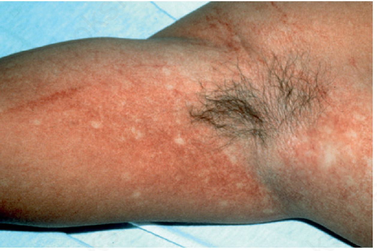
Dengue rash showing the classic pattern. Linear bleeding points are also visible after application of a blood pressure cuff (positive tourniquet test). - Andrews' Diseases of the Skin
Tourniquet test: Inflate BP cuff to midpoint between systolic/diastolic for 5 minutes, wait 2 minutes. ≥10 petechiae per square inch is positive - a useful bedside clue to dengue.
Dengue Hemorrhagic Fever (DHF)
Classic criteria (all four required):
- Fever lasting 2-7 days
- Hemorrhagic tendency - positive tourniquet test, petechiae, ecchymosis, purpura, epistaxis, gingival bleeding, hematemesis, melena
- Thrombocytopenia (platelet count <100,000/mm³)
- Increased vascular permeability - hematocrit rise ≥20%, pleural effusion, ascites, hypoalbuminemia
Dengue Shock Syndrome (DSS): DHF + circulatory shock (narrow pulse pressure ≤20 mmHg, or hypotension). The most severe form - results from sudden massive plasma extravasation into tissues.
Laboratory Findings
| Finding | Notes |
|---|---|
| Leukopenia | Characteristic; helps distinguish from bacterial infection |
| Thrombocytopenia | Platelet <100,000 in ~50% of patients |
| Hemoconcentration | Rising hematocrit signals plasma leakage |
| Elevated LFTs | ~3x normal on average; AST/ALT ≥1000 in severe dengue indicates liver involvement |
| Coagulopathy | Present in DHF/DSS |
| Hyponatremia | Common |
Uncommon/Atypical Manifestations
Less common syndromes include:
- Myocarditis
- Pancreatitis
- Hepatitis (can be severe with fulminant liver failure)
- Hemophagocytic lymphohistiocytosis (HLH)
- Neurological: acute meningoencephalitis, post-dengue ADEM (acute disseminated encephalomyelitis), Guillain-Barre syndrome
- Renal involvement: acute kidney injury (more severe in secondary infections)
Pathophysiology of Severe Dengue
The key mechanism is antibody-dependent enhancement (ADE):
- Infection with one serotype produces lifelong immunity against that serotype only
- Cross-reactive antibodies from a first infection cannot neutralize a different serotype
- These non-neutralizing antibodies enhance uptake of the new serotype into macrophages via Fc receptors - amplifying viral replication and cytokine storm
- Severe dengue predominantly occurs with secondary infection, especially with serotype DENV-2
- Same mechanism explains why dengue is more severe in infants born to dengue-immune mothers (maternal antibodies act similarly)
Key Clinical Pearls
- Helpful bedside predictors: petechiae, skin rash, myalgia, retro-orbital pain, positive tourniquet test, gingival bleeding (validated in a Honduras study)
- Severity risk with secondary infection (especially DENV-2)
- Aspirin and NSAIDs are contraindicated (increase bleeding risk)
- Early aggressive fluid therapy reduces case fatality of severe dengue from 5-10% to <1%
- The critical phase lasts only 24-48 hours - identifying it early is the key to survival
Sources: Red Book 2021 (AAP Committee on Infectious Diseases), p. 520-525 | Rosen's Emergency Medicine, p. 2630 | Robbins & Cotran Pathologic Basis of Disease, p. 336 | Andrews' Diseases of the Skin (Clinical Dermatology), p. 2464-2478
Leptospirosis and it's management and manifestions
Finding Sources
Searching PubMed
"leptospirosis"[MeSH Terms] AND "treatment"
Excellent - strong sources found. Now reading Harrison's, Red Book, the Kidney textbook (for renal manifestations), and Andrews' (skin) in parallel:
Reading File
Reading File
Reading File
Reading File
Reading Article
PMID: 38483092
Excellent - rich data from multiple authoritative sources. Let me get the Goldman-Cecil content for any extra clinical detail:
Reading File
Reading File
I now have comprehensive, multi-source data. Here is the full answer:
Leptospirosis - Manifestations and Management
Overview
Leptospirosis is the most widespread zoonosis globally, caused by pathogenic spirochetes of the genus Leptospira (most notably L. interrogans, species icterohaemorrhagiae). It infects approximately 1 million people per year and causes ~60,000 deaths annually. It is transmitted when leptospires shed in animal urine contaminate water or soil, entering humans through abraded skin, mucous membranes, or conjunctiva.
Reservoir hosts: Rodents (especially brown rat Rattus norvegicus), dogs, cattle, pigs, horses
At-risk groups: Farmers, sewer/abattoir workers, miners, veterinarians, military personnel, adventure travelers, flood-affected populations
Incubation Period
2-26 days (average 10 days; typically 5-14 days)
Clinical Spectrum
~90% of infections are subclinical or mild self-limited illness. About 10% develop severe, potentially fatal disease.
Biphasic Course (Classic Pattern)
Phase 1: Leptospiremic / Septicemic Phase (Days 1-7)
Leptospires are present in the blood and CSF. Features:
- Abrupt-onset high fever with chills and rigors (75-100% of patients)
- Severe myalgias - especially calf and lumbar muscles (40-97%); "myalgia out of proportion to fever" is a hallmark
- Intense headache
- Nausea, vomiting
- Conjunctival suffusion without purulent discharge (28-99%) - a very characteristic finding; non-purulent conjunctival redness
- Pharyngitis, hepatomegaly, splenomegaly, lymphadenopathy
Skin rash:
- Pretibial (Fort Bragg) fever: 1-5 cm erythematous patches/plaques most marked on the shins
- Petechiae and purpura on skin and mucous membranes (in severe/icteric form)
- Facial flushing
After 3-7 days of septicemia, fever may briefly resolve before the second phase.
Phase 2: Immune Phase (Days 7-14+)
The body mounts an IgM antibody response; organisms leave the blood but may persist in aqueous humor, kidney, etc. Features are generally milder:
- Return of fever (often lower-grade)
- Aseptic meningitis - the hallmark of the immune phase (CSF: lymphocytosis)
- Intense headache, photophobia
- Ocular manifestations: conjunctival hemorrhage, uveitis (may appear 4-8 months after illness)
- This phase is less responsive to antibiotics
Severity Categories
1. Mild Anicteric Leptospirosis (~90%)
- Influenza-like illness: fever, headache, myalgias
- Self-limited; resolves in 5-7 days
- Frequently misdiagnosed as dengue, malaria, influenza
2. Weil Syndrome (Severe Icteric Leptospirosis, ~5-10%)
The classic triad:
- Jaundice - marked hyperbilirubinemia (often >30 mg/dL / 512 µmol/L), with only moderately elevated aminotransferases (unlike viral hepatitis where transaminases are proportionally higher)
- Acute Kidney Injury (AKI) - present in 44-67% of hospitalized patients
- Hemorrhage - pulmonary, gastrointestinal, cutaneous, adrenal
Additional features: thrombocytopenia, myocarditis, arrhythmias
Case fatality rate: 5-15%; can rise to >50% in pulmonary hemorrhage syndrome
3. Pulmonary Hemorrhage Syndrome (ARDS-like, "ARDS leptospirosis")
- Most lethal form
- Diffuse alveolar hemorrhage, respiratory failure
- Can occur even without jaundice
- Mortality >50%
- Requires mechanical ventilation with low tidal volumes
4. Meningitis / Meningoencephalitis
- Aseptic meningitis with lymphocytic CSF pleocytosis
- Can mimic bacterial meningitis early on
Organ System Manifestations in Detail
Renal
- Most common severe manifestation; seen in 44-67% of patients
- Mechanism: acute tubulointerstitial nephritis (not glomerulonephritis); leptospires invade tubular cells
- Proximal tubular dysfunction: bicarbonaturia, glycosuria, phosphaturia, magnesuria, uricosuria
- Electrolyte abnormalities: hypokalemia or hyperkalemia, hypomagnesemia (require aggressive correction)
- May be nonoliguric AKI (an important and favorable feature vs. typical ATN)
- Oliguria = poor prognostic sign
- Rhabdomyolysis can further worsen AKI
Hepatic
- Jaundice with markedly elevated bilirubin but only mild-moderate transaminase elevation (unlike viral hepatitis)
- Hyperbilirubinemia >30 mg/dL is characteristic of Weil disease
Pulmonary
- Spectrum: mild cough → pulmonary hemorrhage → ARDS
- Reduced pulmonary compliance; treat with low tidal volume ventilation
Cardiac
- Myocarditis with arrhythmias
- Circulatory collapse / refractory shock
Neurological
- Aseptic meningitis
- Uveitis (late finding, 4-8 months after acute illness)
- Meningoencephalitis
Hematologic
- Thrombocytopenia
- Hemorrhagic tendency (petechiae, ecchymoses, hemoptysis, GI bleeding)
- Leukocytosis (15,000-30,000/mm³) in severe disease
Diagnosis
| Test | Details |
|---|---|
| Microscopic Agglutination Test (MAT) | Gold standard; paired sera 2 weeks apart; 4-fold rise in titer is diagnostic |
| PCR (RT-PCR) | Best in first week (leptospiremic phase); allows earlier diagnosis |
| ELISA (IgM) | More practical than MAT; positive from week 2 |
| Blood/urine cultures | Leptospira can be cultured in EMJH medium; takes weeks |
| Darkfield microscopy | Blood (week 1), urine (week 2); low sensitivity |
| CBC | Leukocytosis, thrombocytopenia |
| LFTs | High bilirubin, mildly elevated transaminases |
| Urinalysis | Proteinuria, hematuria, casts |
| Creatinine | AKI assessment |
Management
Antibiotic Therapy
Treatment should be started as early as possible - early intervention may prevent or reduce organ failure.
| Indication | Regimen | Duration |
|---|---|---|
| Mild leptospirosis | Doxycycline 100 mg PO twice daily | 7 days |
| OR Amoxicillin 500 mg PO three times daily | 7 days | |
| OR Ampicillin 500 mg PO three times daily | 7 days | |
| Moderate/Severe | Penicillin G 1.5 million units IV/IM q6h | 7 days |
| OR Ceftriaxone 2 g IV once daily | 7 days | |
| OR Cefotaxime 1 g IV q6h | 7 days | |
| OR Doxycycline 200 mg IV loading, then 100 mg IV q12h | 7 days |
Notes:
- Doxycycline is contraindicated in pregnant women and children
- In areas where rickettsial diseases are co-endemic, prefer doxycycline or azithromycin
- Azithromycin (3-day course) is an alternative, especially when doxycycline is contraindicated
- No antibiotic resistance reported in clinical isolates to date
- Jarisch-Herxheimer reaction can occur within hours of starting antibiotics (rare)
⚠️ Recent Evidence Note: A 2024 Cochrane systematic review (PMID: 38483092) found that the evidence for antibiotics in leptospirosis remains of very low certainty due to small trial numbers and methodological limitations. The review could not confirm a statistically significant mortality benefit for penicillin vs. placebo. Despite this uncertainty, antibiotics are still recommended given the plausibility of benefit and the severity of the disease. A 2025 Cochrane review (PMID: 40704556) found insufficient evidence to recommend corticosteroids in leptospirosis.
Supportive Care (Essential - Can Be Life-Saving)
Renal:
- Aggressive fluid and electrolyte resuscitation for nonoliguric AKI (prevent progression to oliguria)
- Correct hypokalemia, hypomagnesemia aggressively
- Hemodialysis or peritoneal dialysis for oliguric renal failure - rapid initiation reduces mortality; usually needed only for short periods
- Monitor for fluid overload during recovery
Pulmonary:
- Mechanical ventilation with low tidal volumes (lung-protective strategy, as in ARDS) for pulmonary hemorrhage
- Evidence for glucocorticoids and desmopressin as adjunct therapy is contradictory - not routinely recommended
Hemorrhage:
- Blood product transfusions as needed for severe bleeding
Monitoring:
- Watch for early signs of shock, occult bleeding, plasma leak
- Monitor hematocrit, renal function, electrolytes closely
Chemoprophylaxis
| Setting | Regimen |
|---|---|
| Endemic/epidemic area travelers | Doxycycline 200 mg PO once weekly |
| Alternative (doxycycline contraindicated) | Azithromycin 250 mg PO once or twice weekly |
Key Clinical Pearls
- Conjunctival suffusion (non-purulent) + severe calf myalgias + fever in a flood-exposed or occupationally at-risk patient = leptospirosis until proven otherwise
- AKI in leptospirosis is often nonoliguric and reversible with adequate fluid resuscitation - do not mistake it for a late/terminal finding
- Leptospirosis is frequently misdiagnosed as dengue, malaria, or influenza - a high index of suspicion is needed
- The immune phase is less antibiotic-responsive; supportive care is paramount then
- Uveitis can appear months after apparent recovery - patients should be warned
Sources: Harrison's Principles of Internal Medicine 22E (2025), p. 1487 | Red Book 2021, p. 766-768 | Brenner & Rector's The Kidney (Comprehensive Clinical Nephrology), p. 2993-3012 | Goldman-Cecil Medicine, p. 2416-2434 | Andrews' Diseases of the Skin, p. 1398-1416 | Cochrane 2024 - Antibiotics for Leptospirosis, PMID: 38483092 | Cochrane 2025 - Corticosteroids for Leptospirosis, PMID: 40704556
This is a shared conversation. Sign in to Orris to start your own chat.