
What causes the formation of concentration of urine explain the physiology of ccm in very easy word
Concentration of Urine: Physiology of the Countercurrent Mechanism (CCM)
The Big Picture (Why Does Urine Get Concentrated?)
- A special region in the kidney (the renal medulla) that is kept extremely salty/concentrated - up to 1200-1400 mOsm/L (compared to normal blood at 300 mOsm/L)
- A hormone called ADH (antidiuretic hormone) that opens water channels in the collecting duct
What Does "Countercurrent" Mean?
The Three Players in CCM
1. The Loop of Henle (the Multiplier)
| Part | Water Permeable? | Active Pumping? |
|---|---|---|
| Descending limb | YES - water flows out freely | No |
| Ascending limb (thick) | NO - water cannot escape | YES - pumps Na+, K+, Cl- out |
2. The Vasa Recta (the Exchanger)
3. The Collecting Duct (the Final Concentrator)
How CCM Works Step-by-Step (Very Simple)
The Final Step - ADH and Collecting Duct
- No ADH present → water channels (AQP-2) stay closed → water cannot leave → dilute urine is produced (like when you drink lots of water)
- ADH present (e.g., dehydration) → AQP-2 channels open → water rushes out of the collecting duct into the salty medulla → small volume of concentrated urine is produced
Summary Table
| Structure | Role in CCM |
|---|---|
| Thick ascending limb | Pumps NaCl out, impermeable to water → builds medullary gradient |
| Thin descending limb | Freely permeable to water → fluid concentrates as it descends |
| Vasa recta | Hairpin capillaries that preserve the gradient while removing reabsorbed water |
| Collecting duct | Final site of water reabsorption (only when ADH opens AQP-2 channels) |
| Urea recycling | Contributes ~40-50% of inner medullary osmolarity |
Simple Analogy
- Workers (the ascending limb) keep throwing salt DOWN the hill into the surrounding ground
- Rain water (descending limb fluid) soaks up the salt as it flows down
- The deeper you go, the saltier the ground
- A final tap (collecting duct) opens when the boss (ADH) gives the order, letting water seep into the salty ground and concentrating the remaining urine
- Guyton and Hall Textbook of Medical Physiology, pp. 373-376 (Chapter 29 - Countercurrent Multiplier Mechanism)
- Histology: A Text and Atlas with Correlated Cell and Molecular Biology, p. 1915 (Histophysiology of the Kidney)
H and hv3 ion handling by renal tubules easy words
Explain the role of the kidney in maintaining acid base balance and explain the mechanism of acidification of urine 5 marks invery easy words
Kidney's Role in Acid-Base Balance + Mechanism of Urine Acidification
First - Why Does the Kidney Need to Do This?
- Lungs handle CO₂ (carbonic acid) - fast response (seconds to minutes)
- Kidneys handle the rest - slow but powerful (hours to days), and they are the final defence
The 3 Main Ways the Kidney Controls Acid-Base
1. 🔁 Reclaiming Bicarbonate (HCO₃⁻) - "Save the Base"
Step 1: Tubular cell secretes H⁺ into the tubule lumen via the Na⁺/H⁺ exchanger (NHE3) Step 2: This H⁺ meets the filtered HCO₃⁻ in the lumen Step 3: They combine → H₂CO₃ → then carbonic anhydrase breaks it down into CO₂ + H₂O Step 4: CO₂ drifts back INTO the tubular cell Step 5: Inside the cell, carbonic anhydrase converts CO₂ + H₂O → H₂CO₃ → H⁺ + HCO₃⁻ Step 6: HCO₃⁻ goes back into the blood ✅, H⁺ is secreted again ✅
2. 🧪 Ammonia (NH₃) Production - "Make New Bicarbonate"
Step 1: Tubular cells break down the amino acid glutamine (from muscles/liver) Step 2: This produces NH₃ (ammonia) + α-ketoglutarate Step 3: NH₃ is a gas - it diffuses into the tubule lumen Step 4: In the acidic urine, NH₃ + H⁺ → NH₄⁺ (ammonium) - which is TRAPPED and excreted Step 5: α-ketoglutarate is converted to new HCO₃⁻ which goes back into blood
3. 🧱 Phosphate Buffer - "Titratable Acid"
HPO₄²⁻ + H⁺ → H₂PO₄⁻ (excreted in urine)
Mechanism of Acidification of Urine - Step by Step
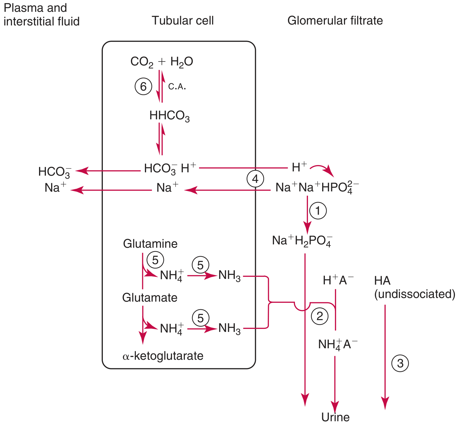
| Step | What Happens | Where |
|---|---|---|
| 1 | Tubular cells make CO₂ + H₂O → H₂CO₃ → H⁺ + HCO₃⁻ (via carbonic anhydrase) | All tubular cells |
| 2 | H⁺ is pumped OUT into the lumen via NHE3 (Na⁺/H⁺ exchanger) or H⁺-ATPase | Proximal + collecting duct |
| 3 | Luminal H⁺ reacts with HCO₃⁻ → CO₂ (reclaimed) OR with NH₃ → NH₄⁺ (excreted) OR with HPO₄²⁻ → H₂PO₄⁻ (excreted) | Lumen |
| 4 | HCO₃⁻ generated inside cell returns to blood via NBC transporter | Basolateral side |
| 5 | Net result: Urine becomes acidic, blood becomes more alkaline | Final urine pH as low as 4.4 |
- Proximal tubule: can maintain up to 1 pH unit gradient
- Distal tubule/collecting duct: can maintain up to 3 pH units gradient
- Minimum urine pH ≈ 4.4
Role Segment by Segment
| Tubule Segment | Job |
|---|---|
| Proximal tubule | Reabsorbs 90% of filtered HCO₃⁻, secretes large amounts of H⁺ via NHE3 |
| Thick ascending limb | Reabsorbs some HCO₃⁻ |
| Distal tubule | Secretes H⁺ via H⁺-ATPase, fine-tunes acid secretion |
| Collecting duct (α-intercalated cells) | Major site of final acidification - secretes H⁺ by H⁺-ATPase pump, generates new HCO₃⁻ |
Summary - "What the Kidney Does in Acidosis vs Alkalosis"
| Body State | Kidney Response |
|---|---|
| Acidosis (blood too acidic) | Secretes more H⁺, produces more NH₃, reclaims all HCO₃⁻ → urine becomes very acidic (pH ~4.5) |
| Alkalosis (blood too basic) | Stops secreting H⁺, lets HCO₃⁻ spill into urine → urine becomes alkaline (pH ~8.0) |
One-Line Memory Aid
"The kidney saves base (HCO₃⁻), dumps acid (H⁺ via phosphate and ammonia), and is the LAST and most powerful controller of blood pH."
- Tietz Textbook of Laboratory Medicine, 7th Edition, pp. 1950-1951 (Renal Mechanisms in Acid-Base Balance)
- Brenner and Rector's The Kidney, p. 365 (Bicarbonate Reabsorption and H⁺ Secretion)
Describe the physiological of micturtion also mention the significance of cytometrogram easyyyy word
Physiology of Micturition + Significance of Cystometrogram
What is Micturition?
Parts of the Bladder You Need to Know
| Structure | Job |
|---|---|
| Detrusor muscle | Smooth muscle of the bladder wall - contracts to push urine out |
| Internal urethral sphincter | Smooth muscle, involuntary - normally CLOSED |
| External urethral sphincter | Skeletal muscle, voluntary - you control this one |
| Stretch receptors | Sensors in the bladder wall that feel how full it is |
Phase 1: FILLING (Storage Phase)
- Urine drips into the bladder from the ureters continuously
- The bladder wall is elastic - it stretches and relaxes to accommodate more urine without much rise in pressure (this is called "compliance")
- Up to about 150 mL - you feel a slight urge
- At 300-400 mL - you feel a definite urge
- At >400 mL - pressure rises sharply and the urge becomes urgent
- Sympathetic nervous system keeps the detrusor relaxed and internal sphincter CLOSED
- Voluntary control keeps the external sphincter CLOSED
- The brain says "not yet!" - it actively inhibits the micturition reflex
Phase 2: THE MICTURITION REFLEX (The Trigger)
Bladder stretch receptors
↓ (pelvic nerves - afferent signals)
Sacral spinal cord (S2, S3, S4) ← THE MICTURITION CENTER
↓ (pelvic nerves - efferent signals)
Parasympathetic activation
↓
Detrusor muscle CONTRACTS
↓
Pressure rises → more stretch → more stretch receptor firing
↓ (self-regenerative cycle)
Strong sustained contraction
Phase 3: VOIDING (Emptying Phase)
Step 1: Cerebral cortex stops inhibiting the sacral micturition center Step 2: Person voluntarily relaxes the external sphincter (you control this) Step 3: Pontine Micturition Center (PMC) in brainstem coordinates everything Step 4: Internal sphincter also relaxes Step 5: A small bit of urine enters the posterior urethra → triggers MORE stretch receptors → makes the detrusor contraction even stronger Step 6: Detrusor contracts powerfully → urine is expelled Step 7: Abdominal muscles may also contract voluntarily to help
Neural Control Summary - "Who Controls What"
| Brain Center | Role |
|---|---|
| Cerebral cortex | Voluntary control - can STOP or START urination |
| Pontine Micturition Center (PMC) | Coordinates detrusor contraction + sphincter relaxation simultaneously |
| Sacral cord (S2-S4) | The actual reflex center - parasympathetic outflow |
| Sympathetic (T10-L2) | Storage phase - relaxes detrusor, closes internal sphincter |
| Pudendal nerve | Controls external sphincter (voluntary) |
Key rule: The detrusor contracts at the SAME TIME the sphincters relax - these must be perfectly coordinated. If they don't coordinate (called "detrusor-sphincter dyssynergia"), urination fails.
What is a Cystometrogram (CMG)?
The Normal Cystometrogram:
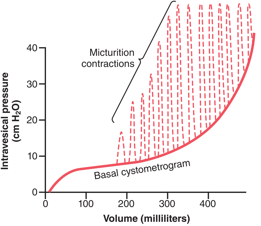
| Volume | What the Graph Shows | What it Means |
|---|---|---|
| 0 - 50 mL | Slight initial pressure rise | Bladder wall is slightly tense at first |
| 50 - 300 mL | Flat/very slowly rising line | Detrusor relaxes, bladder is very compliant - this is the TONIC segment |
| 300 - 400 mL | Pressure starts rising | Bladder is getting full, compliance decreasing |
| >400 mL | Sharp rise in pressure | Bladder is overfull and struggling |
| Dashed spikes | Sudden sharp pressure peaks (micturition waves) | These are the micturition reflex contractions - each spike = one attempted voiding reflex |
Significance of the Cystometrogram
| CMG Finding | What it Means | Clinical Condition |
|---|---|---|
| High pressure at low volumes (steep curve) | Bladder is stiff/non-compliant | Fibrotic bladder, neurogenic bladder |
| Absent micturition spikes | Detrusor cannot contract | Atonic/flaccid bladder (e.g., lower motor neuron lesion, tabes dorsalis) |
| Early and large micturition spikes at low volumes | Detrusor is overactive/irritable | Overactive bladder, upper motor neuron lesion |
| Normal curve but no voiding | Sphincter not relaxing | Detrusor-sphincter dyssynergia |
| Normal CMG | Bladder function intact | Rules out neurogenic bladder |
In one line:
The cystometrogram is the "ECG of the bladder" - it tells us whether the bladder fills normally, feels sensation at the right volume, and contracts at the right time.
One-Line Summary of Micturition
Bladder fills → stretch receptors fire → sacral cord reflex → detrusor contracts + sphincters relax (coordinated by pons) → urine expelled → all under voluntary cortical control.
- Guyton and Hall Textbook of Medical Physiology, pp. 332-334 (Micturition Reflex & Cystometrogram)
- Medical Physiology (Boron & Boulpaep), p. 737 (Cystometrogram & Micturition Reflex Control)
Late distal tubules and collecting tubules contain two types of intercalated cells
Two Types of Intercalated Cells in Late Distal Tubule and Collecting Duct
First - What Are Intercalated Cells?
| Cell Type | Main Job |
|---|---|
| Principal cells | Reabsorb Na⁺ and water, secrete K⁺ (aldosterone-controlled) |
| Intercalated cells | Handle H⁺ and HCO₃⁻ - the acid-base specialists |
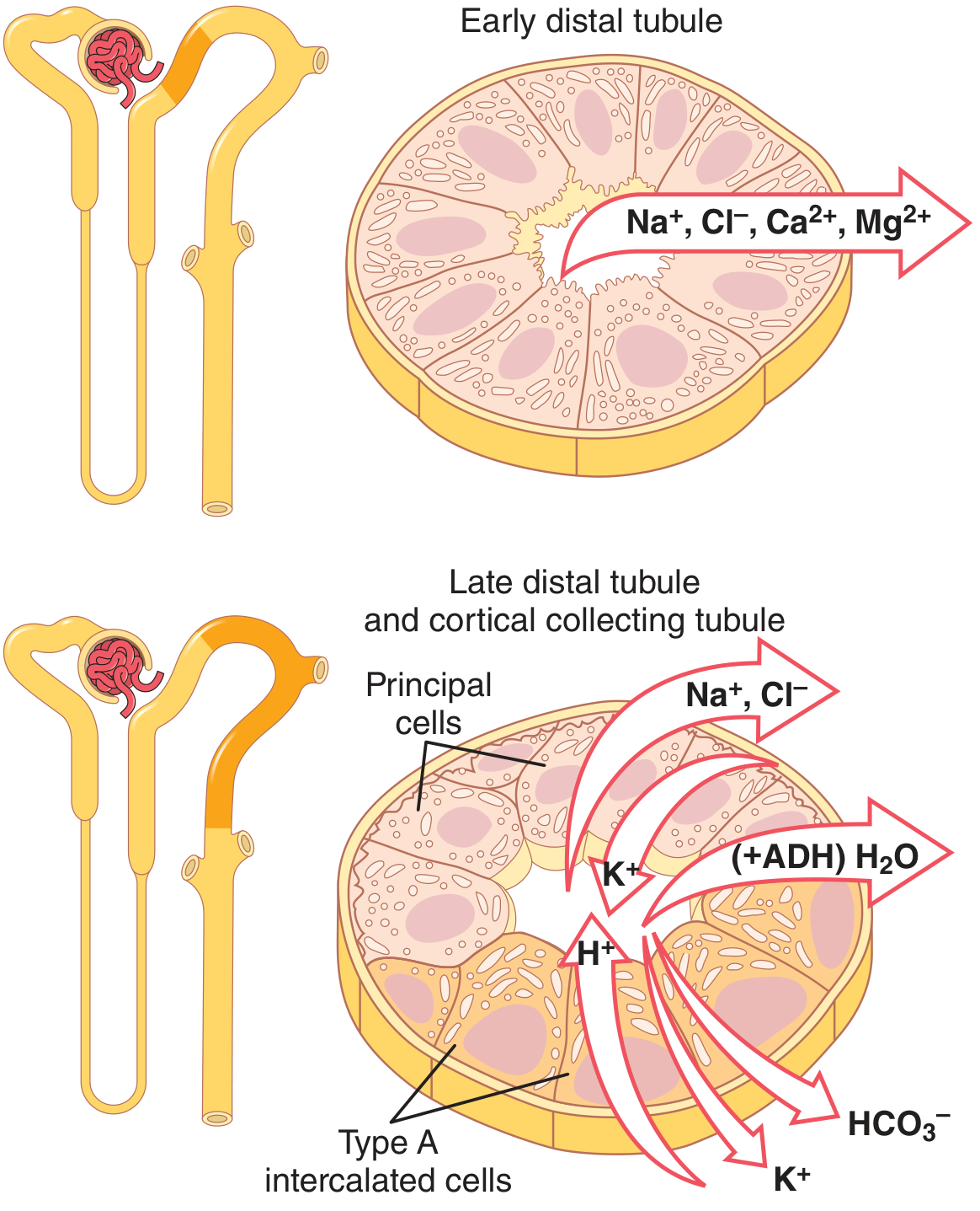
TYPE A Intercalated Cell - "The Acid Secretor"
What it does:
- Secretes H⁺ (acid) INTO the tubular lumen → excreted in urine
- Reabsorbs HCO₃⁻ (base) back into the blood
- Reabsorbs K⁺ from the lumen
How it works (step by step):
Step 1: CO₂ enters the cell from blood Step 2: Inside cell: CO₂ + H₂O → H₂CO₃ (via carbonic anhydrase) → H⁺ + HCO₃⁻ Step 3: H⁺ is pumped OUT into the tubular lumen by two pumps on the APICAL (luminal) side:
- H⁺-ATPase (proton pump) - most important
- H⁺-K⁺-ATPase (exchanges H⁺ out for K⁺ in) Step 4: HCO₃⁻ exits on the BASOLATERAL side (blood side) via a Cl⁻/HCO₃⁻ exchanger → goes back to blood ✅
The Key Pumps: ON THE APICAL (LUMEN) SIDE
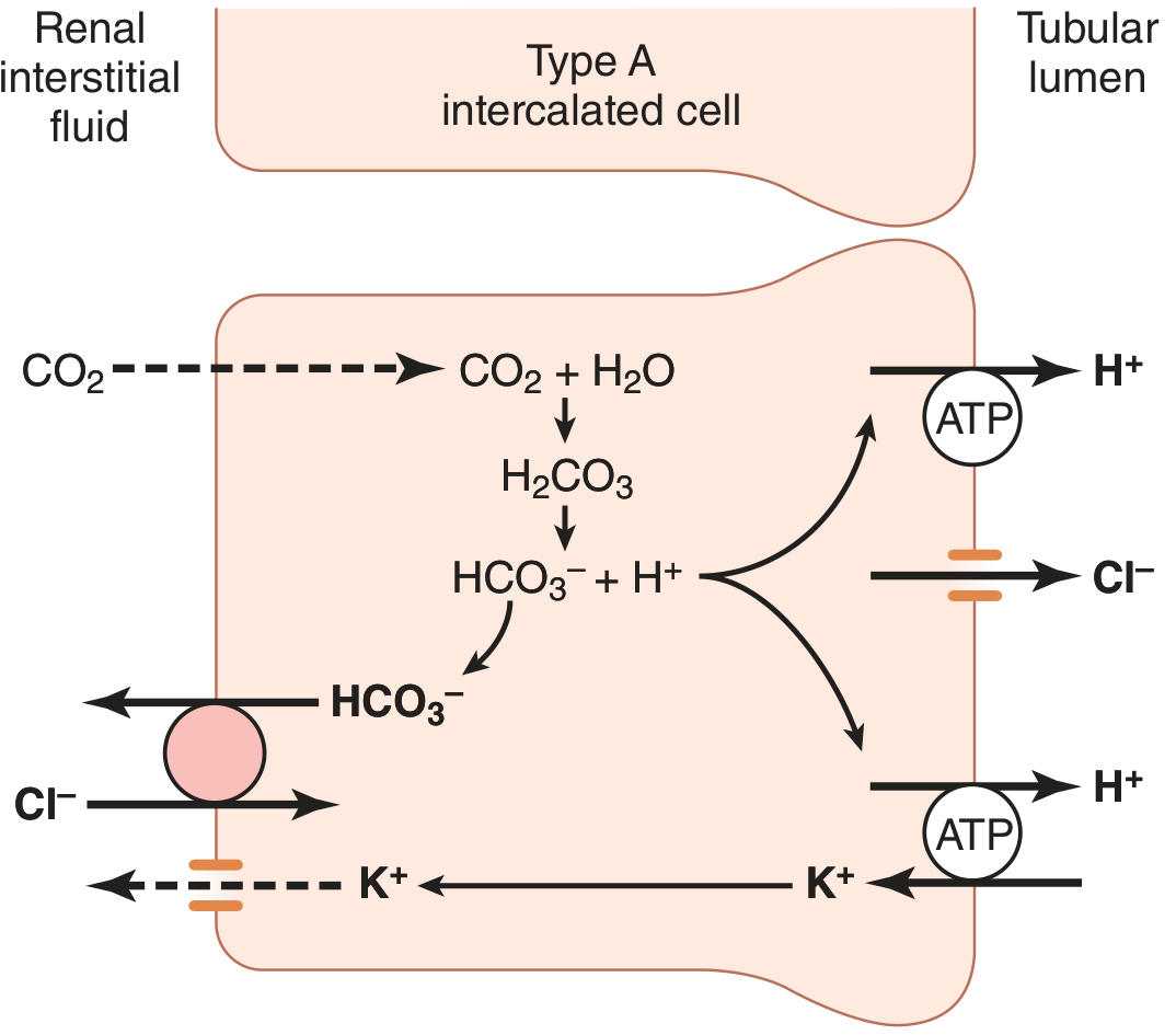
Memory trick for Type A: A = Acid out → H⁺ pumped into LUMEN. "A for Acid secretor, Apical ATPase."
Special feature of Type A:
- Can create a H⁺ concentration gradient of up to 1000:1 between cell and lumen
- This is MUCH more powerful than the proximal tubule (which can only do 4-10:1)
- This allows final maximum acidification of urine (pH as low as 4.4)
TYPE B Intercalated Cell - "The Bicarbonate Secretor"
What it does - EXACTLY OPPOSITE of Type A:
- Secretes HCO₃⁻ (base) INTO the tubular lumen → excreted in urine
- Reabsorbs H⁺ back into the blood
- Secretes K⁺ into the lumen
How it works (step by step):
Step 1: CO₂ enters the cell → same reaction → H⁺ + HCO₃⁻ formed inside Step 2: H⁺-ATPase is on the BASOLATERAL side now (opposite to Type A!) → pumps H⁺ INTO the blood Step 3: HCO₃⁻ is secreted into the LUMEN via a special transporter called PENDRIN (Cl⁻/HCO₃⁻ exchanger on apical membrane) Step 4: Pendrin brings Cl⁻ in and pushes HCO₃⁻ out into the tubule → excreted in alkaline urine ✅
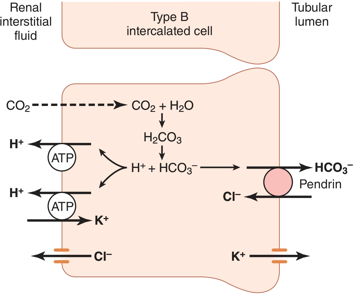
Memory trick for Type B: B = Base (HCO₃⁻) secreted into lumen. B for Bicarbonate secretor, Basolateral ATPase.
Side-by-Side Comparison Table
| Feature | Type A Intercalated Cell | Type B Intercalated Cell |
|---|---|---|
| Active in | Acidosis | Alkalosis |
| H⁺-ATPase location | APICAL (lumen side) | BASOLATERAL (blood side) |
| Cl⁻/HCO₃⁻ exchanger | Basolateral (anion exchanger 1) | Apical (PENDRIN) |
| H⁺ movement | Secreted INTO lumen | Reabsorbed into blood |
| HCO₃⁻ movement | Reabsorbed into blood | Secreted INTO lumen |
| K⁺ movement | Reabsorbed from lumen | Secreted into lumen |
| Net effect on urine | Urine becomes MORE ACIDIC | Urine becomes MORE ALKALINE |
| Net effect on blood | Blood pH rises (corrects acidosis) | Blood pH falls (corrects alkalosis) |
| Numbers (CCD) | ~60% of intercalated cells | ~40% of intercalated cells |
How the Body Adjusts the Numbers
- In chronic acidosis → more Type A cells are made → more H⁺ secretion
- In chronic alkalosis → more Type B cells are made → more HCO₃⁻ secretion
Clinical Relevance
| Disease | Problem |
|---|---|
| Distal RTA (Type 1) | Type A cells CANNOT secrete H⁺ properly → urine can't be acidified below pH 5.5, blood becomes acidic |
| Metabolic alkalosis | Type B cells activated → secrete excess HCO₃⁻ in urine to correct it |
Super Simple Summary
Type A = "Acid out, Base in" → fights ACIDOSIS (H⁺ ATPase on apical/lumen side)Type B = "Base out, Acid in" → fights ALKALOSIS (Pendrin on apical/lumen side, H⁺ ATPase on basolateral/blood side)Both cells generate H⁺ and HCO₃⁻ inside using carbonic anhydrase - they just send them in opposite directions!
- Guyton and Hall Textbook of Medical Physiology, pp. 357-358 (Late Distal Tubules and Collecting Tubules - Figure 28.13)
- Brenner and Rector's The Kidney, pp. 109-110 (Cortical Collecting Duct - Intercalated Cell Subtypes)
Easyyy word
Two Types of Intercalated Cells - Super Easy Version
The Big Idea First
Think of It Like a Water Pump with Two Modes
- In Mode A → pump pushes ACID out into urine
- In Mode B → pump pushes BASE out into urine
🔴 TYPE A Cell - "The Acid Dumper"
When does it turn ON?
What does it do?
It throws H⁺ (acid) out into the urine It pulls HCO₃⁻ (base) back into the blood
How? (Very simple version)
Inside the cell:
CO₂ + H₂O → H⁺ + HCO₃⁻
↓ ↓
PUMPED OUT Goes to BLOOD
into URINE (saves it)
(via H⁺-ATPase
on LUMEN side)
One Line Memory:
"A = Acid out through the Apical (lumen) side"
🔵 TYPE B Cell - "The Base Dumper"
When does it turn ON?
What does it do?
It throws HCO₃⁻ (base) out into the urine It pulls H⁺ (acid) back into the blood
How? (Very simple version)
Inside the cell:
CO₂ + H₂O → H⁺ + HCO₃⁻
↓ ↓
Goes to BLOOD PUMPED OUT
(saves it) into URINE
(via PENDRIN
on LUMEN side)
One Line Memory:
"B = Base (HCO₃⁻) out through the lumen via Pendrin"
The Trick to Remember - They Are Mirror Images!
| Type A | Type B | |
|---|---|---|
| Blood too...? | Acidic | Alkaline |
| Dumps into urine | H⁺ (acid) | HCO₃⁻ (base) |
| Saves for blood | HCO₃⁻ (base) | H⁺ (acid) |
| Pump location | Lumen side (apical) | Blood side (basolateral) |
| Special transporter | Cl⁻/HCO₃⁻ exchanger (basolateral) | Pendrin (lumen side) |
The Simplest Analogy Ever 🎯
- Type A bin collects and throws away acid trash (H⁺) into the urine road
- Type B bin collects and throws away base trash (HCO₃⁻) into the urine road
Both Cells Use the SAME Machine Inside
Which direction they push things - it's the same factory, different exit doors!
Disease Connection (Bonus - Easy!)
- Type A cell broken? → Can't throw away acid → Blood stays acidic → Distal Renal Tubular Acidosis (Type 1 RTA)
- Too much alkalosis? → Type B cells increase in number → dump more base → fixes it
3-Word Summary
| Type A | Type B |
|---|---|
| "Acid → Urine" | "Base → Urine" |
In summary
Summary: Two Types of Intercalated Cells
The One Table You Need
| Type A | Type B | |
|---|---|---|
| Activated by | Acidosis (blood too acidic) | Alkalosis (blood too alkaline) |
| Secretes into urine | H⁺ (acid) | HCO₃⁻ (base) |
| Returns to blood | HCO₃⁻ (base) | H⁺ (acid) |
| H⁺-ATPase pump sits on | Apical (lumen) side | Basolateral (blood) side |
| Special transporter | Cl⁻/HCO₃⁻ exchanger (basolateral) | Pendrin (apical/lumen) |
| Net effect on urine | Acidic urine | Alkaline urine |
| Net effect on blood | Corrects acidosis | Corrects alkalosis |
One Line Each
🔴 Type A = Blood too acidic → dumps H⁺ into urine, saves HCO₃⁻ for blood. Pump on lumen side.
🔵 Type B = Blood too alkaline → dumps HCO₃⁻ into urine via Pendrin, saves H⁺ for blood. Pump on blood side.
The Golden Memory Trick
| A | Acid out, Apical pump |
| B | Base out, Basolateral pump, Pendrin |
Both cells use carbonic anhydrase inside to make H⁺ and HCO₃⁻ from CO₂ - they just send them in OPPOSITE directions.
Renal hypertension easyyy worddd
Renal Hypertension - Super Easy Words
What Is It?
Simple idea: "Kidney feels low pressure → Kidney calls for help → Blood pressure goes UP everywhere"
Why Does the Kidney Cause High BP?
Kidney = a security guard for blood pressure When blood flow to the kidney drops, the guard shouts: "Emergency! Send more blood pressure!"
The Step-by-Step Mechanism (RAAS Explained Simply)
Kidney gets LESS blood flow
↓
Kidney releases RENIN (a chemical alarm)
↓
Renin converts Angiotensinogen → Angiotensin I (in blood)
↓
ACE (in lungs) converts Angiotensin I → Angiotensin II
↓
Angiotensin II does TWO things:
1. SQUEEZES blood vessels → BP rises immediately (within minutes)
2. Tells adrenal gland to release ALDOSTERONE
↓
Aldosterone tells kidneys to HOLD ON to Na+ and water
↓
More water in blood = More blood volume = BP rises more (over days)
The Famous Goldblatt Experiment - Easy Version
He placed a clamp on the renal artery of a dog → Less blood reached the kidney → The dog developed HIGH blood pressure!
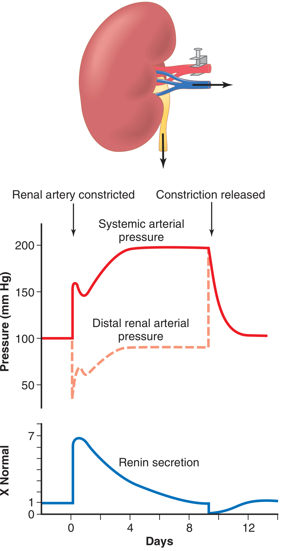
- Red solid line = Systemic (whole body) blood pressure - shoots UP
- Red dashed line = Pressure in the kidney after the clamp - drops first, then slowly recovers
- Blue line = Renin secretion - spikes very high at first, then comes back down
| Phase | Time | Cause |
|---|---|---|
| Phase 1 - Fast | Within hours | Angiotensin II squeezes blood vessels |
| Phase 2 - Slow | Over 5-7 days | Salt and water retention raises blood volume |
Types of Renal Hypertension
1. 🔴 One-Kidney Goldblatt Hypertension
- One kidney is present + its artery is clamped/narrowed
- That single kidney keeps retaining salt and water
- BP stays high permanently until clamp removed
- Clinical example: Single kidney transplant with renal artery stenosis
2. 🟠 Two-Kidney Goldblatt Hypertension
- Both kidneys present, but artery to ONE is narrowed (e.g., atherosclerosis)
- The ischemic kidney releases renin
- Renin and angiotensin II travel to the NORMAL kidney and ALSO make it retain salt
- Both kidneys now hold on to salt → BP rises
- Clinical example: Renal artery stenosis (most common cause of renal hypertension)
3. 🟡 Patchy Kidney Disease
- Small damaged/scarred patches inside one or both kidneys
- These patches feel ischemic → keep releasing renin
- Rest of kidney retains salt
- Common in older people
- Example: Chronic kidney disease, multiple small infarcts
4. 🟢 Coarctation of Aorta
- Aorta is narrowed above the renal arteries (congenital defect in babies)
- Kidneys receive LOW pressure → release renin → BP rises in upper body
- Upper body BP very high, lower body BP normal
- Easy to diagnose: High BP in arms, low BP in legs
Common Real-Life Causes
| Cause | What Happens |
|---|---|
| Renal artery stenosis (atherosclerosis) | Artery to kidney narrows → kidney ischemia → renin release |
| Fibromuscular dysplasia | Artery wall thickens in young women → same effect |
| Chronic kidney disease | Damaged kidney tissue secretes renin |
| Renin-secreting tumor | JGA tumor constantly releases renin |
| Coarctation of aorta | Aorta narrowed → kidneys feel low pressure |
| Polycystic kidney disease | Cysts compress kidney arteries internally |
How Is It Different From Essential Hypertension?
| Essential (Primary) HTN | Renal HTN | |
|---|---|---|
| Cause | Unknown (genetic + lifestyle) | Known - kidney problem |
| Age | Usually middle-aged adults | Any age |
| Response to ACE inhibitors | Partial | Very good - since RAAS is the main driver |
| Curable? | No (manageable) | Often YES - fix the kidney problem → BP normalizes |
Treatment (Easy Version)
| Treatment | How it Helps |
|---|---|
| ACE inhibitors (e.g., enalapril) | Block conversion of Ang I → Ang II → less vasoconstriction |
| ARBs (e.g., losartan) | Block Angiotensin II receptors directly |
| Renal artery stenting/angioplasty | Open up the narrowed artery → restore blood flow → renin falls → BP normalizes |
| Surgery (bypass or nephrectomy) | For severe cases |
One Line to Remember Everything
"Kidney gets less blood → panics → releases Renin → Angiotensin II squeezes vessels + Aldosterone holds salt/water → Blood pressure rises throughout the body = Renal Hypertension"
- Guyton and Hall Textbook of Medical Physiology, pp. 244-246 (Goldblatt Hypertension & RAAS)
- Medical Physiology - Boron & Boulpaep, p. 1255 (Box 40-2 Renal Hypertension)
Enteric nervous system in veryy simple word
Enteric Nervous System (ENS) - Very Easy Words
What Is the ENS?
Fun fact: The ENS has more than 100 million nerve cells - that's more neurons than the entire spinal cord! This is why it is called the "Second Brain" or "Little Brain".
Where Is It?
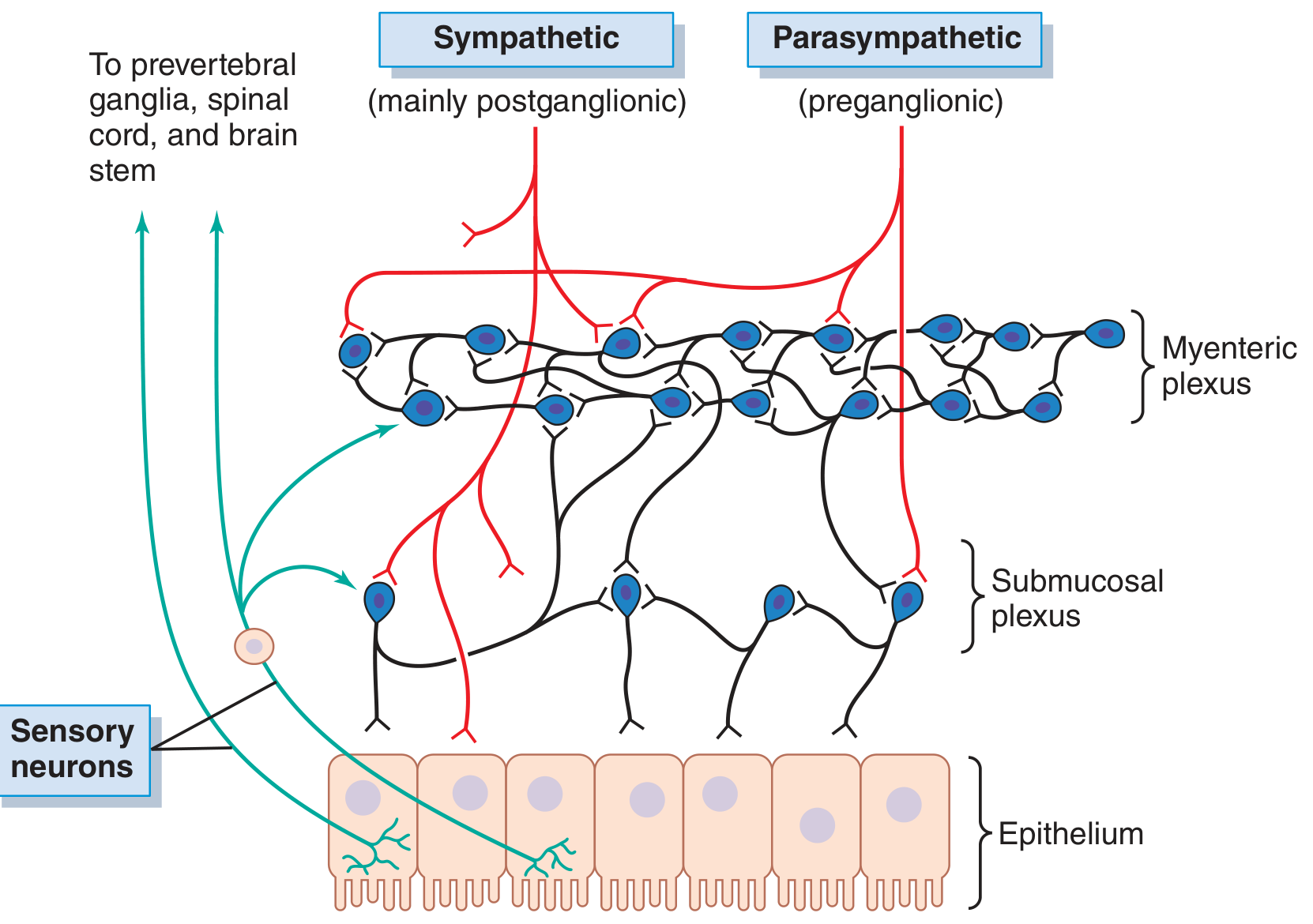
The Two Plexuses - "Two Networks"
1. 🟠 Myenteric Plexus (Auerbach's Plexus)
- Location: Between the two muscle layers (outer longitudinal + inner circular muscle)
- Job: Controls MOVEMENT of the gut - squeezing, pushing food along (peristalsis)
- Simple memory: "Myo = Muscle → Myenteric controls Movement"
- Increases muscle tone (gut stays firm)
- Makes contractions stronger and faster
- Speeds up peristaltic waves (the wave that pushes food forward)
- Also relaxes sphincters (like the pyloric sphincter) so food can pass through
2. 🔵 Submucosal Plexus (Meissner's Plexus)
- Location: In the submucosa (inner layer, close to the gut lining)
- Job: Controls SECRETION - how much juice, enzymes, and mucus the gut produces + local blood flow
- Simple memory: "Sub = underneath the mucosa → controls Secretion"
Summary of the Two Plexuses
| Myenteric Plexus | Submucosal Plexus | |
|---|---|---|
| Other name | Auerbach's plexus | Meissner's plexus |
| Location | Between muscle layers | In submucosa (inner) |
| Main job | Movement / Motility | Secretion + Blood flow |
| Controls | Peristalsis, sphincters | Gland secretion, absorption |
Three Types of Neurons Inside ENS
| Neuron Type | Job | Simple Analogy |
|---|---|---|
| Sensory neurons | Detect stretch, chemicals, temperature in gut | Spies - report what's inside the gut |
| Interneurons | Connect and process the information | Managers - decide what to do |
| Motor neurons | Send commands to muscles and glands | Workers - actually squeeze or secrete |
Does the ENS Need the Brain?
- Move food (peristalsis)
- Secrete digestive juices
- React to food arriving
| System | Effect on Gut | Easy Analogy |
|---|---|---|
| Parasympathetic (Vagus nerve = Rest & Digest) | INCREASES gut activity - more movement, more secretion | "Boss says GO! - speed up digestion" |
| Sympathetic (Fight or Flight) | DECREASES gut activity - slows everything down | "Emergency! Stop digesting, run!" |
Key Neurotransmitters (Chemical Messengers) - Easy List
| Chemical | Effect |
|---|---|
| Acetylcholine | EXCITES gut - more movement and secretion |
| Norepinephrine | INHIBITS gut - slows everything |
| Serotonin (5-HT) | Triggers peristalsis (90% of body's serotonin is in the GUT!) |
| Nitric oxide (NO) | Relaxes smooth muscle - relaxes sphincters |
| VIP (Vasoactive Intestinal Peptide) | Relaxes muscle, increases secretion |
| Substance P | Excites movement |
What Does the ENS Actually Control?
| Function | How ENS Helps |
|---|---|
| Peristalsis | Myenteric plexus coordinates wave contractions to push food forward |
| Sphincter control | Opens/closes pyloric, ileocecal, and anal sphincters at the right time |
| Secretion of enzymes, mucus, water | Submucosal plexus tells glands when and how much to secrete |
| Blood flow to gut | Increases blood flow when digestion is active |
| Absorption | Coordinates how much is absorbed from the gut lining |
| Local reflexes | e.g., distension in one area automatically triggers peristalsis without asking the brain |
The "Gut-Brain Axis" (Bonus Easy Concept)
Brain ←→ Vagus Nerve ←→ ENS (Gut)
- Brain → Gut: Stress makes your gut speed up (nervous diarrhea!) or slow down
- Gut → Brain: Gut produces serotonin, which affects your mood
Diseases When ENS Goes Wrong
| Disease | ENS Problem |
|---|---|
| Hirschsprung disease | No ENS neurons in part of colon → that part cannot relax → severe constipation in babies |
| Irritable Bowel Syndrome (IBS) | ENS hypersensitive → overreacts to normal stimuli |
| Gastroparesis | ENS in stomach damaged (e.g., diabetes) → stomach doesn't empty properly |
| Achalasia | Loss of inhibitory neurons in esophagus → lower esophageal sphincter doesn't open |
One-Line Summary
"The ENS is a complete nervous system (100 million neurons!) inside the gut wall. It has two networks - Myenteric plexus (controls movement) and Submucosal plexus (controls secretion). It works independently but also receives instructions from the brain via parasympathetic (speeds up) and sympathetic (slows down) nerves."
- Guyton and Hall Textbook of Medical Physiology, pp. 778-779 (Neural Control of GI Function - ENS)
- Ganong's Review of Medical Physiology, 26th Edition, p. 274 (Enteric Nervous System)
Fight and flight
Fight or Flight Response - Very Easy Words
What Is It?
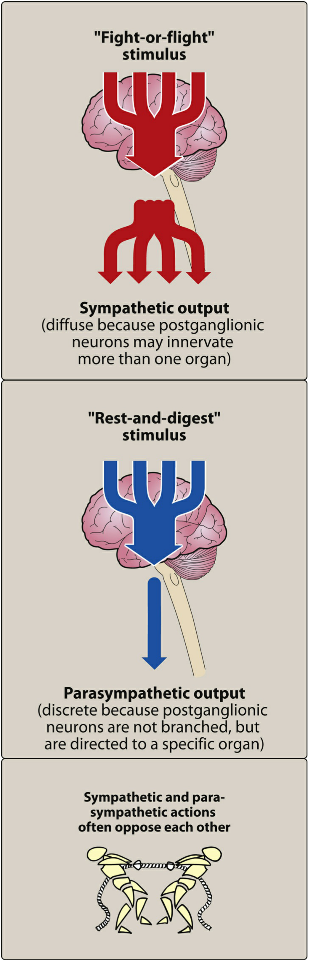
Who Triggers It?
Danger/Fear/Stress
↓
Hypothalamus in brain sends ALARM signal
↓
Sympathetic Nervous System activates EVERYWHERE at once
↓
Adrenal Medulla releases EPINEPHRINE (Adrenaline) into blood
↓
Every organ in your body responds in seconds!
What Happens to Each Organ? (Body-by-Body)
🫀 Heart
| Normal | Fight or Flight |
|---|---|
| 60-80 beats/min | Goes UP - 100-180 beats/min |
| Normal force | Pumps HARDER |
Why? Muscles need more blood to fight or run.
🫁 Lungs
| Normal | Fight or Flight |
|---|---|
| Normal airway | Bronchioles WIDEN (bronchodilation) |
Why? Wider airways = more oxygen in with each breath = more energy available.
👁️ Eyes
| Normal | Fight or Flight |
|---|---|
| Normal pupil | Pupils DILATE (get bigger) |
Why? Bigger pupils = let in more light = see danger better.
💪 Muscles
| Normal | Fight or Flight |
|---|---|
| Normal blood flow | Blood flow INCREASES to skeletal muscles |
Why? Muscles need oxygen and glucose to fight or run.
🩸 Blood Vessels
| Location | What Happens |
|---|---|
| Skin and gut | CONSTRICT (less blood) |
| Heart and muscle | DILATE (more blood) |
Why? In an emergency, blood is redirected from non-essential organs (gut, skin) to essential ones (heart, muscles, brain).
🍬 Blood Sugar (Glucose)
| Normal | Fight or Flight |
|---|---|
| Normal glucose | Glucose RISES in blood |
Why? Liver breaks down glycogen → releases glucose → instant energy for muscles. Also insulin secretion is suppressed so glucose stays in blood longer.
😰 Sweat Glands
| Normal | Fight or Flight |
|---|---|
| Minimal sweating | SWEATING increases |
Why? Cool the body down during intense activity.
💇 Skin
- Piloerection = hair stands up (goosebumps!) - primitive defence mechanism
- Skin blood vessels constrict → skin becomes pale
- Also reduces bleeding if wounded
🧠 Brain
- Alertness increases sharply
- Non-essential thinking (creativity, digestion thoughts) is suppressed
- Focus becomes narrow and sharp
🍽️ Gut (Digestive System)
| Normal | Fight or Flight |
|---|---|
| Active digestion | SHUTS DOWN - motility decreases, secretion decreases |
Why? Digestion is not urgent in an emergency - that energy goes to muscles instead. (That is why you feel nauseous or lose appetite when very stressed!)
🚽 Bladder
- Internal sphincter contracts (holds urine in)
- Detrusor muscle relaxes
You don't want to pee while running from danger!
The Two Chemicals Responsible
| Chemical | Source | How it Works |
|---|---|---|
| Norepinephrine | Sympathetic nerve endings (released directly onto organs) | Fast, local effect |
| Epinephrine (Adrenaline) | Adrenal medulla (released into blood) | Slower but body-wide effect |
Fight or Flight vs. Rest and Digest - Side by Side
| Body Part | Fight or Flight (Sympathetic) | Rest and Digest (Parasympathetic) |
|---|---|---|
| Heart rate | ↑ Increases | ↓ Decreases |
| Airways | Dilate (open up) | Constrict (narrow) |
| Pupils | Dilate (widen) | Constrict (narrow) |
| Gut activity | ↓ Decreases | ↑ Increases |
| Salivation | ↓ Dry mouth | ↑ Increases |
| Blood glucose | ↑ Rises | ↓ Falls (insulin released) |
| Blood to muscles | ↑ Increases | ↓ Decreases |
| Bladder | Holds urine | Empties (micturition) |
Easy Analogy - "The Factory Emergency"
Imagine a factory (your body) running normally. Suddenly the fire alarm goes off:
- All machines speed up (heart, lungs)
- Power is redirected to important areas (muscles, brain)
- Non-essential departments close (gut, skin, kidneys reduce work)
- Emergency exits open (bronchi dilate, blood vessels to muscles open)
- Workers sweat from working hard
- The factory runs at MAXIMUM capacity for a short burst
What Happens Afterward?
- Parasympathetic takes over (Rest and Digest)
- Heart slows down
- Breathing normalizes
- Digestion resumes
- You feel tired, shaky, or relieved (post-adrenaline crash)
Clinical Relevance
| Condition | Connection to Fight or Flight |
|---|---|
| Panic attacks | Fight or flight triggered without real danger |
| Phaeochromocytoma | Adrenal tumor releasing excess epinephrine → constant fight or flight symptoms (hypertension, sweating, racing heart) |
| PTSD | Fight or flight system stuck in "on" mode |
| Asthma | Epinephrine (adrenaline) relieves attacks by dilating bronchi |
| Beta-blockers | Drugs that BLOCK the fight or flight effects on the heart |
One-Line Summary
"Fight or flight = Sympathetic nervous system + Adrenaline activating the WHOLE body at once to prepare for danger: Heart faster, breathing deeper, pupils wider, muscles powered up, digestion OFF, glucose released - all in seconds."
- Medical Physiology - Boron & Boulpaep, p. 520 (Fight-or-Flight Response)
- Lippincott Illustrated Reviews Pharmacology, pp. 125-126 (Sympathetic Nervous System Functions)
In very easy language
Fight or Flight - Simplest Possible Words 🚨
The Story Version
Imagine you are walking alone at night. Suddenly a big dog jumps out and barks at you.In that ONE second - before you even think - your body has ALREADY changed. Your heart is pounding, you are breathing fast, your eyes are wide open, your muscles are tense.That is Fight or Flight.
Who Presses the Alarm Button?
"EMERGENCY! Everyone get ready NOW!"
What Happens to Your Body - Like a Checklist ✅
Things that SPEED UP or OPEN UP (to help you fight/run):
| What | What Happens | Why |
|---|---|---|
| ❤️ Heart | Beats FASTER and HARDER | Pump more blood to muscles |
| 🫁 Lungs | Airways OPEN WIDER | Take in more oxygen |
| 👁️ Eyes | Pupils get BIG | See better in the dark/danger |
| 💪 Muscles | Get MORE blood | Ready to fight or run |
| 🍬 Blood sugar | RISES | Instant energy for muscles |
| 😰 Sweat | INCREASES | Cool the body down |
Things that SLOW DOWN or CLOSE (not needed in emergency):
| What | What Happens | Why |
|---|---|---|
| 🍽️ Stomach/Gut | SHUTS DOWN | Digestion not needed right now |
| 💧 Saliva | DRY MOUTH | Not eating right now |
| 🚽 Bladder | HOLDS urine | Can't stop to pee! |
| 🩸 Skin blood flow | DECREASES | Blood goes to muscles instead |
The Two Chemicals Doing All This
| Chemical | Think of it as... | What it does |
|---|---|---|
| Adrenaline (Epinephrine) | Rocket fuel poured into blood | Affects the whole body at once |
| Noradrenaline (Norepinephrine) | Local fire starters at each organ | Acts directly at each organ |
Super Simple Diagram
🐕 DANGER SEEN
↓
🧠 Brain says "EMERGENCY!"
↓
⚡ Sympathetic nerves fire everywhere
↓
💉 Adrenaline released into blood
↓
❤️ Heart races
🫁 Lungs open
💪 Muscles charged
🍬 Sugar released
👁️ Eyes wide
🍽️ Gut stops
↓
✅ Body ready to FIGHT or RUN
After the Danger is Gone...
- Heart slows down
- Breathing normalizes
- Gut starts working again
- You feel tired and shaky - that is the adrenaline wearing off
One Sentence to Remember Everything
"When danger comes → brain panics → adrenaline floods the body → heart races, lungs open, muscles charged, eyes widen, gut stops → you are ready to FIGHT or RUN."
Compare the Two Systems in 5 Words Each
| Sympathetic = "Fight or Flight" | Parasympathetic = "Rest and Digest" |
|---|---|
| Emergency, speed up, run! | Relax, eat, sleep, recover |
Write thw regulation of gastric secretion in easy word
Regulation of Gastric Secretion - Easy Words
First - What Does the Stomach Secrete?
- HCl (Hydrochloric acid) - kills bacteria, activates enzymes
- Pepsinogen - enzyme that digests proteins
- Mucus - protects stomach wall from its own acid
- Intrinsic factor - helps absorb Vitamin B12
The Big Picture Diagram
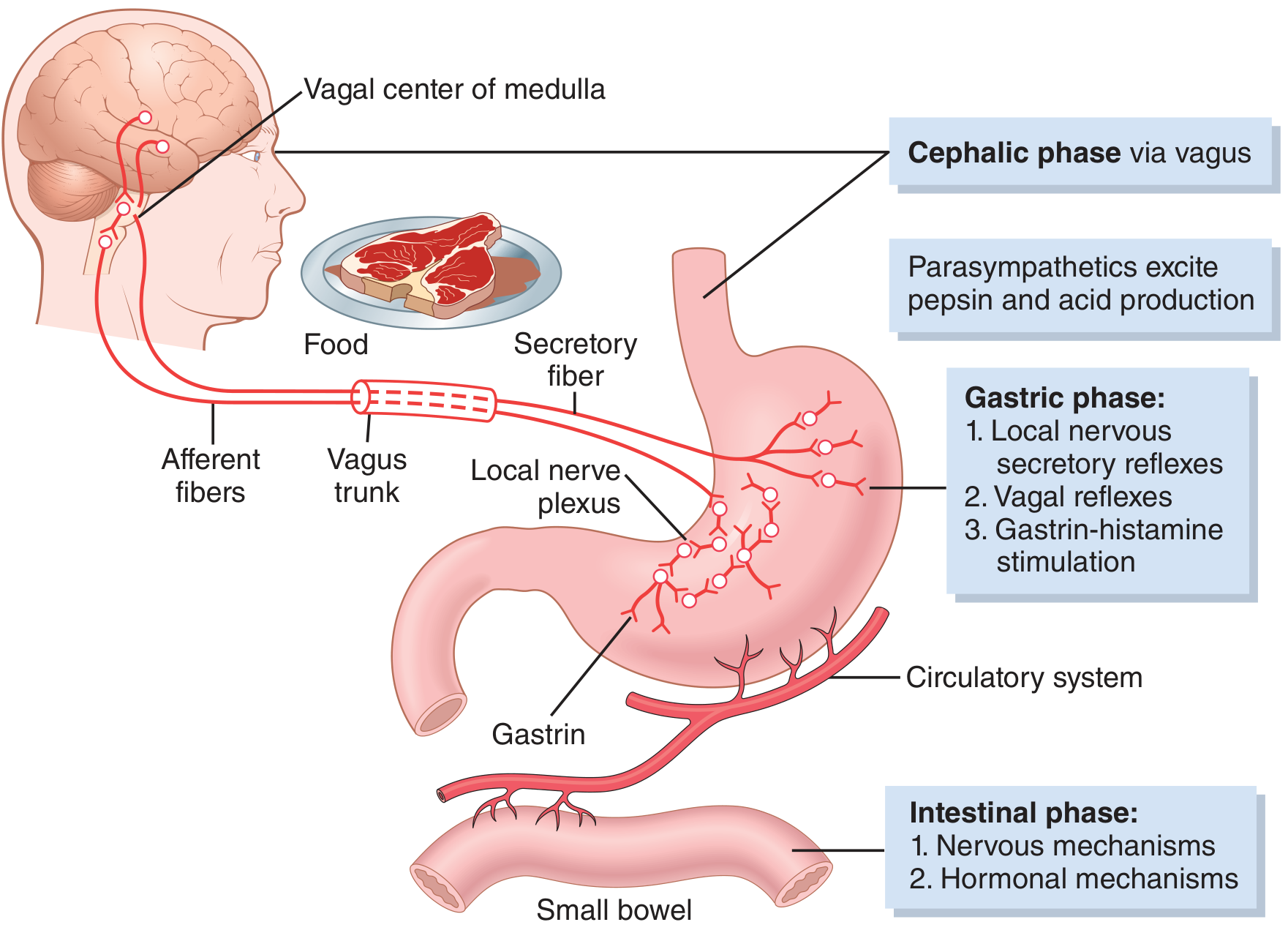
The 3 Phases of Gastric Secretion
🧠 Phase 1 - CEPHALIC PHASE ("Head Phase")
Imagine smelling freshly cooked biryani - your mouth waters and your stomach starts producing acid. That is the cephalic phase!
See/Smell/Think about food
↓
Brain (Cerebral cortex + Hypothalamus) gets excited
↓
Sends signals via VAGUS NERVE (cranial nerve X)
↓
Vagus releases Acetylcholine in the stomach
↓
Stomach wall starts secreting HCl, Pepsinogen and Mucus
Memory: "Cephalic = head thinking about food = vagus gets excited"
🍖 Phase 2 - GASTRIC PHASE ("Stomach Phase")
a) Local Reflex (Enteric nervous system)
Food stretches stomach wall
↓
Local nerve plexus in stomach wall fires
↓
Directly stimulates parietal cells → more HCl
b) Vagovagal Reflex (Brain loop)
Stretch receptors in stomach wall
↓
Signal goes UP to brain via vagus
↓
Brain sends signal back DOWN via vagus
↓
More acid secretion
c) Gastrin-Histamine Mechanism (The most important!) ⭐
Proteins in food stimulate G cells (in antrum of stomach)
↓
G cells release GASTRIN hormone into blood
↓
Gastrin travels to ECL cells (in body of stomach)
↓
ECL cells release HISTAMINE
↓
Histamine stimulates PARIETAL CELLS to secrete HCl
Simple: Protein → Gastrin → Histamine → HCl This is why antacids (H₂ blockers like ranitidine) block histamine to reduce acid!
🫁 Phase 3 - INTESTINAL PHASE
- Duodenum releases a small amount of gastrin → mildly stimulates stomach to keep secreting
- But this phase is mainly important for INHIBITION (see below)
🛑 INHIBITION of Gastric Secretion (How the stomach turns OFF)
- The stomach is empty
- The duodenum is already full / too acidic / has fat
How inhibition happens:
| Trigger | Mechanism | Inhibitor Released |
|---|---|---|
| Acid in duodenum (pH <2) | Enterogastric reflex | Secretin → stops gastrin → reduces acid |
| Fat in duodenum | Hormones from small intestine | GIP (Gastric Inhibitory Peptide) → reduces acid |
| Stomach pH too low (pH <1.5) | Local feedback | Somatostatin from D-cells → shuts off gastrin |
| Duodenum overloaded | Reverse nervous reflex | Sympathetic reflex inhibits stomach |
Somatostatin = the "OFF switch" - when stomach acid gets too high, D-cells release somatostatin → blocks gastrin release → acid stops
The Three Stimulators (Must Know! ⭐)
| Stimulator | Source | Type |
|---|---|---|
| Acetylcholine (ACh) | Vagus nerve | Nerve signal |
| Gastrin | G cells (antrum) | Hormone |
| Histamine | ECL cells | Local chemical (paracrine) |
This is the basis of peptic ulcer drugs:
- Atropine → blocks ACh
- H₂ blockers (ranitidine) → blocks histamine
- Proton pump inhibitors (omeprazole) → blocks the acid pump directly
Summary Table - All 3 Phases
| Phase | When | Trigger | Main Messenger | % of Total Secretion |
|---|---|---|---|---|
| Cephalic | Before food enters stomach | Sight/smell/thought of food | Vagus nerve (ACh) | 30% |
| Gastric | Food in stomach | Protein + distension | Gastrin → Histamine | 60% |
| Intestinal | Food in small intestine | Protein in duodenum | Small gastrin release | 10% |
Super Simple Story Version 📖
One-Line Memory
"Cephalic (brain-vagus, 30%) → Gastric (gastrin-histamine, 60%) → Intestinal (10%) → then INHIBITION by somatostatin, secretin, GIP when gut is full"
Peptic ulcer easy wordd
Peptic Ulcer Disease - Very Easy Words 🔥
What Is a Peptic Ulcer?
Your stomach wall is like a painted wall. The paint (mucus layer) protects the wall from acid. When the paint peels off, acid attacks the wall directly → creates a hole/wound = ULCER.
Where Does It Occur?
| Type | Location | More Common In |
|---|---|---|
| Duodenal ulcer | First part of small intestine (duodenum) | Young men, more common overall |
| Gastric ulcer | Stomach wall | Older people |
What Causes a Peptic Ulcer?
The Balance Concept ⚖️
- Aggressive factors (things that DAMAGE the lining)
- Defensive factors (things that PROTECT the lining)
AGGRESSIVE DEFENSIVE
───────── ─────────
Acid (HCl) vs. Mucus layer
Pepsin vs. Bicarbonate
H. pylori vs. Blood flow
NSAIDs vs. Cell renewal
The Two Big Causes
1. 🦠 Helicobacter Pylori (H. pylori) - 75% of cases!
- It hides in the mucus layer
- It produces urease enzyme → converts urea → ammonia → neutralizes acid around itself
- It slowly destroys the mucus layer that protects the stomach wall
- Triggers inflammation → wall breaks down → ulcer forms
Memory: H. pylori = "acid-loving bacteria that destroys your stomach's shield"
2. 💊 NSAIDs (like Aspirin, Ibuprofen, Diclofenac) - most other cases
- Tell the stomach to make MORE mucus
- Tell the stomach to make MORE bicarbonate
- Maintain good blood supply to the stomach wall
This is why doctors say "don't take painkillers on an empty stomach"!
Other Risk Factors
| Factor | Why It Causes Ulcer |
|---|---|
| Smoking | Reduces bicarbonate, reduces blood flow |
| Stress | Increases acid secretion |
| Alcohol | Directly damages stomach lining |
| Zollinger-Ellison syndrome | Gastrin-secreting tumor → massive acid overproduction |
| Steroids | Reduce mucus production |
Symptoms - How Does It Feel?
Classic Symptoms:
🔥 Burning/gnawing pain in the upper middle abdomen (epigastric pain)
| Duodenal Ulcer | Gastric Ulcer | |
|---|---|---|
| Pain timing | Relieved by food (food buffers acid) | Made WORSE by food |
| Night pain | Common (wakes patient up at 2-3 AM) | Less common |
| Hunger pain | Yes - "hungry pain" | No |
| Nausea/vomiting | Less common | More common |
| Weight | Often normal or gained | Often LOST |
- Nausea
- Bloating
- Belching (burping)
- Loss of appetite (especially gastric ulcer)
Complications - When Things Go Wrong 🚨
1. 🩸 Bleeding (Hemorrhage) - Most common complication
- Ulcer eats into a blood vessel
- Signs: Black tarry stool (melena), vomiting blood (hematemesis)
- Can be life-threatening if large vessel involved
2. 🕳️ Perforation - Most dangerous
- Ulcer burns right THROUGH the stomach/duodenum wall
- Stomach contents spill into abdomen
- Signs: Sudden severe knife-like abdominal pain, rigid abdomen ("board-like rigidity")
- Emergency surgery needed immediately!
3. 🚫 Gastric Outlet Obstruction
- Chronic ulcers cause scarring → narrowing of outlet → food can't leave stomach properly
- Signs: Repeated vomiting of undigested food, fullness, weight loss
4. ➡️ Penetration
- Ulcer burns into an adjacent organ (like pancreas)
- Pain radiates to the back
- Amylase levels rise
Diagnosis - How to Detect It?
| Test | What It Finds |
|---|---|
| Endoscopy (OGD) | Gold standard - directly sees the ulcer |
| Barium meal X-ray | Outline of ulcer crater visible |
| H. pylori tests | Urea breath test, stool antigen test, biopsy |
| Serology | Antibodies to H. pylori in blood |
Treatment - How to Cure It?
Step 1 - Reduce Acid (Antisecretory drugs)
| Drug | How It Works |
|---|---|
| PPI (Proton Pump Inhibitors) e.g., Omeprazole | Block the acid pump (H⁺/K⁺ ATPase) directly - strongest acid suppression |
| H₂ Blockers e.g., Ranitidine | Block histamine → less acid |
| Antacids e.g., Mg(OH)₂ | Neutralize acid already in stomach (quick relief) |
Step 2 - Kill H. pylori (Triple Therapy) ⭐
TRIPLE THERAPY (7-14 days):
PPI (omeprazole) + Clarithromycin + Amoxicillin (or Metronidazole)
Eradication rate = >90%! Once H. pylori is gone, ulcer rarely comes back.
Step 3 - Protect the Mucosa
| Drug | Action |
|---|---|
| Sucralfate | Forms a paste over the ulcer - like a bandage |
| Misoprostol | Synthetic prostaglandin - restores mucus and bicarbonate (used for NSAID ulcers) |
| Bismuth | Coats ulcer + kills H. pylori |
Step 4 - Avoid Triggers
- Stop NSAIDs
- Stop smoking
- Avoid alcohol
- Eat regular meals
Summary Table - Key Points
| Feature | Detail |
|---|---|
| Main causes | H. pylori (75%), NSAIDs |
| Most common site | Duodenum (duodenal ulcer) |
| Key symptom | Epigastric burning pain |
| Duodenal ulcer pain | Relieved by food |
| Gastric ulcer pain | Worsened by food |
| Main complications | Bleeding, perforation, obstruction |
| Gold standard diagnosis | Endoscopy |
| Main treatment | Triple therapy (PPI + 2 antibiotics) |
One-Line Summary
"Peptic ulcer = wound in stomach/duodenum lining caused when H. pylori or NSAIDs break the mucus shield letting acid burn through → treated with PPI + antibiotics to kill H. pylori."
- Mulholland and Greenfield's Surgery, pp. 2249-2259 (Peptic Ulcer Pathophysiology & Treatment)
- Rosen's Emergency Medicine, p. 1217 (Gastritis and Peptic Ulcer Disease)
Movement of small intestines and large intestines
Easyyyy word
Movements of Small & Large Intestines - Easy Words 🌊
Why Does the Intestine Move?
- Mix it with digestive juices so enzymes can break it down
- Push it forward toward the exit
🟡 PART 1: MOVEMENTS OF THE SMALL INTESTINE
Movement 1: SEGMENTATION CONTRACTIONS 🔗
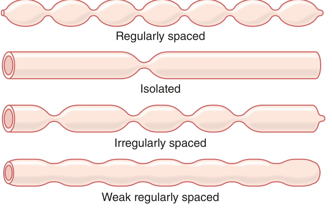
Before: ════════════════════════
After: ═══╬═══╬═══╬═══╬═══╬═══ ← squeeze points
Then squeezes BETWEEN the old points:
══╬═══╬═══╬═══╬═══╬══ ← new squeeze points
- Chops and mixes food with digestive enzymes and bile - like kneading dough
- Exposes food to the intestinal wall for absorption
- Happens 8-12 times per minute
Analogy: Like squeezing a sausage in different places alternately - the contents get thoroughly mixed!
Movement 2: PERISTALSIS 🌊
Direction: MOUTH → ANUS
SQUEEZE → → → → →
════╬══════════════════ ← muscle contracts behind food
══════════════════ ← food pushed forward
- Speed = 0.5 to 2 cm/second (quite slow)
- Each wave travels only 3-5 cm then fades out
- Takes 3-5 hours to move food from stomach to the ileocecal valve (end of small intestine)
- Gets much STRONGER after eating (gastroenteric reflex)
Analogy: Like moving a marble through a rubber tube by squeezing behind it
Movement 3: PERISTALTIC RUSH ⚡ (Emergency only!)
- Massive, rapid, powerful peristalsis occurs
- Sweeps everything through the small intestine in minutes instead of hours
- Dumps contents into colon rapidly
- Result = Diarrhea - the body trying to expel the harmful stuff fast!
Movement 4: VILLOUS MOVEMENTS (Tiny but important)
- They shorten and lengthen repeatedly
- This "milks" lymph fluid through them → helps fat absorption
- Controlled by Meissner's (submucosal) plexus
Quick Summary - Small Intestine
| Movement | Type | Job | Speed |
|---|---|---|---|
| Segmentation | Mixing | Chop and mix food with enzymes | 8-12/min |
| Peristalsis | Propulsive | Push food toward colon | Slow - 1 cm/min net |
| Peristaltic rush | Emergency | Clear intestine rapidly | Very fast |
| Villous movements | Local | Enhance absorption | Continuous |
🟠 PART 2: MOVEMENTS OF THE LARGE INTESTINE
- Absorb water and salts from the remaining waste
- Form and store feces
- Push feces toward the rectum for defecation
Movement 1: HAUSTRAL CONTRACTIONS 🪣
Colon with haustra:
╔═╗ ╔═╗ ╔═╗ ╔═╗ ╔═╗
║ ╚═╝ ╚═╝ ╚═╝ ╚═╝ ║ ← like a string of pouches
- Peak contraction in about 30 seconds, then relaxes in 60 seconds
- Very slow - contents take 8-15 hours to travel through the colon!
- Constantly rolls and turns fecal material - exposing it to the colon wall so water gets absorbed
Analogy: Like someone slowly turning over soil with a spade - turning it around, not pushing it forward
Movement 2: MASS MOVEMENTS 💥
Step 1: A constrictive ring forms in the transverse colon
Step 2: The 20+ cm of colon BELOW it all contract together
Step 3: Feces pushed en masse toward rectum
Step 4: Contraction builds for 30 seconds, then relaxes
Step 5: Another mass movement 2-3 min later
- Occurs only 1-3 times per day
- Usually happens 15 minutes after eating breakfast (gastrocolic reflex!)
- Lasts about 10-30 minutes each episode
- When feces reaches rectum → urge to defecate
That is why many people feel the urge to go to the toilet after breakfast! The stomach filling triggers the gastrocolic reflex → triggers mass movement → pushes feces into rectum.
- Gastrocolic reflex - stomach distension after a meal
- Duodenocolic reflex - food in duodenum
- Irritation of colon (e.g., ulcerative colitis → constant mass movements → chronic diarrhea)
Movement 3: DEFECATION REFLEX 🚽
Feces enters rectum
↓
Rectal wall STRETCHES
↓
Stretch receptors fire → signal to spinal cord (S2-S4)
↓
Parasympathetic reflex:
- Rectum CONTRACTS
- Internal anal sphincter RELAXES (involuntary)
↓
Brain is informed → "Need to go!"
↓
Person voluntarily RELAXES external anal sphincter
↓
Abdominal muscles + diaphragm contract (Valsalva)
↓
DEFECATION ✅
If the time is not right → brain signals external sphincter to stay CLOSED → urge fades temporarily (rectum relaxes)
Quick Summary - Large Intestine
| Movement | Type | Job | Timing |
|---|---|---|---|
| Haustral contractions | Mixing/slow propulsion | Absorb water, form feces | Continuously, slow |
| Mass movements | Propulsive | Push feces toward rectum | 1-3 times/day |
| Defecation reflex | Expulsive | Expel feces | When rectum fills |
The Full Journey - Simple Story 📖
🍚 Food enters small intestine → Segmentation chops and mixes it with enzymes → Peristalsis slowly pushes it forward (3-5 hrs) → Enters large intestine (1500 mL liquid) → Haustral contractions slowly turn it around, absorbing water (8-15 hrs) → After eating, mass movement pushes feces into rectum → Defecation reflex triggers urge → Only 100-200 mL of formed feces exits per day ✅
One-Line Memory
Small intestine: Segmentation (MIX) + Peristalsis (PUSH) Large intestine: Haustrations (MIX + absorb water) + Mass movements (BIG PUSH) + Defecation (OUT)
Deglutination easy language
Deglutition (Swallowing) - Very Easy Words 🍽️
What Is Deglutition?
Important fact: Swallowing is one of the fastest reflexes in the body. Once it starts, you CANNOT stop it - it takes over automatically.
The Control Center
The 3 Phases of Swallowing
MOUTH (Room 1) → THROAT/PHARYNX (Room 2) → FOOD PIPE/ESOPHAGUS (Room 3) → STOMACH
↑ ↑ ↑
VOLUNTARY INVOLUNTARY REFLEX INVOLUNTARY REFLEX
(you control it) (automatic!) (automatic!)
🟢 PHASE 1: ORAL PHASE (Mouth Phase)
- You chew food and mix it with saliva → forms a soft ball called a BOLUS
- Your tongue pushes the bolus backward toward the throat
- Tongue tip presses against the hard palate and sweeps backward
- Once the bolus touches the back of the throat → automatic reflex kicks in - you can no longer stop it!
Analogy: You roll a ball to the top of a slide. Once it goes over the edge, gravity takes over - you can't stop it. That is exactly what happens when food touches the pharynx!
🟡 PHASE 2: PHARYNGEAL PHASE (Throat Phase)
| Step | What Happens | Why |
|---|---|---|
| 1 | Soft palate lifts UP | Closes off the nose - food can't go into the nose |
| 2 | Larynx (voice box) moves UP and FORWARD | Creates space for food to pass |
| 3 | Epiglottis flips DOWN like a lid | Covers the windpipe entrance - food can't enter lungs |
| 4 | Vocal cords close tightly | Extra protection for airway |
| 5 | Breathing STOPS temporarily | Can't breathe and swallow at same time! |
| 6 | Upper esophageal sphincter OPENS | Door to food pipe opens |
| 7 | Pharynx muscles contract in a wave | Push food down into esophagus |
Analogy: It is like a traffic signal system at a junction. When the food-road opens, the air-road gets a RED LIGHT (epiglottis closes, breathing stops). No collisions allowed!
- Food enters the windpipe = aspiration → choking, coughing, or pneumonia
- Common in elderly, stroke patients, or unconscious patients
🔵 PHASE 3: ESOPHAGEAL PHASE (Food Pipe Phase)
Upper esophageal sphincter closes (food locked in)
↓
Ring of CONTRACTION forms above the bolus
↓
RELAXATION below the bolus
↓
Wave moves downward → pushes food toward stomach
↓
Takes about 8-10 seconds to reach stomach
- If the first wave doesn't fully clear the food, a second wave starts from wherever the food is stuck
- Triggered by stretching of the esophagus wall
- Controlled by the enteric nervous system (gut's own brain)
- Lower Esophageal Sphincter (LES) normally stays CLOSED (stops acid from stomach coming back up)
- When food arrives, LES RELAXES → food enters stomach ✅
- Then LES CLOSES again to prevent reflux
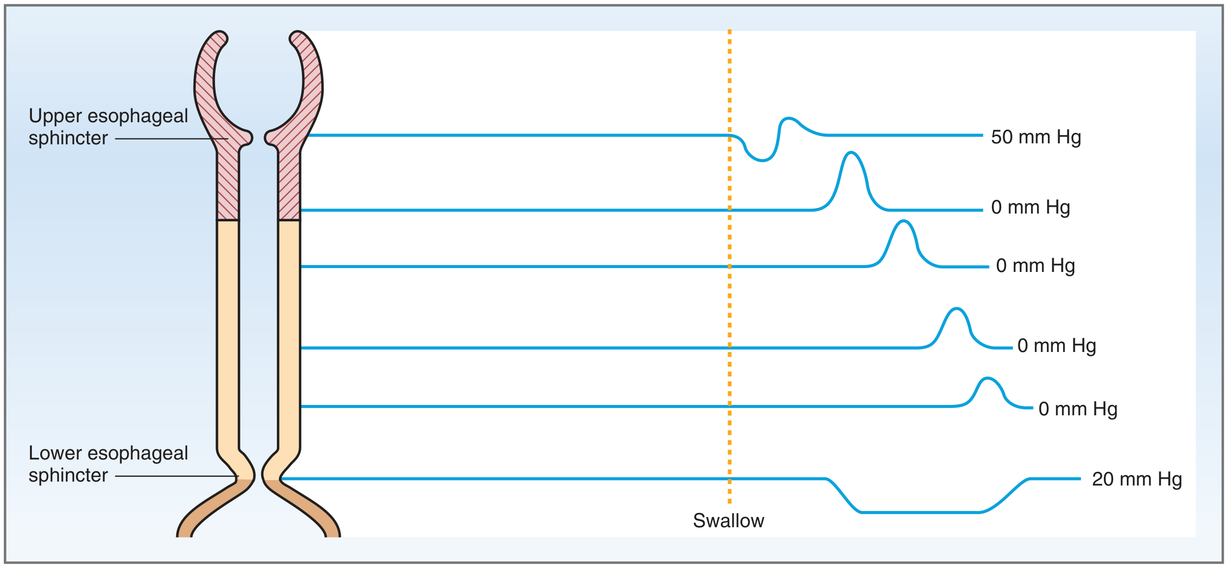
- Upper sphincter - brief high pressure spike then drops to 0 (opens)
- Middle esophagus - peristaltic wave moves down progressively
- Lower sphincter - drops to 0 (relaxes) to let food in, then goes back up (closes)
Summary of All 3 Phases
| Phase | Location | Control | Key Events | Duration |
|---|---|---|---|---|
| Oral | Mouth | Voluntary | Chew, form bolus, tongue pushes back | ~1 sec |
| Pharyngeal | Throat | Involuntary reflex | Nose closes, epiglottis covers windpipe, upper sphincter opens | <1 sec |
| Esophageal | Food pipe | Involuntary (peristalsis) | Waves push food to stomach, lower sphincter opens | 8-10 secs |
The Full Journey - Simple Story 📖
🥗 You chew food → tongue rolls it into a ball (bolus) → tongue pushes it to the back of throat → AUTOMATIC reflex fires - nose closes, epiglottis flips down, breathing stops → food shoots into the esophagus → peristaltic wave pushes it down like squeezing a tube → lower sphincter opens → food enters stomach ✅ → breathing resumes → whole thing done in 10 seconds!
Clinical Conditions When Swallowing Goes Wrong
| Condition | Problem | Cause |
|---|---|---|
| Dysphagia | Difficulty swallowing | Stroke, tumor, achalasia |
| Aspiration | Food goes into lungs | Epiglottis/reflex failure (stroke, unconscious patients) |
| Achalasia | Lower sphincter doesn't relax | Nerve degeneration → food stuck in esophagus |
| GERD | Acid refluxes back up | Lower sphincter too weak/relaxed |
| Globus | Feeling of lump in throat | Muscle tension, anxiety |
Cranial Nerves Involved (Quick List)
| Nerve | Role |
|---|---|
| V (Trigeminal) | Sensation from mouth, chewing muscles |
| VII (Facial) | Facial muscles, sensation from tongue |
| IX (Glossopharyngeal) | Sensation from pharynx - TRIGGERS the reflex |
| X (Vagus) | Motor to pharynx and esophagus |
| XII (Hypoglossal) | Controls the tongue |
One-Line Summary
"Deglutition = 3 phases: Oral (voluntary - tongue pushes food back), Pharyngeal (automatic - epiglottis closes airway, food goes into esophagus), Esophageal (automatic - peristaltic waves push food to stomach in 10 seconds)."
Explain yhe major events that are associated with the motility of git describe in brief about the migratory motor complex easyyy language
Make a presentation on GIT motility and MMC