
Placenta accreta spectrum
Reading File
Finding Sources
Finding Sources
Searching PubMed
"placenta accreta" AND management
Searching the Web
ACOG placenta accreta spectrum obstetric care consensus management antenatal diagnosis
Reading File
Reading File
Searching Images
placenta accreta spectrum ultrasound signs placental lacunae loss clear zone diagram
Reading Web Page
https://www.acog.org/clinical/clinical-guidance/obstetric-ca…
Placenta accreta spectrum (PAS)
Placenta accreta spectrum is abnormal placental attachment or invasion into the uterine wall, usually at a previous uterine scar. It can cause major obstetric hemorrhage, transfusion, adjacent-organ injury, hysterectomy, and preterm birth.
Types
| Type | Depth of invasion |
|---|---|
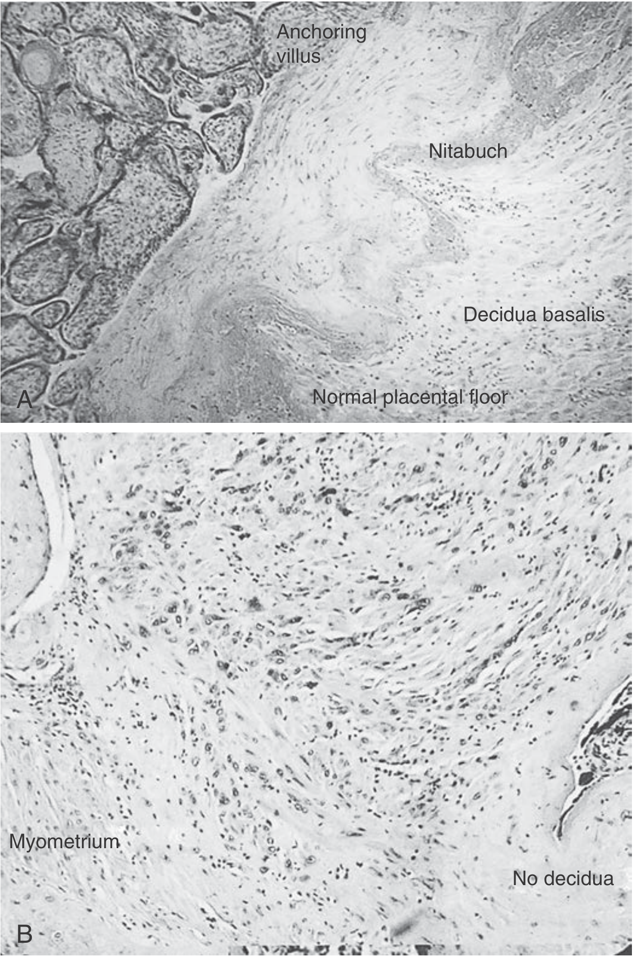
| Placenta accreta | Chorionic villi attach directly to the superficial myometrium because the normal decidual layer is absent or deficient. |
| Placenta increta | Villi invade into the myometrium. |
| Placenta percreta | Villi extend through the uterine serosa, sometimes into the bladder or other pelvic structures. |
Pathogenesis and risk factors
The leading model is defective decidualization at a scarred endometrial-myometrial interface, enabling abnormally deep placental anchoring.
Important risk factors:
- Placenta previa with a prior cesarean delivery, especially multiple prior cesareans
- Other uterine surgery: myomectomy, curettage, endometrial ablation
- Prior PAS
- IVF conception
- Advanced maternal age, multiparity, short interpregnancy interval, smoking, and pelvic radiation
The combination of placenta previa and three or more prior cesareans carries a particularly high risk, reported at over 60% in a maternal-fetal medicine textbook.
Antenatal diagnosis
Targeted obstetric ultrasound with color Doppler is the main investigation. Typical findings include:
- Multiple irregular placental lacunae, often with turbulent high-velocity flow
- Loss of the retroplacental hypoechoic “clear zone”
- Marked myometrial thinning
- Abnormal uterine or placental bulge
- Bridging vessels across the uteroplacental or uterovesical interface
- Bladder-wall irregularity or hypervascularity, raising concern for percreta
MRI is not routinely required, but can help define posterior placentation, depth, or possible bladder/parametrial involvement when ultrasound is uncertain. A normal ultrasound does not rule out PAS when major clinical risk factors are present.
Management principles
Suspected PAS requires referral to a specialist maternal-fetal medicine center. The ACOG-SMFM consensus recommends planned delivery at a Level III or IV maternal care facility, before labor or significant bleeding, with an experienced multidisciplinary team.
Standard definitive management
- Planned cesarean delivery.
- Avoid incising through the placenta, often using a uterine incision away from placental implantation.
- Deliver the fetus.
- Leave the placenta in situ. Do not attempt manual placental removal.
- Proceed directly to cesarean hysterectomy when PAS is confirmed or strongly suspected.
Attempting to detach the placenta can trigger catastrophic hemorrhage.
Timing
For a stable patient, planned cesarean delivery with possible hysterectomy is generally scheduled at 34+0 to 35+6 weeks, with antenatal corticosteroids if preterm delivery is expected. Earlier delivery may be necessary for bleeding, labor, membrane rupture, preeclampsia, fetal compromise, or maternal deterioration, as outlined by ACOG guidance.
Preparation
- Obstetric anesthesiology, MFM, pelvic surgeon or gynecologic oncologist, urology if bladder involvement is possible, blood bank, ICU, neonatal team
- Massive-transfusion readiness, large-bore vascular access, cell salvage where available
- Counseling about likely hysterectomy, blood transfusion, ICU admission, urinary tract injury, prematurity, and loss of fertility
Conservative, fertility-preserving options
In highly selected patients who strongly desire future fertility, the placenta may be left in situ with close follow-up, or focal resection may be attempted in selected localized disease. These are nonstandard, high-risk approaches and require an expert PAS center. They can result in delayed hemorrhage, infection, secondary hysterectomy, recurrent PAS, and serious complications.
Recent-evidence caution: systematic reviews published in 2024-2025 compare conservative approaches with cesarean hysterectomy, but the data are mostly observational and affected by selection bias. They do not replace planned cesarean hysterectomy as the usual recommended approach for typical PAS. Relevant reviews: PMIDs 39884567, 38969992, and 37918506.
Key exam point
PAS = placenta previa + prior cesarean until proved otherwise.
The central management rule is: planned multidisciplinary cesarean hysterectomy with the placenta left in situ, rather than attempting placental separation.
The central management rule is: planned multidisciplinary cesarean hysterectomy with the placenta left in situ, rather than attempting placental separation.
Histopathology reporting
Reading File
Finding Sources
Finding Sources
Searching PubMed
"placenta accreta" AND pathology
Searching the Web
placenta accreta spectrum pathology histopathology reporting guideline FIGO PAS reporting histology
Reading Article
PMID: 32415266
Reading Article
PMID: 40109065
Reading Web Page
https://pubmed.ncbi.nlm.nih.gov/32415266/
Finding Sources
Histopathology reporting of placenta accreta spectrum (PAS)
Histopathology should use PAS terminology with a descriptive grade, rather than relying only on the older terms accreta, increta, and percreta. The report must state the specimen type and whether the uterus, serosa, bladder, or other tissues are available for assessment. This is essential because a delivered placenta alone usually cannot establish the full depth of invasion.
The recommended approach is based on the expert pathology reporting consensus, designed to align with FIGO terminology.
Histological diagnostic criteria
| PAS grade | Histopathologic finding | Traditional correlate |
|---|---|---|
| PAS grade 1 | Chorionic villi are directly apposed to superficial myometrium, with absent or markedly deficient intervening decidua. No myometrial invasion. | Placenta accreta |
| PAS grade 2 | Chorionic villi infiltrate into the myometrium. | Placenta increta |
| PAS grade 3A | Villi extend through the full thickness of myometrium to, or just beneath, the uterine serosa. | Percreta, serosal involvement |
| PAS grade 3B | Villi invade the urinary bladder wall. | Percreta with bladder invasion |
| PAS grade 3C | Villi invade other pelvic tissues or organs, such as broad ligament, bowel, or pelvic sidewall. | Percreta with other-organ invasion |
What to examine and document
1. Clinical information
Include, where provided:
- Prior cesarean section(s), myomectomy, curettage, or other uterine surgery
- Placenta previa or low-lying placenta
- Prenatal ultrasound or MRI suspicion of PAS
- Operative findings: adherent placenta, hemorrhage, bladder involvement, hysterectomy
- Procedure performed: delivered placenta, partial hysterectomy, total hysterectomy, placental bed curettage, bladder resection
2. Gross examination
For hysterectomy specimens:
- Uterine size and external serosal irregularity or placental bulge
- Site of placental implantation, especially lower uterine segment and cesarean scar
- Placental adherence and whether it separates from the uterine wall
- Myometrial thickness at the implantation site
- Disruption, defect, dehiscence, or scar at the implantation site
- Serosal involvement, adhesions, or suspected extrauterine extension
- Bladder or other attached organs/tissues, if present
- Map and extensively sample the entire placental bed, especially the deepest or most abnormal-looking region
For a delivered placenta, document:
- Completeness and membrane/cord features
- Basal plate appearance
- Adherent fragments of myometrium, if present
- Whether the specimen is fragmented or disrupted
3. Microscopy
Describe:
- Decidua basalis: present, attenuated, or absent
- Direct villous contact with myometrium
- Depth and extent of villous invasion into myometrium
- Involvement of serosa, bladder wall, or other tissue
- Cesarean-scar change, uterine dehiscence, or scar defect, if identified
- Associated placental lesions as appropriate, for example infarction, chorioamnionitis, fetal vascular malperfusion, or maternal vascular malperfusion
Important limitation: delivered placenta alone
A delivered placenta with basal plate myometrial fibers may support abnormal adherence, but usually cannot prove the depth of invasion. Therefore, do not diagnose increta or percreta unless relevant uterine tissue is present and adequately sampled.
Use wording such as:
“Basal plate myometrial fibers are present. As no intact placental bed/myometrium is submitted, the findings cannot assess or exclude placenta accreta spectrum or determine depth of invasion.”
A pathology diagnosis must also be interpreted alongside the operative findings. PAS can be clinically obvious but not demonstrable microscopically if the implantation site is unsampled, disrupted, or the placenta has been manually removed.
Suggested final-report format
Example 1: PAS grade 1
Final diagnosis
Uterus and placenta, cesarean hysterectomy:
- Placenta accreta spectrum, PAS grade 1, at the lower uterine segment/previous cesarean scar.
- Chorionic villi directly appose superficial myometrium with absent intervening decidua.
- No histologic evidence of myometrial invasion.
- No serosal or bladder involvement identified in sampled tissue.
Example 2: PAS grade 2
Final diagnosis
Uterus and placenta, cesarean hysterectomy:
- Placenta accreta spectrum, PAS grade 2.
- Chorionic villi infiltrate the myometrium at the lower uterine segment cesarean-scar site.
- No extension to uterine serosa identified in the sections examined.
- Surgical margins/attached bladder tissue, if submitted: negative for villous invasion.
Example 3: PAS grade 3B
Final diagnosis
Uterus, placenta, and partial bladder wall, cesarean hysterectomy:
- Placenta accreta spectrum, PAS grade 3B.
- Chorionic villi extend through the myometrium and uterine serosa with invasion of the urinary bladder wall.
- Background lower uterine segment scar/dehiscence.
- [State whether villi reach lamina propria or muscularis propria of bladder, if assessable.]
Sample microscopic description
Sections from the lower uterine segment demonstrate chorionic villi directly apposed to and infiltrating between smooth-muscle bundles of the myometrium. Intervening decidua basalis is absent at the implantation site. The deepest foci of villous invasion extend to [outer myometrium/serosa/bladder wall]. No villi are identified in [bladder muscularis propria or other specified tissue], in the sections examined.
Practical points
- Use “PAS grade” plus a description of the actual tissue involvement.
- State the anatomic site: lower uterine segment, anterior wall, cesarean scar, fundus, etc.
- State the deepest confirmed extent, not the presumed intraoperative extent alone.
- Include a limitation if sampling is incomplete or no placental bed is present.
- Correlate with operative findings, imaging, and clinical severity.
The standardization is particularly important because pathology reporting distinguishes what is actually confirmed in tissue from the clinical or surgical diagnosis. The 2020 consensus guideline recommends separate terminology and reporting considerations for hysterectomy specimens, delivered placentas, and curettage specimens.
This is a shared conversation. Sign in to Orris to start your own chat.