
Medulla
Reading File
The query "Medulla" is broad - it could refer to the medulla oblongata (brainstem), adrenal medulla, renal medulla, or something else. Let me ask before doing deep research.
Asking for Preferences
Which medulla are you asking about?
Can u generate me 3d image of arachnoid cisterns
Generating Image
3D anatomical illustration of arachnoid cisterns of the brain, showing the major cisterns including cisterna magna, pontine cistern, interpeduncular cistern, ambient cistern, quadrigeminal cistern, chiasmatic cistern, and sylvian cistern. The subarachnoid space is filled with cerebrospinal fluid shown in translucent blue. The brainstem, cerebellum, and base of the brain are visible underneath. Detailed medical anatomy style, cross-sectional 3D view, labeled diagram, high resolution.
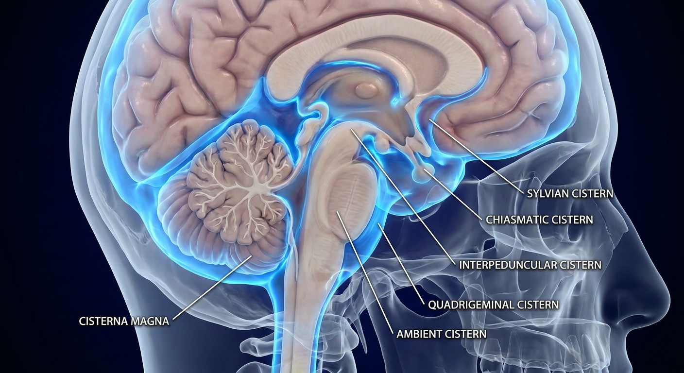
Here's a 3D anatomical illustration of the major arachnoid cisterns. The key cisterns shown include:
- Cisterna magna (cerebellomedullary cistern) - largest, located between the cerebellum and medulla
- Pontine cistern - anterior to the pons, contains the basilar artery
- Interpeduncular cistern - between the cerebral peduncles, contains the circle of Willis
- Ambient cistern - wraps around the midbrain laterally
- Quadrigeminal cistern - posterior to the midbrain (also called the cistern of the great cerebral vein)
- Chiasmatic cistern - around the optic chiasm
- Sylvian cistern - along the lateral sulcus
The arachnoid cisterns are expansions of the subarachnoid space where CSF pools. They are clinically important in neurosurgery (surgical corridors), neuroradiology (herniation patterns), and infectious/hemorrhagic conditions like subarachnoid hemorrhage and meningitis.
Concise me spino cerebellar tracts
Finding Sources
Finding Sources
Reading File
Reading File
Loading Image
Here is a concise breakdown of the spinocerebellar tracts, sourced from Neuroanatomy through Clinical Cases, 3rd Ed.
Spinocerebellar Tracts - Concise Summary
There are 4 spinocerebellar tracts, grouped by limb and function:
Lower Extremity Tracts
| Feature | Dorsal SCT | Ventral SCT |
|---|---|---|
| Input | Proprioception, touch, pressure from legs | Spinal cord interneuron activity (leg) |
| 1st neuron origin | Dorsal root ganglion cells | Spinal border cells + intermediate zone neurons |
| Relay nucleus | Nucleus dorsalis of Clark (C8-L3) | No dedicated relay - direct from cord |
| Crossing | Does NOT cross (ipsilateral) | Crosses once in ventral commissure, then crosses AGAIN at pons → double-crossed = ipsilateral |
| Peduncle | Inferior cerebellar peduncle | Superior cerebellar peduncle |
| Location in cord | Dorsolateral funiculus | Ventrolateral funiculus (just anterior to dorsal SCT) |
Upper Extremity Tracts
| Feature | Cuneocerebellar tract | Rostral SCT |
|---|---|---|
| Input | Proprioception from arms & neck | Spinal cord interneuron activity (arm) |
| Origin | Dorsal root ganglion cells (C2-T1) | Spinal cord neurons (cervical) |
| Relay nucleus | External cuneate nucleus (medulla) | None |
| Crossing | Does NOT cross (ipsilateral) | Poorly characterized; enters via both inferior and superior peduncles |
| Peduncle | Inferior cerebellar peduncle | Both inferior + superior cerebellar peduncles |
Key Concept
All spinocerebellar inputs reach the IPSILATERAL cerebellum (either uncrossed OR double-crossed). This is why cerebellar lesions cause ipsilateral limb ataxia.
What Information Reaches the Cerebellum?
| Type | Tracts | Purpose |
|---|---|---|
| Afferent sensory feedback | Dorsal SCT + Cuneocerebellar | Limb position and movement (proprioception) |
| Efference copy / motor command activity | Ventral SCT + Rostral SCT | Reflects activity of descending motor pathways via interneurons |
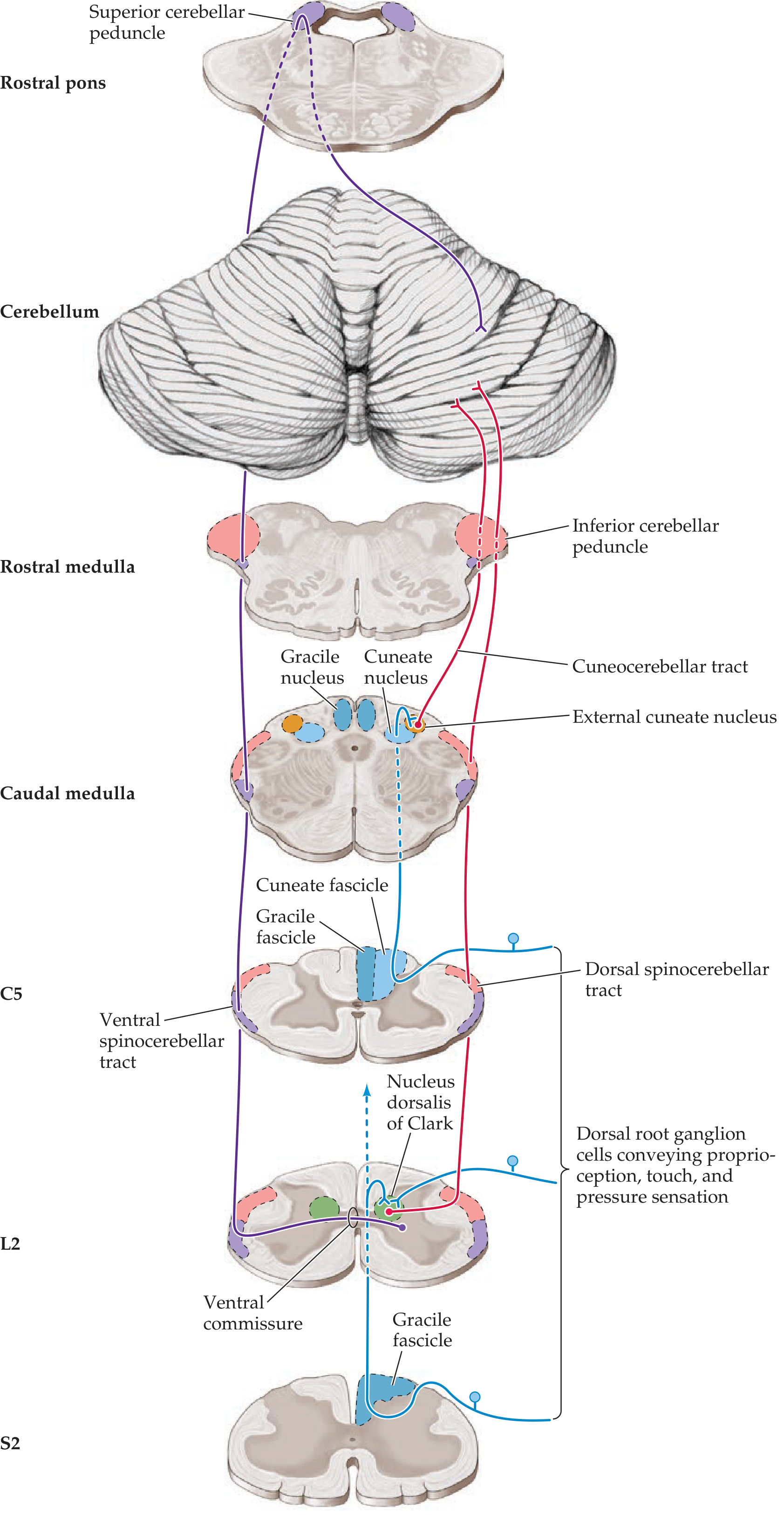
Memory trick:
- Dorsal = Direct (ipsilateral, no cross) = Does legs
- Ventral = crosses Vice versa (double cross) = also legs
- Cuneocerebellar = arms, via Cuneate nucleus
- Rostral = arms, Reflects interneuron activity
Umn vs lmn facial. Palsy
The key distinguishing feature is that UMN facial palsy spares the forehead (bilateral cortical representation of the upper face), while LMN facial palsy affects the entire face including forehead.
Searching sources...
Finding Sources
Finding Sources
Finding Sources
Finding Sources
Reading File
Reading File
Loading Image
Here is a concise, high-yield comparison of UMN vs LMN facial palsy, sourced from Rosen's Emergency Medicine and Bradley and Daroff's Neurology in Clinical Practice.
UMN vs LMN Facial Palsy
The Anatomy Behind the Difference
The upper face (forehead, orbicularis oculi) receives bilateral cortical innervation - both hemispheres send UMN fibers to the facial nucleus that controls forehead muscles. The lower face receives only contralateral UMN input.
So if the UMN is damaged (e.g., stroke), the contralateral hemisphere compensates for the forehead → forehead is SPARED. The LMN (CN VII itself) is damaged below the nucleus, so everything is lost.
Quick Comparison Table
| Feature | UMN Facial Palsy | LMN Facial Palsy |
|---|---|---|
| Forehead | SPARED (bilateral cortical supply) | INVOLVED (entire face weak) |
| Weakness location | Lower face only (contralateral) | Entire ipsilateral face |
| Eye closure | Intact | Impaired - Bell's phenomenon visible |
| Nasolabial fold | Flattened (lower face) | Flattened (entire side) |
| Emotional vs voluntary | Dissociation possible (one may be worse than other) | Both affected equally |
| Site of lesion | Contralateral motor cortex or corticobulbar tract | Facial nucleus (pons) or CN VII peripherally |
| Lesion side | Contralateral to weakness | Ipsilateral to weakness |
| Taste | Normal | May be impaired (chorda tympani involvement) |
| Hyperacusis | Absent | May be present (stapedius branch) |
| Lacrimation | Normal | May be reduced (greater superficial petrosal nerve) |
| Typical cause | Stroke, cortical tumor | Bell's palsy, parotid tumor, skull base fracture |
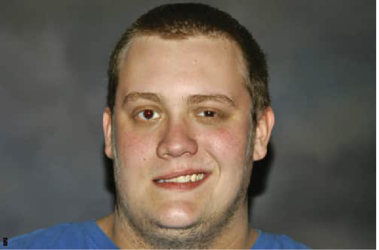
Clinical Photo - LMN (Bell's Palsy)
Key Clinical Points
UMN (Central) - Think STROKE:
- Forehead sparing is the hallmark
- Other neuro deficits usually present (hemiplegia, aphasia, sensory loss)
- Contralateral lower face weakness
LMN (Peripheral) - Think Bell's Palsy:
- Forehead involved = diagnostic of peripheral lesion
- Associated features by level of lesion:
| Level | Additional Features |
|---|---|
| Proximal to geniculate ganglion | Reduced lacrimation + taste loss + hyperacusis |
| Proximal to stapedius branch | Taste loss + hyperacusis (no lacrimal involvement) |
| Proximal to chorda tympani | Taste loss only |
| Distal to chorda tympani (stylomastoid foramen) | Pure motor palsy, no taste/hyperacusis |
Memory Hook
"UMN = Upper face Unaffected" "LMN = Loses everything (whole face)"
- Rosen's Emergency Medicine, Table 91.4
- Bradley and Daroff's Neurology in Clinical Practice, Ch. 103
This is a shared conversation. Sign in to Orris to start your own chat.