I now have all the material needed. Let me update the todos and compile the comprehensive BHMS-style note.---
BRONCHIAL ASTHMA
BHMS Study Notes | Medicine & Surgery Paper
DEFINITION
Bronchial asthma is a chronic inflammatory disorder of the airways characterized by episodic and reversible airflow obstruction, bronchial hyperresponsiveness, and underlying airway inflammation. It manifests clinically as recurrent episodes of wheezing, breathlessness, chest tightness, and cough - particularly at night or in the early morning.
"Asthma has considerable heterogeneity in its pathways, but inflammation is the underlying common feature." - Swanson's Family Medicine Review
EPIDEMIOLOGY
- Affects approximately 300 million people worldwide
- Prevalence increasing in Westernized/urban populations
- Can begin at any age; commonly begins in childhood
- Higher prevalence in females adults; males more affected in childhood
- Strong association with socioeconomic and environmental factors (indoor pollutants, tobacco smoke, allergen exposure)
- Heritability estimated at 35-95% for asthma; 30-66% for bronchial hyperresponsiveness
ETIOPATHOGENESIS
A. Predisposing Factors (Risk Factors)
| Category | Examples |
|---|
| Genetic | Family history of asthma/atopy; chromosome 17q21 locus; multiple GWAS-identified susceptibility genes |
| Atopy | Allergic rhinitis, eczema, urticaria |
| Sex | Males predominate in childhood; females in adulthood |
| Obesity | BMI strongly associated with asthma severity |
| Early life | Respiratory infections in infancy, prematurity, low birth weight |
| Hygiene hypothesis | Reduced early microbial exposure → failure to suppress atopic phenotype |
B. Precipitating/Triggering Factors
- Allergens - pollens, dust mites, cockroaches, animal dander, mold spores
- Respiratory infections - viral URTIs (most common trigger for exacerbations)
- Exercise - airway dehydration from hyperventilation → increased osmolarity → mediator release
- Cold, dry air
- Air pollutants - ozone, SO₂, NO₂, tobacco smoke
- Drugs - NSAIDs/aspirin (COX-1 inhibition → leukotriene excess), beta-blockers (including ophthalmic), ACE inhibitors (cough)
- Emotional stress / psychosocial factors
- Occupational exposures - isocyanates, flour dust, latex (> 400 agents recognized)
- GERD - microaspiration triggers bronchoconstriction
CLASSIFICATION
A. Based on Etiology
| Type | Features |
|---|
| Allergic (Extrinsic) Asthma | IgE-mediated; onset in childhood; positive skin tests; associated with atopy; identifiable allergen triggers |
| Non-Allergic (Intrinsic) Asthma | No IgE antibodies to environmental antigens; adult onset; triggered by infections, aspirin, exercise |
| Occupational Asthma | Triggered by workplace sensitizers |
| Aspirin-Exacerbated Respiratory Disease (AERD) | Asthma + nasal polyposis + aspirin sensitivity; leukotriene overproduction |
| Exercise-Induced Bronchoconstriction (EIB) | Triggered purely by exercise |
| Cough-Variant Asthma | Chronic cough as the sole manifestation |
B. Based on Severity (GINA Classification)
| Severity | Symptoms | Nighttime symptoms | FEV₁/PEF | Variability |
|---|
| Intermittent | < 1/week | ≤ 2/month | ≥ 80% predicted | < 20% |
| Mild Persistent | > 1/week but < 1/day | > 2/month | ≥ 80% predicted | 20-30% |
| Moderate Persistent | Daily | > 1/week | 60-80% predicted | > 30% |
| Severe Persistent | Continuous | Frequent | < 60% predicted | > 30% |
C. Molecular Phenotypes (Current Understanding)
- T2-High (Eosinophilic) - overexpression of IL-13 target genes (periostin, CLCA1, serpinB2); higher IgE; sputum eosinophilia; responds well to ICS
- T2-Low (Non-Eosinophilic) - does not respond to ICS as well; may benefit from azithromycin or tiotropium
PATHOPHYSIOLOGY
Key Cellular Players
| Cell | Role |
|---|
| Mast cells | Release histamine, prostaglandins, leukotrienes, TNF-α on allergen exposure |
| Eosinophils | Major role in chronic airway inflammation; release toxic proteins causing epithelial damage |
| Th2 lymphocytes | Release IL-4, IL-5, IL-13 → drive IgE production, eosinophil recruitment, goblet cell hyperplasia |
| Basophils | IgE-mediated mediator release |
| Dendritic cells | Antigen presentation; bridge innate and adaptive immunity |
| Airway epithelial cells | Barrier dysfunction; produce TSLP, IL-33 (alarmins); activate innate immune responses |
| Airway smooth muscle cells | Hypertrophy/hyperplasia → bronchoconstriction; key in thermoplasty |
| Neutrophils | Prominent in severe, non-eosinophilic, and smoking-associated asthma |
Sequence of Events
ALLERGEN EXPOSURE
       ↓
Antigen presentation by Dendritic cells
       ↓
Th2 cell activation → IL-4, IL-5, IL-13 release
       ↓
B cell class switching → IgE production
       ↓
IgE binds to mast cells & basophils (sensitization)
       ↓
RE-EXPOSURE TO ALLERGEN
       ↓
Mast cell degranulation (EARLY PHASE, within minutes)
→ Histamine, prostaglandins, leukotrienes
→ Bronchospasm, mucosal edema, mucus hypersecretion
       ↓
LATE PHASE RESPONSE (2-24 hours)
→ Eosinophil, T cell, basophil infiltration
→ Chronic inflammation → airway remodeling
Pathological Changes (Airway Remodeling)
- Subbasement membrane thickening (tenascin, type III collagen deposition)
- Goblet cell hyperplasia → increased mucus production
- Mucous plugging of airways
- Airway smooth muscle hypertrophy and hyperplasia
- Angiogenesis (increased vascularity)
- Loss of epithelial integrity → denuded epithelium
- Submucosal inflammatory cell infiltration (eosinophils, mast cells, neutrophils)
Remodeling leads to irreversible airflow limitation and increased asthma mortality in severe cases.
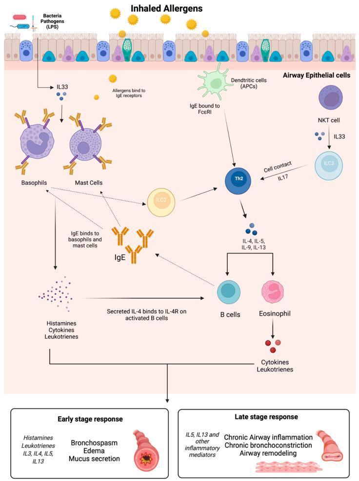
Early and Late phase asthma responses showing cellular mediators and structural changes
CLINICAL FEATURES
Symptoms (Classic Triad)
- Episodic wheeze (high-pitched musical rhonchi, more prominent on expiration)
- Dyspnea / breathlessness
- Chest tightness (often described as a band around the chest)
- Cough - typically dry, worse at night and early morning
Signs
Between attacks: May be completely normal.
During an attack:
- Tachypnea, tachycardia
- Use of accessory muscles (sternocleidomastoid, scalene)
- Nasal flaring, intercostal/subcostal retractions (especially in children)
- Hyperresonant percussion (air trapping)
- Prolonged expiratory phase (expiratory:inspiratory ratio > 2:1)
- Diffuse bilateral expiratory wheeze (inspiratory wheeze in severe cases)
- Pulsus paradoxus (>10 mmHg fall in systolic BP during inspiration) in severe attacks
- Barrel chest in chronic severe cases
WARNING SIGNS of Severe/Life-Threatening Attack
- Silent chest (no wheeze - no air movement!)
- Inability to speak in full sentences
- Cyanosis
- Bradycardia / hypotension
- Altered consciousness / confusion
- SpO₂ < 92%
Onset Patterns
- Slow-onset (>80% of cases): >6 hours, female predominance, triggered by URI, slower response to treatment
- Sudden-onset (<20% of cases): <6 hours, male predominance, triggered by allergens/exercise, faster response to treatment
INVESTIGATIONS
Pulmonary Function Tests (PFTs) - Most Important
| Test | Findings in Asthma |
|---|
| Spirometry | ↓ FEV₁, ↓ FEV₁/FVC ratio (< 0.70), ↑ RV, ↑ TLC (air trapping) |
| Bronchodilator reversibility | ≥ 12% and ≥ 200 mL improvement in FEV₁ after SABA = diagnostic |
| Peak Expiratory Flow Rate (PEFR) | Reduced; morning dip pattern (diurnal variation > 20%) |
| Methacholine challenge | PD₂₀ ≤ 400 μg = airway hyperresponsiveness (used when PFTs non-confirmatory) |
| Exercise challenge | ≥ 10% drop in FEV₁ from baseline |
Blood Tests
- CBC - eosinophilia (> 300-500 cells/μL suggests type 2/eosinophilic asthma)
- Total serum IgE - elevated in allergic asthma; >1000 IU/mL → consider ABPA or omalizumab eligibility
- ABG - in severe attack: initial ↓ PaCO₂, ↑ pH (respiratory alkalosis); rising PaCO₂ = impending respiratory failure
Allergy Tests
- Skin prick test - to identify specific allergens
- RAST (Radioallergosorbent Test) - serum-specific IgE against individual allergens
Inflammatory Biomarkers
- Fractional exhaled nitric oxide (FeNO):
-
35-40 ppb in untreated = type 2 airway inflammation
-
20-25 ppb despite ICS = inadequate control or persistent T2 inflammation
- Induced sputum eosinophils - guides biologic therapy selection
- Blood eosinophil count - markers for biologic eligibility
Imaging
- Chest X-ray - usually normal; during attack may show hyperinflation, flattened diaphragm; rules out pneumothorax/consolidation
- Chest HRCT - for bronchiectasis, structural abnormalities; not routine
Other
- Sinus CT - if rhinosinusitis/polyps suspected
- GERD workup - if reflux is suspected comorbidity
- α₁-antitrypsin level - if non-reversible disease suspected
THERAPEUTICS / MANAGEMENT
Goals of Treatment
- Minimize symptoms and maintain normal activity levels
- Maintain normal/near-normal lung function
- Prevent asthma exacerbations
- Avoid adverse effects of medications
- Prevent asthma mortality
NON-PHARMACOLOGICAL MEASURES
- Allergen avoidance - impermeable mattress covers (dust mites), pet removal, pest control, mold remediation
- Environmental control - avoid smoke, pollutants, strong odors
- Smoking cessation (patient and household members)
- Vaccination - yearly influenza, pneumococcal (regardless of age), COVID-19, RSV vaccines
- Occupational exposure reduction - removal from offending environment may produce remission
- Written asthma action plan - individualized plan for patients
- Weight reduction in obese patients
- Allergen immunotherapy (AIT) - subcutaneous or sublingual; useful in mild-moderate allergic asthma; risk of anaphylaxis requires controlled asthma
- Exercise warm-up and face mask/nasal breathing to warm/humidify cold air
PHARMACOLOGICAL TREATMENT
I. BRONCHODILATORS
A. Beta-2 Agonists
Short-Acting Beta-2 Agonists (SABAs)
- Drug: Salbutamol (Albuterol) - most commonly used
- Mechanism: Activate β₂ receptors → activate adenyl cyclase → ↑ cAMP → relaxation of airway smooth muscle
- Onset: 3-5 minutes | Duration: 4-6 hours
- Route: Metered-dose inhaler (MDI), nebulization, dry powder inhaler
- Use: Rescue/reliever therapy for acute symptoms; pre-exercise prophylaxis (5-10 min before)
- Caution: Regular monotherapy use → tachyphylaxis, increased airway reactivity, increased asthma mortality; always combine with ICS
Long-Acting Beta-2 Agonists (LABAs)
- Drugs: Salmeterol, Formoterol
- Duration: ~12 hours
- Formoterol has rapid onset (similar to SABA) + long duration
- Use: Add-on to ICS in moderate-severe persistent asthma; NEVER as monotherapy without ICS
- Fixed combinations: Salmeterol/fluticasone, Formoterol/budesonide
B. Anticholinergics (Muscarinic Antagonists)
Short-Acting (SAMA): Ipratropium bromide
- Less effective than SABAs for acute asthma; useful as add-on in severe exacerbations
Long-Acting (LAMA): Tiotropium
- Add-on therapy in severe asthma (≥12 years)
- Useful in T2-low asthma phenotype
- Delivered via Respimat inhaler
C. Theophylline (Methylxanthine)
- Mechanism: Phosphodiesterase inhibitor → ↑ cAMP → bronchodilation; also has anti-inflammatory effects
- Narrow therapeutic index (therapeutic range: 10-20 μg/mL); requires serum level monitoring
- Side effects: Tachycardia, palpitations, nausea/vomiting, seizures at toxic levels
- Use: Third-line bronchodilator; low-dose theophylline has anti-inflammatory benefits
- Now largely superseded by inhaled therapies
II. ANTI-INFLAMMATORY AGENTS
A. Corticosteroids
Inhaled Corticosteroids (ICS) - CORNERSTONE OF TREATMENT
| Drug | Example Doses |
|---|
| Beclomethasone | Low: ≤250 μg/day; Medium: 251-500 μg/day; High: >500 μg/day |
| Budesonide | (first-line in pregnancy and children) |
| Fluticasone | |
| Mometasone | |
| Ciclesonide | (prodrug - lower oropharyngeal side effects) |
- Mechanism: Reduce airway hyperresponsiveness; reduce inflammatory cell infiltration; prevent exacerbations; improve lung function
- Onset of chronic benefit: weeks; NOT curative
- Local side effects: Oropharyngeal candidiasis, dysphonia (hoarseness) → prevent by gargling, using spacer
- Systemic side effects at high doses: Adrenal suppression, osteoporosis, cataracts, glaucoma, growth suppression in children
- KEY POINT: ICS are "controllers" - effective only while being taken; return of symptoms within weeks of stopping
Systemic / Oral Corticosteroids (OCS)
- Drug: Prednisolone/Prednisone, Methylprednisolone, Hydrocortisone IV (in acute severe asthma)
- Use: Short "bursts" for acute exacerbations; maintenance only in severe refractory asthma
- Long-term side effects: Diabetes, osteoporosis, cataracts, Cushing's features, hypertension, peptic ulcer, adrenal suppression, immunosuppression, growth retardation in children
B. Leukotriene Modifiers
CysLT1 Receptor Antagonists (LTRAs)
- Drugs: Montelukast (once daily, oral), Zafirlukast (twice daily, oral)
- Mechanism: Block LTD₄ receptor on airway smooth muscle → prevent bronchoconstriction, mucus hypersecretion, edema
- Uses:
- Mild persistent asthma (alternative to low-dose ICS)
- Add-on therapy in poorly controlled moderate asthma
- Aspirin-Exacerbated Respiratory Disease (AERD) - particularly effective
- Exercise-induced bronchoconstriction (without tachyphylaxis)
- Prevention of exercise-induced symptoms in children
- Allergic rhinitis comorbidity
- Pediatric: Montelukast approved from 12 months of age
- Safety: Montelukast associated with neuropsychiatric effects including suicidal ideation (FDA black box warning)
5-Lipoxygenase Inhibitor
- Drug: Zileuton (1200 mg SR twice daily)
- Mechanism: Prevents leukotriene synthesis
- Side effects: Elevation of transaminases (3% of patients); inhibits CYP1A2
C. Cromolyn Sodium (Sodium Cromoglycate)
- Mechanism: Mast cell stabilizer; inhibits mediator release from sensitized mast cells
- Route: Nebulization (2-4 times daily)
- Use: Exercise-induced bronchospasm; primarily in pediatrics; largely replaced by ICS
- Very safe profile - suitable where ICS side effects are a concern
III. BIOLOGICAL / TARGETED THERAPIES (Severe Refractory Asthma)
| Drug | Target | Use Criteria | Route |
|---|
| Omalizumab | Anti-IgE (Fc portion) | IgE ≥30 IU/mL + positive skin test/RAST + poorly controlled on ICS/LABA; reduces exacerbations by 25-50% | SC every 2-4 weeks |
| Mepolizumab | Anti-IL-5 | Severe eosinophilic asthma (eosinophils ≥150 cells/μL) | SC monthly |
| Reslizumab | Anti-IL-5 | Severe eosinophilic asthma | IV monthly |
| Benralizumab | Anti-IL-5Rα | Severe eosinophilic asthma | SC every 4-8 weeks |
| Dupilumab | Anti-IL-4Rα (blocks IL-4 and IL-13) | Moderate-severe eosinophilic asthma; also for atopic dermatitis and CRS | SC every 2 weeks |
| Tezepelumab | Anti-TSLP | Severe asthma (broadest indication; not limited to eosinophilic) | SC every 4 weeks |
These agents target the upstream type 2 inflammatory cascade and significantly reduce exacerbation rates in selected patients.
IV. STEPWISE MANAGEMENT (GINA Framework)
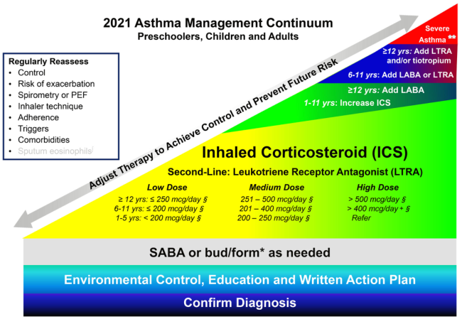
GINA 2021 Asthma Management Continuum - stepwise approach from mild to severe asthma
| Step | Treatment |
|---|
| Step 1 (Intermittent) | SABA as needed (PRN); consider low-dose ICS-formoterol PRN |
| Step 2 (Mild Persistent) | Low-dose ICS daily + SABA PRN; alternative: LTRA |
| Step 3 (Moderate Persistent) | Low-dose ICS + LABA; or medium-dose ICS + SABA PRN |
| Step 4 (Severe Persistent) | Medium-to-high dose ICS + LABA; + LTRA and/or tiotropium |
| Step 5 (Very Severe) | High-dose ICS + LABA + tiotropium; add biologic therapy (anti-IgE, anti-IL-5, anti-IL-4/13, anti-TSLP) |
Key Principle: Regular reassessment of control, spirometry, inhaler technique, adherence, triggers, and comorbidities at every visit. "Step up" therapy if poorly controlled; "Step down" if well controlled for ≥3 months.
V. MANAGEMENT OF ACUTE SEVERE ASTHMA (Status Asthmaticus)
- Oxygen - high-flow, target SpO₂ 94-98%
- Nebulized SABA - salbutamol 2.5-5 mg every 20 minutes x 3 doses, then as needed
- Ipratropium bromide - nebulized, add to SABA in severe attacks
- Systemic corticosteroids - prednisolone 40-60 mg oral OR methylprednisolone 125 mg IV
- IV Magnesium sulfate (2 g IV over 20 min) - in life-threatening attacks not responding to above
- IV Aminophylline - second-line; requires serum level monitoring
- Heliox (helium-oxygen mixture) - reduces airway resistance, considered in refractory cases
- Non-invasive ventilation (NIV) - BIPAP in selected cases
- Mechanical ventilation - for respiratory failure (permissive hypercapnia strategy)
- Antibiotics - only if evidence of bacterial infection
DIFFERENTIAL DIAGNOSIS
- COPD (chronic bronchitis/emphysema)
- Cardiac asthma (left ventricular failure - "cardiac asthma")
- Vocal cord dysfunction / Inducible laryngeal obstruction
- Foreign body aspiration (especially in children)
- Anaphylaxis
- Pulmonary embolism
- Bronchiectasis
- Bronchiolitis obliterans
- GERD with pulmonary manifestations
- α₁-antitrypsin deficiency
COMPLICATIONS
- Status asthmaticus (life-threatening prolonged attack)
- Acute respiratory failure / hypercapnic respiratory failure
- Pneumothorax / pneumomediastinum
- Subcutaneous emphysema
- Cor pulmonale (in chronic severe disease)
- Allergic Bronchopulmonary Aspergillosis (ABPA) - fungal sensitization complicating asthma
- Chronic airflow limitation / irreversible airway remodeling
- Respiratory failure → death
PROGNOSIS
- Childhood asthma: ~50% achieve remission in adulthood
- Adult-onset asthma: Less likely to remit; more likely chronic
- Poor prognosis markers: history of intubation, frequent exacerbations, chronic OCS use, non-adherence, eosinophilia on high-dose ICS, psychosocial problems
- Asthma mortality risk factors: near-fatal episode, >1 hospitalization/year, SABA overuse (>1 canister/month), lack of ICS, poor adherence, food allergy in asthmatic
HOMOEOPATHIC PERSPECTIVE (BHMS Context)
In Homoeopathy, bronchial asthma is treated constitutionally (miasmatic basis - Psora + Sycosis) along with totality of symptoms. Important remedies include:
| Remedy | Keynotes for Asthma |
|---|
| Arsenicum Album | Worse 1-3 AM; anxiety; restlessness; burning sensation; better sitting upright; thirst for small sips |
| Ipecacuanha | Constant nausea; rattling mucus; cannot breathe; spasmodic cough |
| Antimonium Tartaricum | Rattling, loose cough; inability to raise mucus; drowsiness; better sitting up |
| Natrum Sulphuricum | Worse in damp weather; green expectoration; holds chest during cough |
| Pulsatilla | Worse in warm rooms, better in open air; variable symptoms; non-thirsty; mild disposition |
| Nux Vomica | Worse early morning; constriction of chest; dyspnea after eating; associated with gastric disorders |
| Spongia Tosta | Dry, barking, croupy cough; sawing sound; better eating and drinking warm fluids |
| Blatta Orientalis | Specific for asthma with obesity; purulent expectoration |
| Lobelia Inflata | Vagal asthma; nausea with respiration; faintness; weakness of respiratory muscles |
| Drosera | Spasmodic, prolonged attack; whooping-like; worse after midnight |
Miasmatic Correlation:
- Acute/intermittent presentation → Psoric background
- Chronic with structural remodeling → Sycotic or Syphilitic elements
- Recurrent + hereditary tendency → Psora (Hahnemann)
QUICK REVISION SUMMARY
| Feature | Key Point |
|---|
| Definition | Chronic inflammatory airway disease with reversible obstruction and hyperresponsiveness |
| Cardinal symptoms | Wheeze, dyspnea, chest tightness, cough |
| Hallmark finding | Reversibility of obstruction: ≥12% + ≥200 mL FEV₁ improvement |
| Key cells | Mast cells, eosinophils, Th2 cells, airway smooth muscle |
| Key mediators | Histamine, leukotrienes, IL-4/5/13, IgE |
| Cornerstone of treatment | Inhaled corticosteroids (ICS) |
| Rescue drug | Salbutamol (SABA) |
| Biological therapy anchor | Omalizumab (anti-IgE); Mepolizumab (anti-IL-5) |
| Dangerous sign | "Silent chest" = no air movement = emergency |
| Best investigation | Spirometry with bronchodilator reversibility test |
Sources: Harrison's Principles of Internal Medicine 22E (McGraw Hill, 2025); Katzung's Basic & Clinical Pharmacology 16th Ed; Rosen's Emergency Medicine; Murray & Nadel's Textbook of Respiratory Medicine; Fishman's Pulmonary Diseases and Disorders