
hemodynamic disorders, thromboembolic disease an shock
Reading File
This is a broad pathology topic. Let me search the library for all three areas simultaneously.
Finding Sources
Finding Sources
Finding Sources
The main Robbins chapter covers all three topics. Let me read the full chapter content in parallel sections.
Reading File
Finding Sources
Finding Sources
The full Robbins chapter is in block1.md starting at line 2139. Let me read the full content in large parallel chunks.
Reading File
Reading File
Reading File
I have extensive content. Let me read the shock section and the beginning of the chapter (edema/hyperemia) in parallel.
Reading File
Reading File
I now have comprehensive content from Robbins. Let me read the remaining shock stages and fetch key images to include.
Reading File
Reading File
I now have all the content needed for a comprehensive response. Let me compile the full, detailed answer from Robbins & Kumar Basic Pathology.
Hemodynamic Disorders, Thromboembolic Disease, and Shock
Based on Robbins & Kumar Basic Pathology (Robbins Pathology)
1. HYPEREMIA AND CONGESTION
Both refer to increased blood volume within a tissue, but differ in mechanism:
| Feature | Hyperemia | Congestion |
|---|---|---|
| Mechanism | Active - arteriolar dilation, increased inflow | Passive - impaired venous outflow |
| Occurs in | Inflammation, exercising muscle | Cardiac failure (systemic), venous obstruction (local) |
| Color | Red (oxygenated blood) | Blue-red/cyanotic (deoxygenated Hb) |
| Consequences | Usually transient, beneficial | Parenchymal cell death, fibrosis, focal hemorrhages |
Morphology:
- Acute pulmonary congestion: blood-engorged alveolar capillaries, alveolar septal edema, intraalveolar hemorrhage
- Chronic pulmonary congestion: thickened fibrotic septa; alveolar macrophages laden with hemosiderin = "heart failure cells"
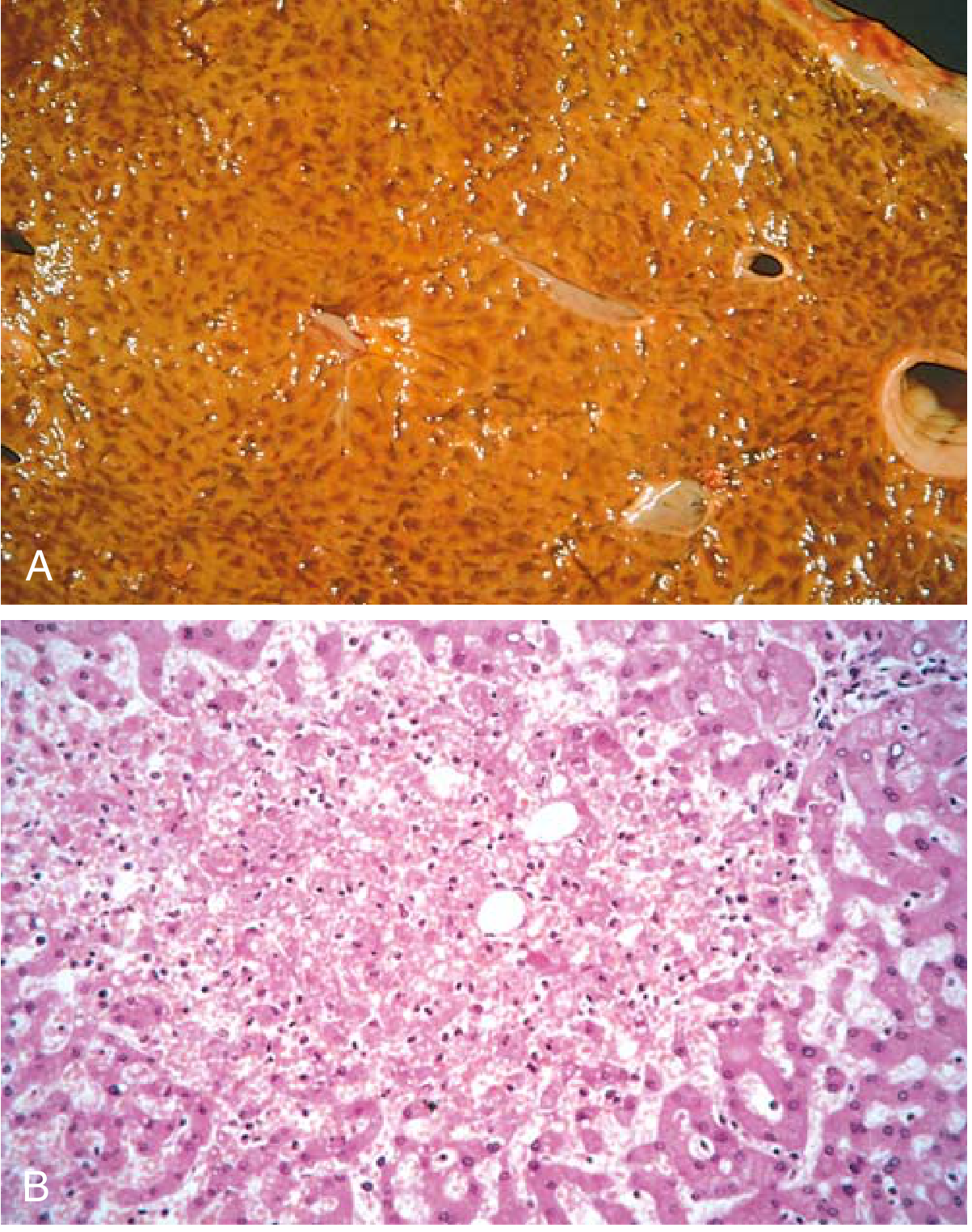
- Acute hepatic congestion: central vein and sinusoids distended with blood; centrilobular hepatocyte necrosis
- Chronic passive liver congestion ("nutmeg liver"): congested red-brown central zones surrounded by tan, fatty periportal hepatocytes
2. EDEMA
Edema = accumulation of interstitial fluid in tissues from net movement of water into extravascular spaces.
- ~60% of lean body weight is water; 2/3 intracellular, 1/3 interstitial; only 5% in plasma
- Cavity collections: hydrothorax (pleural), hydropericardium (pericardial), hydroperitoneum/ascites (peritoneal)
- Fluid types: transudate (protein-poor, low specific gravity, non-inflammatory) vs. exudate (protein-rich, inflammatory)
Causes of Non-Inflammatory Edema
1. Increased Hydrostatic Pressure
- Left heart failure → pulmonary edema
- Right heart failure → peripheral and dependent edema (legs, presacral region when supine)
- Venous obstruction (DVT, hepatic vein obstruction in Budd-Chiari syndrome)
2. Reduced Plasma Osmotic Pressure (Hypoalbuminemia)
- Decreased synthesis: liver disease (cirrhosis), protein malnutrition (kwashiorkor)
- Increased loss: nephrotic syndrome (protein lost in urine), protein-losing enteropathy
- Result: generalized edema (anasarca)
3. Lymphatic Obstruction
- Causes: fibrosis (post-irradiation), tumors, parasitic infection (filariasis)
- Results in lymphedema - hard, non-pitting
4. Sodium and Water Retention
- Activation of RAAS (low renal perfusion → aldosterone → Na+ retention)
- Occurs in heart failure, renal failure
Inflammatory edema is caused by increased vascular permeability (from mediators like histamine, prostaglandins).
3. HEMORRHAGE
Hemorrhage = escape of blood from blood vessels.
- Hematoma: blood accumulates within tissue
- Petechiae (<1-2 mm): tiny pin-point bleeds (e.g., thrombocytopenia, vitamin C deficiency)
- Purpura (3-5 mm): slightly larger
- Ecchymoses (>1-2 cm): larger subcutaneous bleeds ("bruises")
- Hemothorax, hemopericardium, hemarthrosis: blood in body cavities
Clinical Significance:
- Loss of up to 20% blood volume: well tolerated in healthy adults
- Greater loss → hemorrhagic (hypovolemic) shock
- Location matters: minor subcutaneous bleed vs. fatal intracranial bleed
- Chronic external blood loss (e.g., peptic ulcer, menorrhagia) → iron deficiency anemia
- Internal bleeding (hematoma) does NOT cause iron deficiency - iron is recycled from phagocytosed RBCs
4. HEMOSTASIS AND THROMBOSIS
Normal Hemostasis
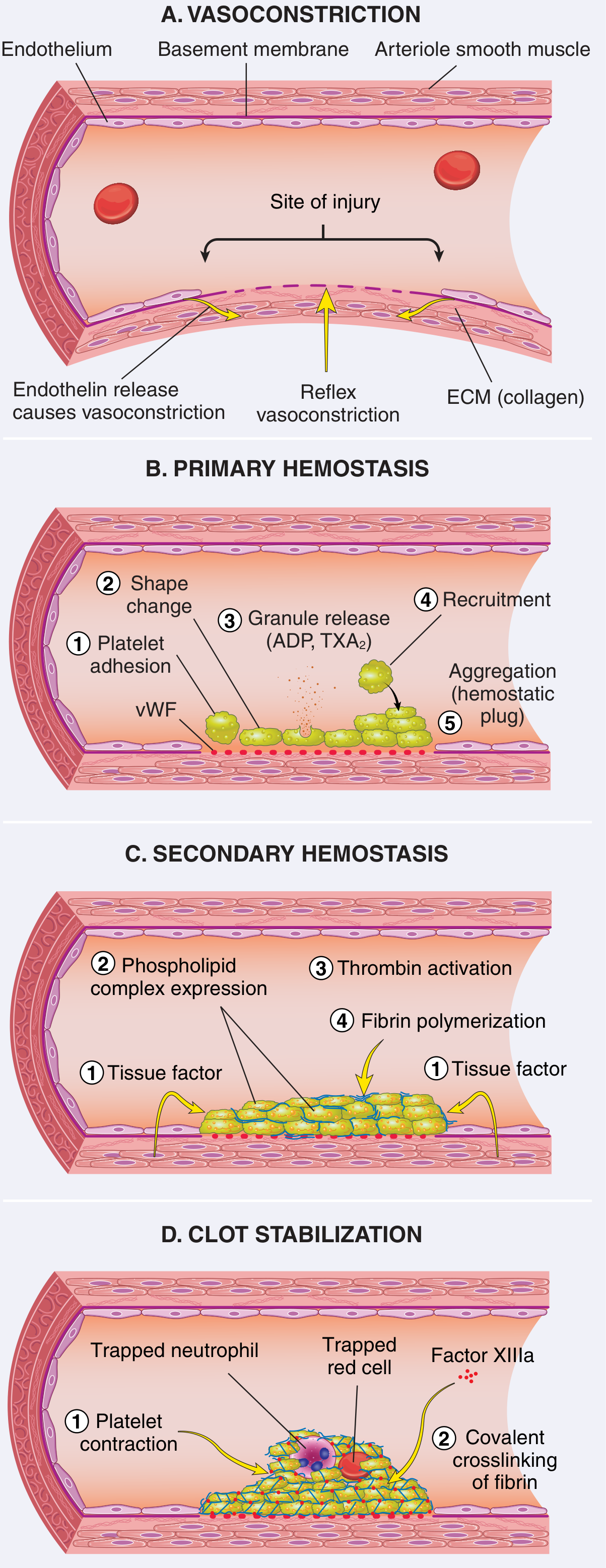
Hemostasis involves three overlapping events after vascular injury:
Step 1 - Arteriolar Vasoconstriction
- Immediate, mediated by neurogenic reflexes and local endothelin release
- Transient - bleeding resumes without platelet/coagulation activation
Step 2 - Primary Hemostasis (Platelet Plug)
- Endothelial injury exposes subendothelial ECM (especially collagen and vWF)
- von Willebrand factor (vWF) bridges collagen and platelet GpIb receptors → platelet adhesion
- Adhesion triggers platelet activation: shape change, secretion of granules (ADP, TXA2, serotonin, fibronectin)
- Secreted ADP and TXA2 recruit more platelets → aggregation via conformational change in GpIIb-IIIa receptors that bind fibrinogen
- Result: primary hemostatic plug (loose)
Step 3 - Secondary Hemostasis (Coagulation Cascade)
- Tissue factor (TF/factor III) is exposed at injury site and initiates coagulation
- Most important factors: VII, IX, X, II (prothrombin), and fibrinogen; cofactors V and VIII
- Thrombin (factor IIa) is the central enzyme - it:
- Converts fibrinogen → fibrin
- Activates factor XIII (crosslinks fibrin)
- Promotes platelet contraction
- Result: secondary hemostatic plug (solid, stable fibrin mesh)
Step 4 - Counter-Regulatory Mechanisms (prevent excessive clotting)
- Washout of activated factors; hepatic removal
- Requirement for phospholipid surfaces (only on activated platelets)
- Antithrombin III: inhibits thrombin and factors IXa, Xa, XIa, XIIa (heparin enhances this)
- Proteins C and S: inactivate factors Va and VIIIa
- Tissue plasminogen activator (t-PA): activates plasminogen → plasmin, which degrades fibrin clots
- Thrombomodulin: on intact endothelium, binds thrombin and shifts it to activate protein C
5. THROMBOSIS
Thrombosis = pathologic intravascular clot formation. Governed by Virchow's Triad:
Virchow's Triad
| Component | Mechanism | Examples |
|---|---|---|
| Endothelial injury | Exposes pro-coagulant ECM; releases TF | Atherosclerosis, hypertension, vasculitis, trauma, smoking |
| Abnormal blood flow (stasis or turbulence) | Prevents dilution of activated factors; disrupts laminar flow | Atrial fibrillation, aneurysms, atherosclerotic plaques, MI, DVT |
| Hypercoagulability | Alters coagulation factors or anticoagulant pathways | See table below |
Hypercoagulable States
Primary (Genetic):
- Factor V Leiden (Arg506Glu): most common; factor V becomes resistant to protein C inactivation; ~3-fold increased venous thrombosis risk; heterozygous frequency >1% in USA
- Prothrombin G20210A variant: elevated prothrombin levels
- Antithrombin III, Protein C, Protein S deficiencies (rare; present in young adults with venous thromboembolism)
- Elevated homocysteine: associated with both arterial and venous thrombosis
Secondary (Acquired) - High Risk:
- Prolonged bed rest / immobilization
- Myocardial infarction, atrial fibrillation
- Surgery, fracture, burns
- Cancer (especially mucinous adenocarcinomas - Trousseau syndrome)
- Prosthetic cardiac valves
- DIC, Heparin-induced thrombocytopenia (HIT)
- Antiphospholipid antibody syndrome (APS): antibodies against phospholipid-binding proteins → recurrent arterial/venous thrombosis, recurrent miscarriages; can be primary or secondary (e.g., in SLE)
Secondary - Elevated Risk:
- Cardiomyopathy, nephrotic syndrome
- Pregnancy, oral contraceptive use
- Sickle cell anemia, smoking
Morphology of Thrombi
- Lines of Zahn: pale platelet/fibrin layers alternating with darker RBC-rich layers (indicates thrombus formed in flowing blood - distinguishes antemortem from postmortem clot)
- Mural thrombi: attached to wall of vessels/heart chambers (e.g., post-MI left ventricular thrombus, aortic aneurysm thrombi)
- Occlusive thrombi: completely obstruct vessel lumen (common in smaller arteries and veins)
- Venous thrombi: form in areas of stasis; propagate toward heart; more prone to embolize
Fates of Thrombi
- Propagation: enlargement by accumulation of more platelets/fibrin
- Embolization: dislodgement and travel to distant sites
- Dissolution: by fibrinolytic activity (most effective with fresh thrombi)
- Organization and recanalization: fibroblasts and smooth muscle cells grow in; new capillary channels form over weeks, restoring some flow
DIC (Disseminated Intravascular Coagulation)
- Widespread simultaneous activation of coagulation throughout microvascular system
- Causes: obstetric complications (abruptio placentae, septic abortion), sepsis, malignancy, severe trauma, massive transfusions
- Paradoxical combination: thrombosis AND hemorrhage - clotting factors consumed ("consumption coagulopathy")
- Lab: elevated PT, PTT; elevated D-dimers; low fibrinogen, low platelets, fragmented RBCs (schistocytes)
6. EMBOLISM
An embolus = detached intravascular solid, liquid, or gaseous mass carried by blood to a distant site.
Pulmonary Thromboembolism (PTE)
- Source: >95% from deep veins of the leg (popliteal vein or above)
- Frequency: very common; 2-4 per 1000 hospital patients/year; underdiagnosed
- Most small emboli are clinically silent; 60-80% resolve through fibrinolysis
- Massive emboli (>60% of pulmonary circulation): sudden death, right heart failure
- Multiple smaller emboli over time: pulmonary hypertension, right heart strain
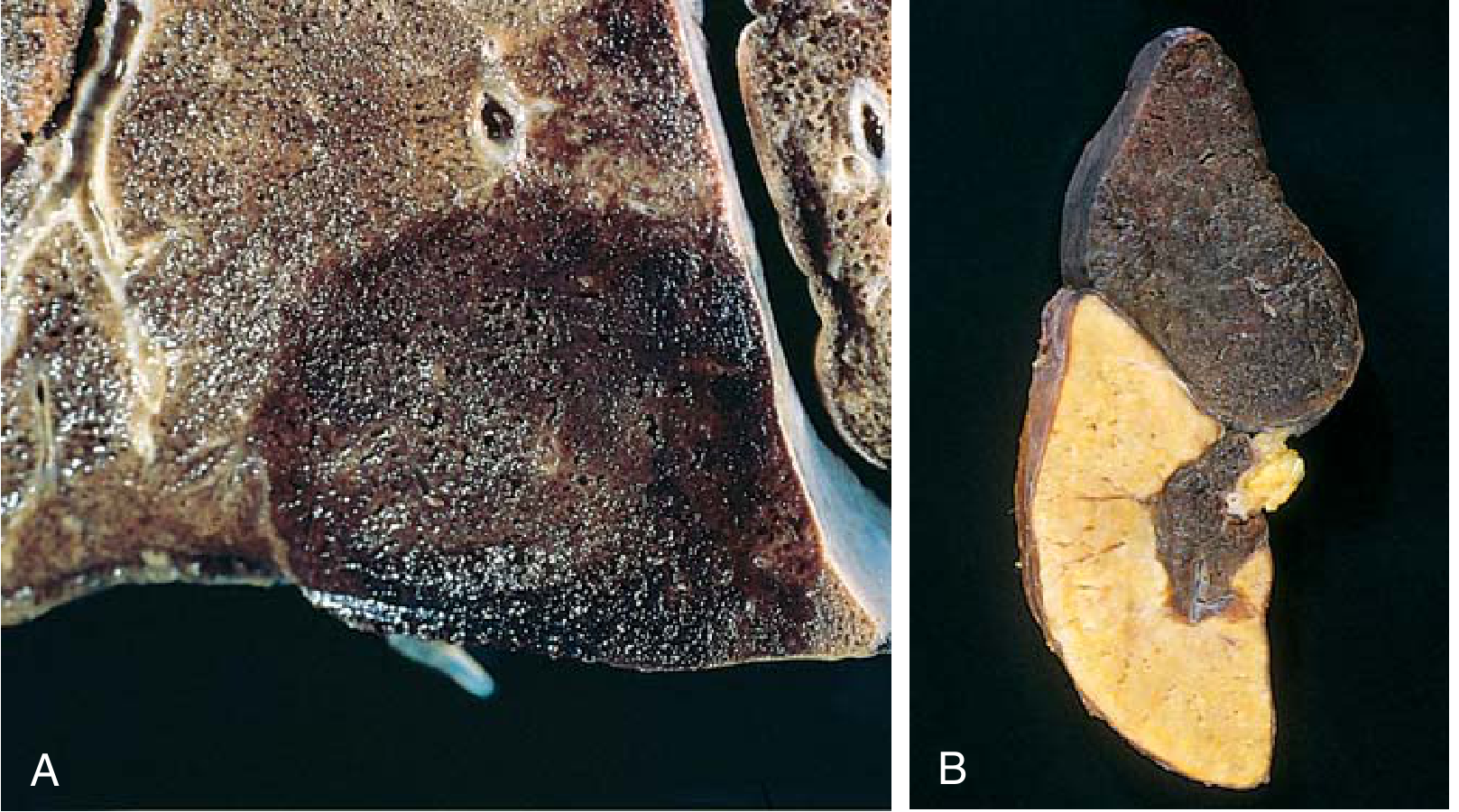
- Hemorrhagic pulmonary infarction: when emboli occur in patients with pulmonary congestion (underlying cardiac or respiratory disease); produces wedge-shaped hemorrhagic infarct
Systemic Thromboembolism
- Source: left heart (80%) - mural thrombi after MI, atrial thrombi in atrial fibrillation, valvular disease
- Also: aortic aneurysm thrombi, paradoxical embolism (through patent foramen ovale)
- Target organs: lower extremity arteries (75%), brain (also common), intestines, kidneys, spleen
- Most result in infarction
Fat Embolism
- Source: fat globules from bone marrow after fractures of long bones or pelvis; also after liposuction, burns
- Fat Embolism Syndrome (1-3% of severe fractures): 1-3 days after injury; triad of:
- Pulmonary insufficiency (hypoxemia, tachypnea)
- Neurological symptoms (confusion, irritability, coma)
- Petechial rash (from thrombocytopenia/fat emboli in skin vessels)
- Mechanism: mechanical obstruction + free fatty acid release → toxic endothelial injury, platelet activation
Amniotic Fluid Embolism
- Rare but catastrophic (~1/40,000 deliveries); mortality ~60%
- Amniotic fluid enters maternal circulation via uterine vein tears at placental site
- Onset: sudden severe dyspnea, cyanosis, hypotension, neurological symptoms, DIC
- Pathology: fetal squamous cells, mucin, lanugo hair in pulmonary vasculature
Air Embolism
- Gas bubbles obstruct flow; 100 mL needed to cause cardiovascular compromise (small amounts tolerated)
- Decompression sickness: rapid pressure decrease (divers, aviators) - dissolved nitrogen forms bubbles
- Joints/muscles: "the bends"
- Pulmonary: "the chokes"
- Chronic form: caisson disease - multifocal ischemic necrosis (especially femoral heads)
- Treatment: hyperbaric oxygen (re-compresses nitrogen bubbles)
7. INFARCTION
Infarct = area of ischemic necrosis caused by obstruction of blood supply.
Red (Hemorrhagic) vs. White (Anemic) Infarcts
| Type | Color | Location | Mechanism |
|---|---|---|---|
| Red infarct | Dark red, hemorrhagic | Lung, small bowel, ovary, liver, brain | Dual blood supply OR venous occlusion OR reperfusion after arterial occlusion |
| White infarct | Pale, anemic | Heart, spleen, kidney | Single (end-arterial) blood supply |
Histology
- Main finding: ischemic coagulative necrosis (except brain → liquefactive)
- Inflammatory response at margins: 1-2 hours, well defined by 1-2 days
- Followed by repair: regeneration at periphery or scar formation
- Remote infarcts: replaced by scar tissue
Factors Influencing Infarct Development
- Anatomy of vascular supply: dual supply (lung, liver) → more protected; end-arterial organs (heart, kidney, spleen) → more vulnerable
- Rate of occlusion: slow occlusion → time for collaterals to develop (e.g., gradual coronary stenosis in atherosclerosis)
- Tissue vulnerability to hypoxia:
- Neurons: irreversible after 3-4 minutes
- Myocardium: irreversible after 20-30 minutes
- Fibroblasts: viable for many hours
8. SHOCK
Shock = state of diminished cardiac output or reduced effective circulating blood volume that impairs tissue perfusion → cellular hypoxia.
Types of Shock
| Type | Mechanism | Causes | Skin |
|---|---|---|---|
| Cardiogenic | Low cardiac output - pump failure | MI, arrhythmia, tamponade, massive PE | Cool, clammy, cyanotic |
| Hypovolemic | Low cardiac output - loss of blood/fluid | Hemorrhage, severe burns | Cool, clammy, cyanotic |
| Septic | Massive vasodilation, vascular leakage | Gram+ bacteria (most common), gram-, fungi | Warm, flushed (early) |
| Neurogenic | Loss of vascular tone | Anesthesia, spinal cord injury | Warm, flushed |
| Anaphylactic | Systemic vasodilation, vascular permeability | IgE-mediated hypersensitivity | Warm, urticarial |
Pathogenesis of Septic Shock
- Incidence: >750,000 cases/year in USA; 2% of all hospital admissions; 50% need ICU
- Mortality: 20-30% despite modern care
- Most common trigger: gram-positive bacteria > gram-negative > fungi
Key Mediators:
- Pattern recognition receptors (TLRs, NOD-like receptors) on macrophages, neutrophils, dendritic cells, endothelium recognize:
- LPS (gram-negative endotoxin) + LPS-binding protein → CD14/TLR4 complex
- Gram-positive cell wall components (peptidoglycan, lipoteichoic acid)
- Trigger massive release of: TNF, IL-1, IL-6, IL-12, IFN-γ, IL-17 and other mediators
- Result: widespread endothelial activation, vasodilation, increased vascular permeability, procoagulant state, metabolic derangements
- Severe septic shock → multi-organ failure → death
Stages of Shock
Stage 1 - Non-Progressive (Compensated) Shock:
- Compensatory mechanisms maintain perfusion to vital organs
- Baroreceptor reflexes, catecholamine release, RAAS activation, ADH release
- Peripheral vasoconstriction, tachycardia, fluid conservation by kidneys
Stage 2 - Progressive Shock:
- Compensatory mechanisms fail
- Widespread tissue hypoxia → anaerobic glycolysis → lactic acidosis
- Acidosis blunts vasoconstriction response; decreased cardiac output
- Renal conservation fails (oliguria)
- Progressive organ dysfunction
Stage 3 - Irreversible Shock:
- Severe cellular and tissue injury
- Myocardial depression, lysosomal enzyme leakage, gut microbe translocation
- Damage to vital organs becomes irreversible despite restoration of perfusion
- Multi-organ failure (kidney → renal tubular necrosis; lungs → ARDS; GI → ischemic enterocolitis)
Morphologic Changes in Shock
- Brain: ischemic encephalopathy
- Heart: foci of coagulative necrosis; subendocardial hemorrhage
- Kidneys: acute tubular necrosis (ATN) - most common cause of acute kidney injury in shock
- Lungs (in septic/traumatic shock): diffuse alveolar damage → ARDS
- Adrenals: lipid depletion (cortical cells secrete all stored lipid)
- GI: ischemic enterocolitis; stress ulcers
Clinical Features
Hypovolemic/Cardiogenic shock:
- Hypotension, weak rapid pulse, tachypnea
- Cool, clammy, cyanotic skin (due to peripheral vasoconstriction)
Septic shock:
- Skin may be warm and flushed early (vasodilation)
- Later stages mirror hypovolemic shock as decompensation occurs
Prognosis:
- Hypovolemic shock in young healthy patients: >90% survival with appropriate treatment
- Septic or cardiogenic shock: substantially worse outcomes even with optimal care
Summary Table: Rapid Review
| Topic | Key Points |
|---|---|
| Edema | Transudate (non-inflammatory) vs. exudate; causes: ↑hydrostatic pressure, ↓oncotic pressure, lymphatic obstruction, Na+ retention |
| Hemorrhage | Petechiae, purpura, ecchymoses; chronic external loss → iron deficiency anemia |
| Hemostasis | Vasoconstriction → platelet plug (1°) → fibrin clot (2°) → anticoagulant mechanisms |
| Thrombosis | Virchow's Triad: endothelial injury + stasis/turbulence + hypercoagulability |
| Embolism | PTE from DVT; systemic from left heart; fat, amniotic fluid, air emboli |
| Infarction | Red (dual supply/venous) vs. white (end-arterial); coagulative necrosis (except brain) |
| Shock | Cardiogenic, hypovolemic, septic (most common in hospitals), neurogenic, anaphylactic |
Source: Robbins & Kumar Basic Pathology (Robbins Pathology), Chapter 3 - Hemodynamic Disorders, Thromboembolism, and Shock
This is a shared conversation. Sign in to Orris to start your own chat.