
peptic ulcer disease vs gastrinoma
"gastrinoma" AND "peptic ulcer"
peptic ulcer disease gastric duodenal ulcer
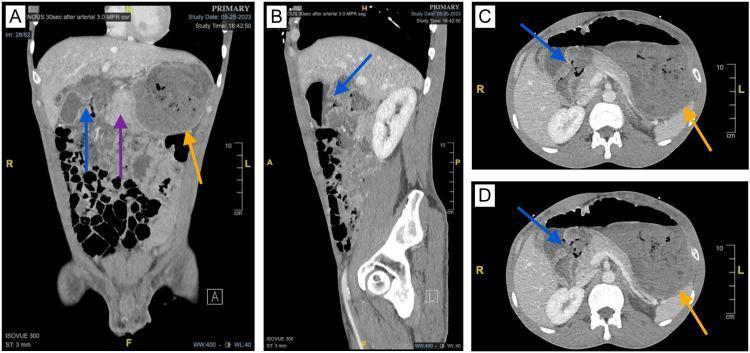
Diagnostic CT imaging of the abdomen and pelvis presented in coronal (A), sagittal (B), and axial (C, D) planes, demonstrating complications of peptic ulcer disease. The visual findings include severe edema of the gastric pylorus and duodenum (purple arrow), which has led to significant gastric outlet obstruction. This obstruction is evidenced by a markedly distended stomach containing retained gastric contents (orange arrows). Additionally, there is a localized area of increased soft tissue attenuation with focal gas and central necrosis, indicated by blue arrows, which suggests a contained or walled-off perforation of the duodenal wall. These images highlight the radiological features of complicated duodenal ulcers, including secondary obstructive changes and focal inflammatory masses associated with chronic perforation. This content is suitable for intermediate to advanced medical learners studying gastrointestinal pathology and radiology.
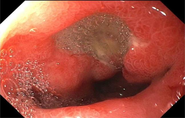
This endoscopic clinical photograph displays a 7 mm cratered ulcer located in the duodenal bulb, a common manifestation of peptic ulcer disease. The ulcer is characterized by a central, depressed crater filled with a brownish-gray fibrinous base and mucus. The surrounding duodenal mucosa is intensely erythematous and edematous, indicating active inflammation (duodenitis). Visible mucosal changes include a granular texture and prominent mucosal folds. Bubbles from gastric secretions are present in the foreground, indicating the fluid-rich environment of the upper gastrointestinal tract. This visual is representative of H. pylori-associated ulceration or NSAID-induced mucosal injury, providing a classic example for gastroenterology and internal medicine education regarding diagnostic upper endoscopy (esophagogastroduodenoscopy) and the morphological evaluation of duodenal lesions.
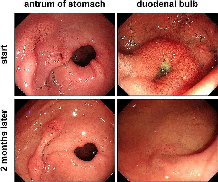
This diagnostic comparison chart displays four gastroscopic images documenting the healing progression of peptic ulcers. The images are arranged in a 2x2 grid, categorized by anatomical location (antrum of stomach and duodenal bulb) and timeline (start vs. 2 months later). The 'start' images reveal significant mucosal injury: the gastric antrum shows a deep, large ulcer with surrounding mucosal erosions, while the duodenal bulb demonstrates a prominent ulcer with central green slough and erythematous, inflamed surrounding tissue. The '2 months later' images, following treatment with a proton pump inhibitor (omeprazole), illustrate substantial healing. In the gastric antrum, the ulcer has markedly decreased in size with smoothing of the adjacent mucosa. In the duodenal bulb, the ulcer has completely resolved, replaced by a red linear scar and healthy regenerative epithelium. This visual evidence highlights the efficacy of pharmacologic intervention in treating NSAID-related (aspirin-induced) peptic ulcer disease and the capacity for mucosal recovery over a two-month period.
gastrinoma Zollinger-Ellison syndrome pancreatic neuroendocrine tumor
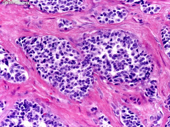
Histopathology image of a gastroenteropancreatic neuroendocrine tumor (gastrinoma) evaluated on hematoxylin and eosin stain. The tumor is composed of uniform nests and cords of neuroendocrine cells embedded in dense fibrous stroma with conspicuous fibrous septa. Cells are polygonal with round to oval nuclei and granular (salt-and-pepper) nuclear chromatin; cytoplasm is moderate to scant, with low mitotic activity. Architectural pattern shows cohesive clusters separated by desmoplastic stroma rather than mucosal nodules with tightly packed trabeculae and acini typical of Type 2 gastrinomas. In this case the desmoplastic reaction may contribute to firm, fibrous tissue surrounding tumor nodules. Immunohistochemistry (not shown) would commonly be positive for chromogranin A and synaptophysin, supporting neuroendocrine differentiation, and gastrin production in clinically suspected Zollinger-Ellison syndrome or MEN1. Clinically, gastrin-secreting tumors cause hypergastrinemia, peptic ulcers, and refractory ulcers; they are part of the MEN1 spectrum. Pathologic differential includes well-differentiated neuroendocrine tumor versus poorly differentiated neuroendocrine carcinoma and pancreatic ductal adenocarcinoma with desmoplasia; architectural and cytologic features favor NET. The image highlights important diagnostic clues: neuroendocrine cytology, uniform cell morphology, nesting patterns, and desmoplastic stroma, guiding clinical correlation and management. This histologic pattern informs prognosis, surgical planning, and consideration of somatostatin receptor-targeted therapies in NETs. Moreover, it guides clinical management.
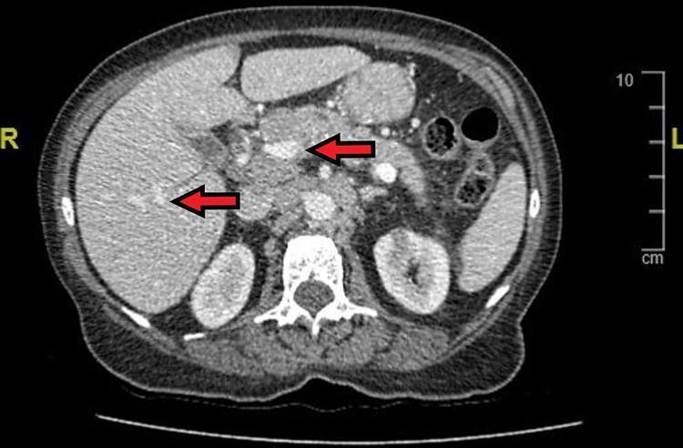
This diagnostic image is a contrast-enhanced axial (transverse) computed tomography (CT) scan of the upper abdomen. The visualization shows a cross-section of the abdominal cavity with anatomical landmarks including the liver (right), pancreas (central), kidneys (bilateral posterior), and the vertebral body of the spine. Red arrows highlight significant clinical findings: one arrow indicates a hyperenhancing mass located within the pancreatic head, identified as a primary gastrinoma (Zollinger-Ellison syndrome). A second arrow identifies a focal lesion within the hepatic parenchyma, consistent with metastatic disease. The image demonstrates the relationship between primary neuroendocrine tumors and their secondary spread. The surrounding soft tissues, retroperitoneal space, and bowel loops are also visible, providing context for tumor localization and staging. This visual is intended for medical education regarding the diagnostic imaging of pancreatic neuroendocrine tumors and their common metastatic patterns to the liver.
Peptic Ulcer Disease vs. Gastrinoma (Zollinger-Ellison Syndrome)
Overview
Pathophysiology Comparison
| Feature | PUD | Gastrinoma (ZES) |
|---|---|---|
| Core mechanism | Imbalance: mucosal defense vs. acid/pepsin damage | Autonomous gastrin hypersecretion → unregulated acid overproduction |
| Gastrin level | Normal or mildly elevated | Markedly elevated (often >10x ULN) |
| Parietal cell mass | Normal or mildly increased | Greatly increased (gastrin is trophic to parietal cells) |
| Acid secretion | Normal to moderately elevated | Massively elevated; BAO often >15 mEq/h |
| Feedback inhibition | Gastrin secretion normally suppressed by luminal acid | Tumor-derived gastrin NOT inhibited by acid - secretion continues unabated |
| H. pylori role | Major cause (>70% of cases); damages mucosa, inhibits somatostatin → increased gastrin → more acid | Can coexist but is not the primary driver |
| NSAID role | Major cause (especially gastric ulcers); inhibit prostaglandin E2, impair mucosal barrier | Not a primary driver |
Physiology of the Defect
Clinical Features Comparison
| Feature | PUD | Gastrinoma (ZES) |
|---|---|---|
| Prevalence | Very common; lifetime risk ~10% (M), ~4% (F) | Rare; <0.5% of peptic ulcers |
| Age of onset | Any age; increasing >60 yrs | Typically 30-50 years |
| Sex | M slightly > F | Men 60% |
| Ulcer number | Usually solitary (>80% of cases) | Often multiple |
| Ulcer location | Gastric antrum / proximal duodenum (within a few cm of pyloric valve) | Multiple in duodenum AND stomach; can involve jejunum (atypical - red flag) |
| Response to therapy | Good response to PPIs + H. pylori eradication | Refractory to standard therapy - a key red flag |
| Diarrhea | Uncommon | Present in 70% of patients as initial symptom (secretory + osmotic, from acid inactivating pancreatic enzymes) |
| Steatorrhea | Uncommon | Yes - low duodenal pH inactivates pancreatic lipase; fat malabsorption |
| Gastric folds | Normal | Thickened gastric folds on endoscopy (94% of cases) |
| Complications | Bleeding, perforation, obstruction | Same + hepatic metastases (>50% malignant at diagnosis) |
| Associated syndromes | - | MEN-1 (Werner syndrome) in ~25%; multifocal tumors |
Morphology
PUD
- Solitary (>80%), round to oval, "punched-out" defect with smooth, clean base (peptic digestion of exudate)
- Sharp margins, level with surrounding mucosa (heaped-up margins suggest cancer)
- Most common: proximal duodenum (a few cm from pylorus) and gastric lesser curvature (body-antrum junction)
- Histology: fibrinoid debris + neutrophils acutely; granulation tissue + fibrous scar chronically
- Robbins, Cotran & Kumar Pathologic Basis of Disease
Gastrinoma
- Tumor itself: uniform nests/cords of neuroendocrine cells with "salt-and-pepper" chromatin; bland histology; desmoplastic stroma; IHC positive for chromogranin A, synaptophysin, and gastrin
- More than half are locally invasive or metastatic at diagnosis; 60% are malignant
- Sporadic gastrinomas: usually single; MEN-1 associated: frequently multifocal
- Located in the "gastrinoma triangle": pancreas, duodenum, peripancreatic soft tissues
- Robbins, Cotran & Kumar Pathologic Basis of Disease, p. 1028
Diagnosis
When to Suspect Gastrinoma Over PUD
- Ulcers refractory to standard PPI therapy
- Multiple or recurrent ulcers
- Ulcers in atypical locations (distal duodenum, jejunum)
- Unexplained secretory diarrhea or steatorrhea
- Prominent gastric rugal folds on endoscopy/imaging
- PUD + family history of MEN-1 (pituitary, parathyroid, pancreatic tumors)
- Hypercalcemia (from MEN-1 hyperparathyroidism stimulating gastrin)
Diagnostic Workup for ZES
| Test | Details | Interpretation |
|---|---|---|
| Fasting serum gastrin | First-line; must stop PPI for ≥7 days before testing (substitute H2 blocker, stop 24h prior) | Normal <150 pg/mL; ZES typically >150-200 pg/mL; >10x ULN (e.g., >1000 pg/mL) + acid hypersecretion = virtually diagnostic |
| Gastric pH / BAO | Gastric aspirate pH; Basal Acid Output | pH <3 suggestive; BAO >15 mEq/h + hypergastrinemia = pathognomonic of ZES |
| Secretin stimulation test | Gold standard provocative test; IV secretin 2 U/kg bolus, gastrin measured before and after | In ZES: paradoxical rise in gastrin ≥120 pg/mL above baseline (normally secretin inhibits gastrin); sensitivity and specificity superior to calcium infusion test |
| Calcium infusion test | Older test; less sensitive/specific than secretin | Less preferred |
| Endoscopy | Look for multiple ulcers, thickened folds, jejunal ulcers | 94% of ZES have thickened gastric folds |
| Imaging (CT/MRI/EUS/Octreoscan) | Localize tumor | Somatostatin receptor scintigraphy (Octreoscan) most sensitive for metastases |
| H. pylori testing | Urea breath test, stool antigen, biopsy CLO test | Positive in PUD; can coexist in ZES but not primary driver |
Treatment Comparison
| Aspect | PUD | Gastrinoma (ZES) |
|---|---|---|
| Acid suppression | PPI (first-line); H2 blockers | High-dose PPI (H+/K+-ATPase inhibitors) - often require doses 2-3x standard |
| H. pylori eradication | Triple/quadruple therapy if H. pylori positive; essential | Treat if present; does not cure ZES |
| Surgery | Rarely needed (for complications: perforation, bleeding, obstruction) | Resection of the tumor when localized; total gastrectomy historically (now replaced by medical acid suppression) |
| Tumor-targeted therapy | N/A | Surgical resection (potentially curative in sporadic, non-metastatic disease); somatostatin analogs (octreotide) for symptom control; chemotherapy/targeted therapy for metastatic disease |
| Prognosis | Excellent with treatment | Depends on tumor extent; hepatic metastases = shortened life expectancy (often <10 years with progressive liver failure) |
Key Distinguishing Summary Table
| Feature | PUD | Gastrinoma (ZES) |
|---|---|---|
| Frequency | Very common | Rare |
| Etiology | H. pylori, NSAIDs | Gastrin-secreting neuroendocrine tumor |
| Fasting gastrin | Normal | Markedly elevated |
| Ulcer locations | Duodenal bulb, gastric antrum | Multiple; distal duodenum, jejunum |
| Response to PPIs | Good | Refractory / requires high-dose |
| Diarrhea/steatorrhea | Uncommon | Common (70%); due to acid-inactivated pancreatic enzymes |
| Secretin test | Gastrin falls/unchanged | Paradoxical gastrin rise ≥120 pg/mL |
| Malignancy | Ulcer itself is not malignant (but gastric ulcers need biopsy to exclude cancer) | Tumor itself: >60% malignant |
| Association | H. pylori, NSAIDs, smoking | MEN-1 (25% of cases) |