
General Medicine MBBS Pedal Edema Practical theory
Reading File
Now let me search the medical library comprehensively for pedal edema content.
Finding Sources
Finding Sources
Searching the Web
pedal edema MBBS general medicine clinical examination causes pathophysiology
Finding Sources
Finding Sources
Excellent - I found rich content. Let me now read the key sections from Harrison's, Frameworks for Internal Medicine, and Robbins Pathology in parallel.
Reading File
Reading File
Finding Sources
Reading File
Reading File
Reading File
Reading File
Reading File
Finding Sources
Finding Sources
Finding Sources
Reading File
Loading Image
Loading Image
Finding Sources
I now have comprehensive information from multiple authoritative textbooks. Let me compile the full detailed answer on Pedal Edema for MBBS General Medicine.
Pedal Edema - General Medicine MBBS: Complete Practical Theory
1. Definition
Pedal edema (also called peripheral or dependent edema) is the accumulation of excess fluid (interstitial fluid) in the tissues of the feet, ankles, and lower legs. Broadly, edema refers to an excess of salt and water in the extracellular, particularly interstitial, space.
-
Anasarca = severe generalized edema with profound subcutaneous swelling + fluid in body cavities
-
Effusions = fluid in pleural (hydrothorax), pericardial (hydropericardium), or peritoneal (ascites/hydroperitoneum) cavities
-
Robbins & Kumar Basic Pathology, p. 2223
2. Pathophysiology - Starling Forces
Fluid movement between vascular and interstitial spaces is governed by four Starling forces:
| Force | Direction of Action |
|---|---|
| Capillary hydrostatic pressure (Pc) | Pushes fluid OUT of capillary |
| Plasma oncotic pressure (πc) | Pulls fluid INTO capillary |
| Interstitial hydrostatic pressure (Pi) | Pushes fluid INTO capillary |
| Interstitial oncotic pressure (πi) | Pulls fluid OUT of capillary |
Normally, a small net outflow at the arterial end is drained by lymphatics back to the thoracic duct. Edema forms when this balance tips toward excess filtration OR when lymphatic drainage is overwhelmed.
- Medical Physiology (Boron & Boulpaep), p. 3719
- Robbins & Kumar Basic Pathology, p. 2227
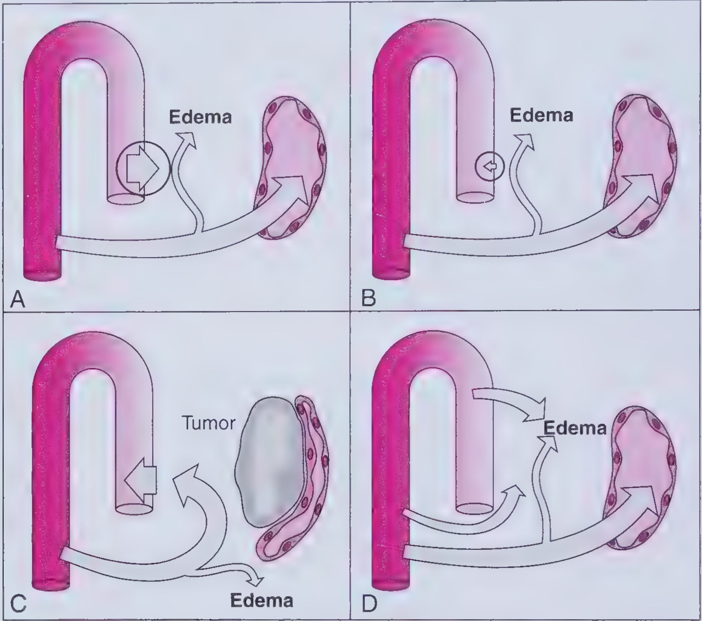
3. Classification of Causes (Mechanistic Framework)
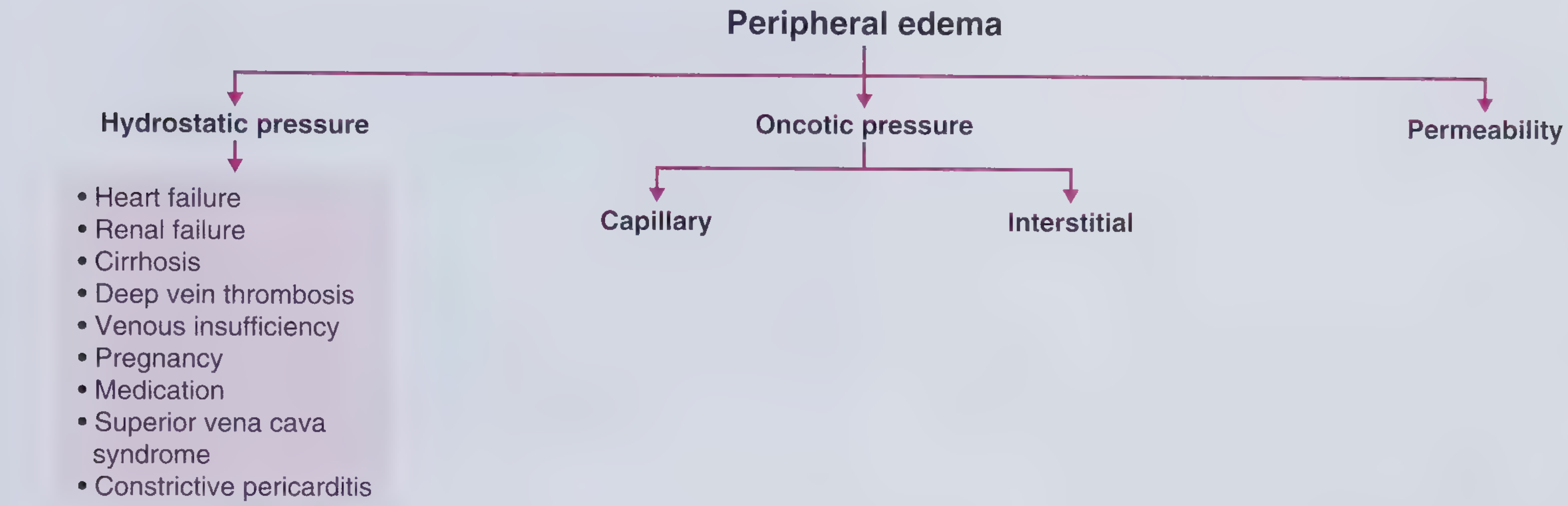
A. Increased Capillary Hydrostatic Pressure
Results in bilateral, dependent, pitting, gradual-onset edema.
| Mechanism | Causes |
|---|---|
| Impaired venous return | Congestive heart failure (CHF), constrictive pericarditis, liver cirrhosis |
| Venous obstruction/compression | Deep vein thrombosis (DVT), external mass compression |
| Lower extremity dependency | Prolonged standing, immobility |
| Arteriolar dilation | Heat, CCB drugs (DHP - amlodipine), pregnancy |
Note: Systemic arterial hypertension does NOT cause peripheral edema because precapillary sphincter autoregulation prevents transmission of arterial pressure to capillaries.
- Frameworks for Internal Medicine, p. 5713-5720
B. Decreased Capillary Oncotic Pressure (Hypoalbuminemia)
Results in generalized, dependent, pitting edema; often with ascites and pleural effusions (but NOT pulmonary edema).
Plasma albumin <2 g/dL is associated with clinically evident edema.
| Mechanism | Cause |
|---|---|
| Decreased albumin synthesis | Liver disease, malnutrition, cirrhosis |
| Increased albumin loss | Nephrotic syndrome (proteinuria), protein-losing enteropathy |
- Frameworks for Internal Medicine, p. 5885-5896
C. Increased Interstitial Oncotic Pressure (Lymphatic Obstruction)
Results in localized, dependent, NON-pitting edema (early pitting may occur; over time skin becomes thickened, darkened, with warty projections = lymphostatic verrucosis).
| Cause | Details |
|---|---|
| Lymphedema | Malignant obstruction (lymphoma), post-surgical (mastectomy), radiation, infection (filariasis) |
| Myxedema (hypothyroidism) | Bilateral non-pitting lower extremity edema + weight gain, constipation, bradycardia, dry hair |
- Frameworks for Internal Medicine, p. 5962-6015
D. Increased Capillary Permeability
Variable phenotype - pitting or non-pitting, localized or generalized.
| Cause | Key Features |
|---|---|
| Local inflammation | Trauma, cellulitis, burns, insect stings - localized, non-pitting |
| Preeclampsia | Hypoalbuminemia + endothelial dysfunction; lower extremity ± pulmonary/cerebral edema |
| ACE inhibitor-induced angioedema | Non-dependent, non-pitting; face/lips/tongue/hands/feet |
| Systemic capillary leak syndrome | Rare; episodic hypotension, hemoconcentration, peripheral edema |
| Idiopathic edema | Unexplained episodic edema in women; no systemic cause found |
- Frameworks for Internal Medicine, p. 6052-6105
4. Pitting vs. Non-Pitting Edema
| Feature | Pitting Edema | Non-Pitting Edema |
|---|---|---|
| Definition | Pressing leaves a "pit" (depression) | No pit on pressing |
| Mechanism | High water content; fluid displaced temporarily | Protein/fibrosis in interstitium |
| Causes | CHF, nephrotic syndrome, cirrhosis, DVT, hypoalbuminemia | Lymphedema, myxedema |
| Fluid type | Transudate (protein-poor) | Protein-rich (lymphatic) or mucopolysaccharide-rich |
5. Grading of Pitting Edema (Clinical Scale)
| Grade | Pit Depth | Rebound Time |
|---|---|---|
| 1+ | 2 mm | Immediate |
| 2+ | 4 mm | <15 seconds |
| 3+ | 6 mm | 15-30 seconds |
| 4+ | 8 mm (severe) | >30 seconds |
6. Clinical Approach to a Patient with Pedal Edema
History
- Onset: Acute (DVT, allergy) vs. chronic (CHF, cirrhosis, nephrotic)
- Bilateral vs. unilateral: Bilateral = systemic cause; Unilateral = DVT, venous insufficiency, local pathology
- Aggravating factors: Worsens with standing/evening (venous), position independent (lymphedema)
- Associated symptoms:
- Dyspnea, orthopnea, PND → CHF
- Jaundice, alcohol use → cirrhosis
- Frothy urine, facial puffiness → nephrotic syndrome
- Weight gain, cold intolerance, constipation → hypothyroidism
- Unilateral calf pain/erythema → DVT
- Drug history: Amlodipine, NSAIDs, steroids, thiazolidinediones, minoxidil
Physical Examination
- Inspect: Location, extent, skin changes (hyperpigmentation, varicosities, ulcers near medial malleoli suggest venous disease)
- Palpation: Pitting test - press over the dorsum of foot or pretibial area for 5 seconds; assess depth and recovery
- JVP assessment: Elevated JVP → CHF or constrictive pericarditis; Normal JVP → cirrhosis, nephrotic syndrome
- Abdomen: Ascites + pedal edema → cirrhosis or right heart failure
- Systemic signs:
- Spider angiomas, palmar erythema, Terry's nails, caput medusae → liver disease
- S3 gallop, displaced apex → CHF
- Periorbital edema in morning → nephrotic syndrome (note: periorbital at awakening, pedal at end of day reflects gravitational redistribution)
- Brenner and Rector's The Kidney, p. 1545
7. Investigations
| Investigation | What it tells you |
|---|---|
| Urinalysis / 24-hr urine protein | Nephrotic syndrome (>3.5 g/day proteinuria) |
| Serum albumin | <2 g/dL → hypoproteinemic edema |
| LFTs / PT-INR | Liver disease |
| RFTs / serum creatinine | Renal failure |
| TSH / T3, T4 | Hypothyroidism / myxedema |
| ECG + Echo / BNP/NT-proBNP | Heart failure |
| Compression Doppler USS | DVT (gold standard) |
| Chest X-ray | Pulmonary edema, pleural effusion |
| Urine protein : creatinine ratio | Quick screen for nephrotic range proteinuria |
8. Bilateral vs. Unilateral Edema - Differential at a Glance
| Bilateral | Unilateral |
|---|---|
| CHF (right-sided) | DVT |
| Nephrotic syndrome | Cellulitis / local infection |
| Liver cirrhosis | Lymphedema (post-mastectomy, filariasis) |
| Renal failure | Chronic venous insufficiency |
| Hypoalbuminemia | Ruptured Baker's cyst |
| Hypothyroidism/Myxedema | Compartment syndrome |
| Drugs (amlodipine, NSAIDs) | SVC syndrome (upper limb) |
| Pregnancy | Venous obstruction by pelvic mass |
9. Treatment Principles
Treatment targets the underlying cause, supplemented by:
| Approach | Details |
|---|---|
| Diuretics | Loop diuretics (furosemide) for CHF, nephrotic syndrome, cirrhosis; avoid if severely hypoalbuminemic (poor gut absorption - use IV route) |
| Sodium restriction | <2 g/day dietary sodium in CHF/cirrhosis |
| Leg elevation | Reduces hydrostatic pressure |
| Compression stockings | Chronic venous insufficiency, lymphedema |
| Treat underlying cause | ACE inhibitors/ARBs for CHF; steroids/immunosuppression for nephrotic; thyroid replacement for myxedema; anticoagulation for DVT |
| Lymphedema | Diuretics usually ineffective; pneumatic compression, manual lymphatic drainage |
- Frameworks for Internal Medicine, p. 6175-6186
- Goodman & Gilman's Pharmacological Basis of Therapeutics, p. 2216
10. High-Yield MBBS Exam Points
- Transudate (protein-poor) = high hydrostatic OR low oncotic pressure. Exudate (protein-rich) = increased permeability / inflammation.
- Periorbital edema in the morning + pedal edema by evening = hypoproteinemia (gravitational redistribution).
- JVP is the key discriminator: Raised JVP + edema = CHF/constrictive pericarditis. Normal JVP + edema = nephrotic/cirrhosis.
- Non-pitting + bilateral = myxedema until proven otherwise.
- Unilateral + acute + painful = DVT; Doppler USS is diagnostic.
- Amlodipine (dihydropyridine CCB) is the most common drug cause of pedal edema.
- Lymphedema does NOT respond to diuretics.
- Plasma albumin <2 g/dL is the threshold for clinical hypoproteinemic edema.
- Filariasis (Wuchereria bancrofti) = classic cause of lymphedema / elephantiasis in tropical countries - highly relevant for India-based MBBS.
- In cirrhosis: ascites typically appears before pedal edema; JVP is normal (unlike CHF where it is raised).
Sources: Frameworks for Internal Medicine (9781496359308); Robbins & Kumar Basic Pathology (9780323790185); Medical Physiology - Boron & Boulpaep (9780323319737); Brenner and Rector's The Kidney (9780323532655); Harrison's Principles of Internal Medicine 22E (9781265977061); Goodman & Gilman's Pharmacological Basis of Therapeutics (9781264258079)
This is a shared conversation. Sign in to Orris to start your own chat.