
Reading File
This is an orthopaedics pre-university exam paper from MVASMC Basti with 5 questions. Let me search all topics simultaneously from the medical library.
Finding Sources
Finding Sources
Finding Sources
Finding Sources
Finding Sources
Reading File
Reading File
Finding Sources
Finding Sources
Reading File
Reading File
Finding Sources
Finding Sources
Reading File
Reading File
Reading File
Reading File
I now have comprehensive content from the textbooks to write full model answers. Let me compile the complete answers.Here are model answers for your Pre-University Orthopaedics examination (MVASMC Basti):
Pre University Examination - Orthopaedics: Model Answers
Q1. Clostridial Gas Gangrene (Clostridial Myonecrosis) [10 marks]
Definition
Gas gangrene is a rapidly progressive, life-threatening soft-tissue infection characterised by widespread muscle necrosis, gas production in tissues, and severe systemic toxaemia. It is most commonly caused by Clostridium perfringens.
Etiopathogenesis
Causative Organisms:
- Clostridium perfringens - most common (80-90%)
- Clostridium septicum, C. bifermentans, C. sporogenes
- Non-clostridial gas-producing organisms (coliforms) are co-isolated in 60-85% of cases
Predisposing Conditions:
- Crushing injuries, missile/gunshot wounds
- Compound fractures with devitalised muscle
- Ischaemic/vascular injuries
- Bowel surgery (spontaneous gas gangrene from C. septicum)
- Premature wound closure
Pathogenesis:
- Devitalised (ischaemic) tissue creates the anaerobic environment needed for spore germination
- Incubation period: usually <24 hours (range: 1 hour to 6 weeks)
- C. perfringens produces multiple exotoxins:
- Alpha-toxin (lecithinase) - most important; destroys RBCs, WBCs, platelets, fibroblasts, and muscle cells
- Phi-toxin - causes myocardial suppression
- Kappa-toxin - destroys connective tissue and blood vessels
- A vicious cycle ensues: bacteria multiply -> exotoxins cause local necrosis and vascular thrombosis -> further anaerobic spread -> gas (CO2, H2, N2) tracks along muscle planes
- Systemic exotoxins cause severe haemolysis, leading to hypotension, shock, AKI, and ARDS
Clinical Features
Local Signs:
- Pain - earliest symptom; rapidly increases in severity (disproportionate to wound appearance)
- Tense, woody swelling of the limb
- Wound exudes serosanguineous (brownish, "dishwater") discharge
- Skin turns bronze/brown, then blue-black
- Haemorrhagic bullae (blisters)
- Soft-tissue crepitus on palpation (gas in tissue planes)
- Characteristic sickly-sweet odour
Systemic Signs:
- High-grade fever, tachycardia, tachypnoea
- Altered mental status (restlessness, anxiety, then delirium)
- Hypotension progressing to septic shock
- Jaundice (from haemolysis)
- Oliguria/anuria (AKI)
Investigations
- X-ray/CT scan - feathery gas shadows within muscle planes
- Gram stain of exudate - large Gram-positive bacilli, notably WITHOUT neutrophils (neutrophils are destroyed by the toxins)
- Blood tests - haemolytic anaemia, leucocytosis, metabolic acidosis, raised creatinine
- Culture - anaerobic culture of wound discharge confirms organism
Differential Diagnosis
| Condition | Distinguishing Features |
|---|---|
| Necrotising fasciitis | Fascia necrosis, not primarily muscle; mixed organisms; less rapid gas |
| Cellulitis | No gas, no crepitus, less systemic toxicity |
| Non-clostridial crepitant myositis | Coliforms, slower; less toxaemia |
| Clostridial cellulitis | Gas in subcutaneous tissue, muscle not involved; less toxic |
| Streptococcal myonecrosis | S. pyogenes; incubation 3-4 days; less rapid |
| Diabetic foot infection | Gas from facultative anaerobes; less acute onset |
Management
1. Resuscitation (ICU admission):
- IV fluid resuscitation, inotropes for shock
- Monitor urine output, ABG, renal function
- Blood transfusion for haemolytic anaemia
2. Antibiotics (IV, high-dose):
- Penicillin G (high dose) - first-line
- Clindamycin - inhibits toxin production (key adjunct)
- Third-generation cephalosporins added for polymicrobial cover
3. Surgery - EMERGENCY:
- Radical surgical debridement - all necrotic muscle must be excised
- Amputation - in established gas gangrene with systemic toxicity, amputation is life-saving and must not be delayed
- Wound is left OPEN (no closure); packed with saline-soaked gauze
- Re-exploration at 24-48 hours
4. Hyperbaric Oxygen (HBO):
- Recommended in severe cases if facilities available
- Inhibits bacterial growth in high O2 environment; may limit extent of debridement needed
5. Anti-gas gangrene serum - of limited evidence; rarely used now
Prognosis: Mortality 20-30%; higher with truncal/bowel involvement
Q2A. Classical Hand Deformities in Rheumatoid Arthritis [5 marks]
The inflamed rheumatoid synovium (pannus) destroys ligaments, tendons, and articular cartilage, producing characteristic deformities.
1. Ulnar Deviation/Drift at MCPJs
- Most characteristic deformity of RA
- Pannus destroys the MCPJ capsule and collateral ligaments
- Flexor tendons bowstring towards the ulnar side
- Results in ulnar deviation of fingers at the MCPJs with radial deviation of the wrist (zig-zag deformity)
2. Swan Neck Deformity
- Hyperextension at PIPJ + Flexion at DIPJ
- Caused by: MCPJ synovitis -> intrinsic muscle tightness -> PIP hyperextension; or FDS tendon rupture
- Resembles a swan's neck in profile
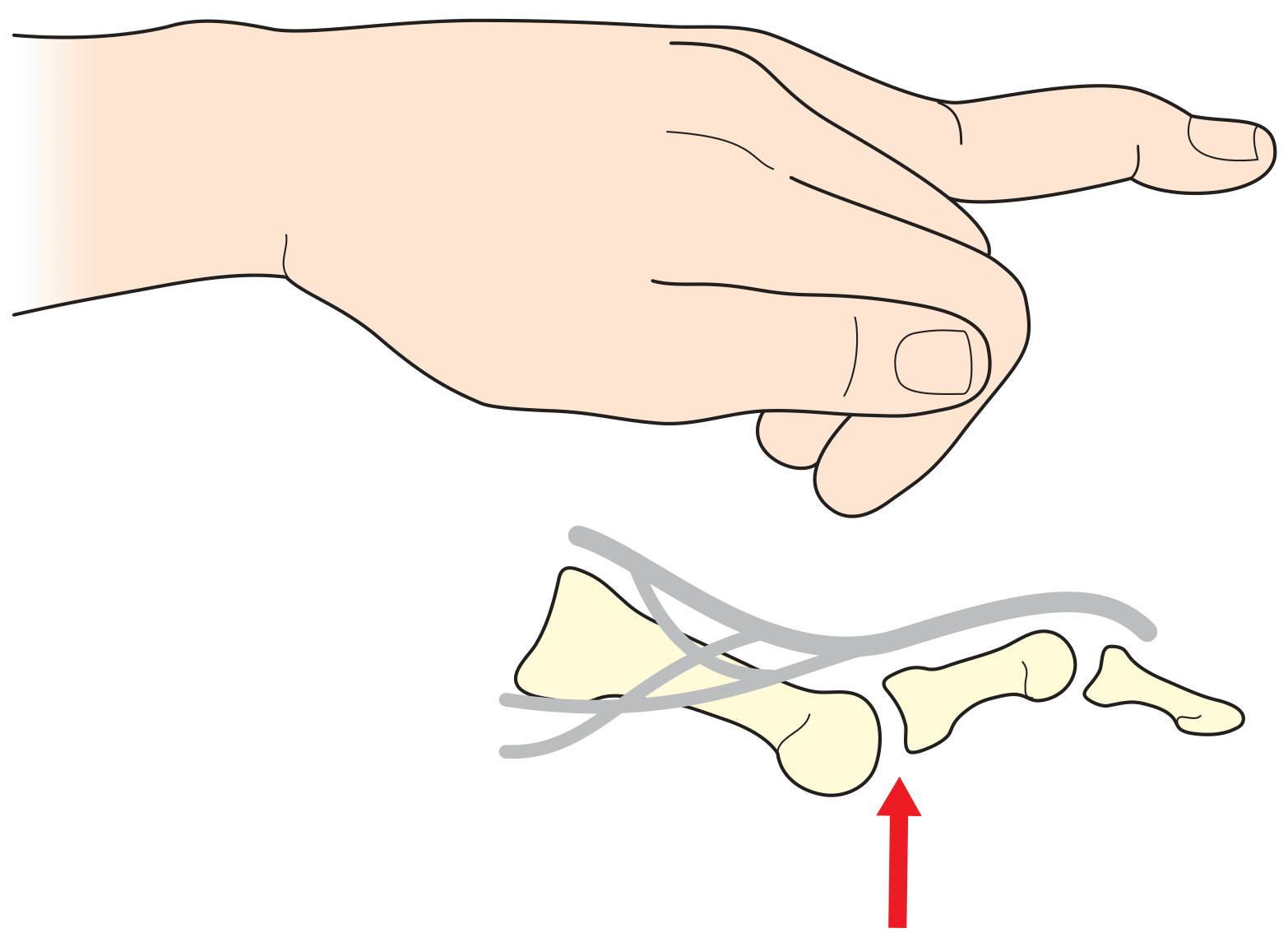
3. Boutonniere Deformity
- Flexion at PIPJ + Hyperextension at DIPJ
- Caused by: attenuation/rupture of the central slip of extensor tendon -> lateral bands sublux volar to PIPJ axis -> PIPJ stuck in flexion, DIPJ hyperextends
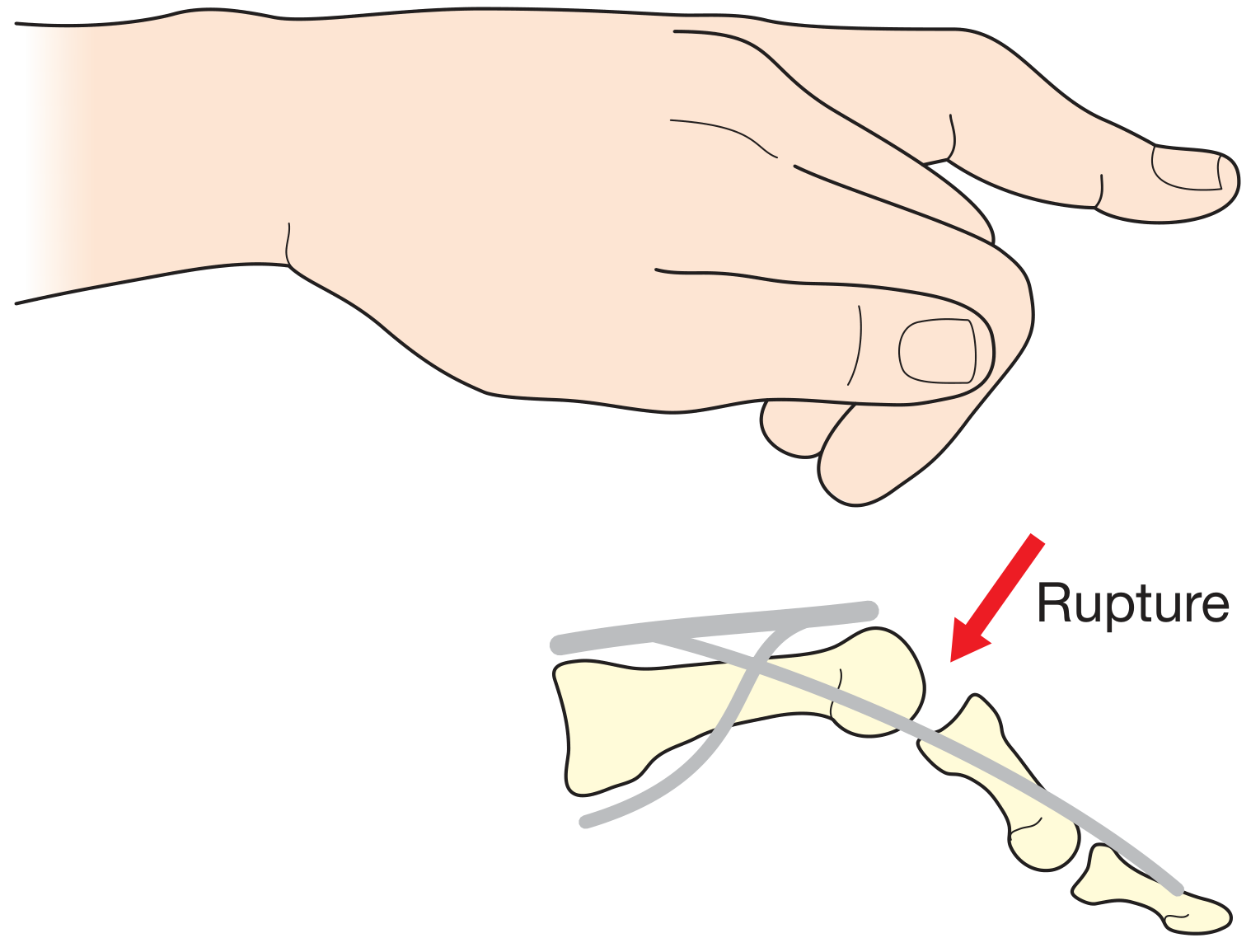
4. Z-deformity of Thumb (Hitchhiker's Thumb)
- Flexion of IPJ + Hyperextension of MCPsJ (or vice versa)
- Three types (Nalebuff classification)
5. Caput Ulnae Syndrome
- Dorsal subluxation of the distal ulna due to DRUJ destruction
- The prominent ulnar head ("caput ulnae") can rupture the extensor tendons of the little finger first, progressing radially (Vaughan-Jackson syndrome)
6. Extensor Tendon Ruptures
- Begin with the little finger (EDM, EDC5), progressing radially (Vaughan-Jackson)
- Unable to actively extend the involved fingers
7. Mallet Finger
- Loss of active DIP extension due to extensor tendon attrition over a dorsal osteophyte
Q2B. Bursitis Around the Knee [5 marks]
A bursa is a synovium-lined sac that reduces friction between structures. Multiple bursae surround the knee joint.
1. Prepatellar Bursitis ("Housemaid's Knee")
- Location: Subcutaneous, in front of the lower patella and upper patellar ligament
- Cause: Repeated kneeling (carpet layers, roofers, clergy)
- Features: Tender, fluctuant swelling directly over the patella; knee flexion is painful; joint itself is not involved
- Treatment: Rest, aspiration, anti-inflammatory drugs; surgical excision for chronic/recurrent cases
2. Infrapatellar Bursitis ("Clergyman's Knee")
- Superficial infrapatellar bursa: Between skin and patellar ligament below the tibial tuberosity
- Deep infrapatellar bursa: Between patellar ligament and the tibia
- Cause: Kneeling in a more upright position (prayer)
- Features similar to prepatellar bursitis but lower in position
3. Suprapatellar Bursa
- Located between the femur and the quadriceps tendon
- Normally communicates with the knee joint cavity
- Becomes distended in any knee joint effusion; best detected as a horseshoe-shaped swelling above the patella
4. Semimembranosus Bursa (Baker's Cyst precursor)
- Located posteromedially, between semimembranosus and medial head of gastrocnemius
- Becomes prominent and tense on knee extension
- More common in children; usually resolves spontaneously
5. Baker's Cyst (Popliteal Cyst)
- A herniation of the posterior synovial membrane through the oblique popliteal ligament
- Located in the midline of the popliteal fossa, slightly below the joint line
- Prominent on extension; disappears on flexion
- Often associated with intra-articular pathology (OA, RA, TB)
- On examination: transilluminates; Foucher's sign (becomes tense on extension, soft on flexion)
- Rupture can mimic DVT (pseudothrombophlebitis)
6. Pes Anserinus Bursitis
- Located medially, deep to the conjoined insertion of sartorius, gracilis, and semitendinosus
- Common in obese women with OA; pain on the medial side of the knee
7. Iliotibial Band Bursa
- Between the iliotibial band and the lateral femoral condyle
- Common in runners ("runner's knee")
Q2C. Hangman's Fracture [5 marks]
Definition
A hangman's fracture is a traumatic spondylolisthesis of the axis (C2) - bilateral fractures through the pars interarticularis (isthmus) of C2, with forward displacement of C2 on C3.
Mechanism
- Primary mechanism: Hyperextension + axial loading of the cervical spine
- Classically described in judicial hanging (the noose forces hyperextension)
- In modern practice: high-energy trauma - road traffic accidents, diving injuries, falls
- Some patterns involve flexion-distraction
Classification (Levine-Edwards - Most Widely Used)
| Type | Displacement | Angulation | Disc/Ligament | Stability |
|---|---|---|---|---|
| Type I | <3 mm | <11° | Intact | Stable |
| Type Ia | Atypical; distraction | Angulation | Variable | Unstable |
| Type II | >3 mm | >11° | C2/3 disc disrupted | Unstable |
| Type IIa | Minimal | Severe angulation | Disrupted | Very unstable |
| Type III | >3 mm + facet dislocation | Marked | Fully disrupted | Very unstable |
- Starr-Eismont variant: Fracture extends into the posterior body of C2; higher rate of neurological injury; needs halo vest or surgery
Clinical Features
- Neck pain, tenderness over C2
- Torticollis
- Neurological deficit is LESS common than expected because the bilateral pars fracture actually decompresses the spinal canal (the vertebral body shifts forward, widening the canal)
- However, severe displacement or disc herniation can cause cord injury
Investigations
- Plain X-ray: lateral view shows forward displacement of C2 body on C3; fracture through the pars
- CT scan (gold standard): precise fracture characterisation
- MRI: disc/ligament injury, cord signal changes
Management
Non-operative:
- Type I and Type II without severe angulation: Rigid cervical collar or halo vest immobilisation for 8-12 weeks
- Halo vest complications (especially in elderly): pin site infection, loosening, dysphagia
Operative:
- Type IIa, Type III, and Starr-Eismont variant: Surgical stabilisation
- Posterior C2-C3 fusion (most common)
- Anterior C2-C3 discectomy and fusion (for disc disruption)
Prognosis: Generally good; most patients achieve bony union with non-operative treatment
Q2D. Pathoanatomy of CTEV (Congenital Talipes Equinovarus) Foot [5 marks]
Definition
Congenital Talipes Equinovarus (CTEV / Club Foot) is a complex three-dimensional congenital foot deformity that is fixed (not postural). The deformity has four components, remembered by the mnemonic CAVE or CAVE-S:
The Four Components of CTEV Deformity (CAVE)
| Component | Deformity | Level |
|---|---|---|
| C - Cavus | High arch of medial foot | Midfoot |
| A - Adductus | Forefoot adduction/medial deviation | Tarsometatarsal joints |
| V - Varus | Hindfoot inversion/varus | Subtalar joint |
| E - Equinus | Plantarflexion of hindfoot | Ankle joint |
Detailed Pathoanatomy
1. Bony Changes:
- Talus: The head and neck of the talus are deviated medially and plantarward (the talar neck is short and internally rotated)
- Calcaneus: In equinus (plantarflexed) and varus (inverted) position; the posterior part is pulled up by the shortened tendo-Achilles, making the heel feel "empty"
- Navicular: Displaced medially with respect to the talar head (talonavicular subluxation) - the most consistent pathological finding
- Cuboid: Displaced medially with respect to the calcaneus
2. Soft-Tissue Changes (Medial and Posterior):
- Ligaments: Calcaneofibular ligament and plantar calcaneonavicular (spring) ligament are shortened and thickened
- Tendons and sheaths: Posterior tibial tendon sheath, tendo-Achilles, tibialis posterior, flexor hallucis longus, and flexor digitorum longus are contracted
- Tendon sheaths contain contractile myofibroblasts (fibroblastic component)
- The lateral structures (peroneal tendons and their sheaths) are elongated
3. Muscular Changes:
- Gastrocsoleus and posterior tibial muscles are smaller than normal
- Reduced myofibrils, increased connective tissue (possibly due to neuromuscular abnormality)
- Evertors (peronei) are weak
4. Vascular Changes:
- The dorsalis pedis artery may be diminished in calibre
- Contributes to difficulty with healing post-surgery
5. Skin:
- Deep medial crease and single posterior crease are characteristic
- All children have a smaller calf and foot on the affected side
Key Pathological Distinction
- In postural CTEV (intrauterine moulding): deformity is correctable and fully reducible; no structural joint/bone change
- In structural idiopathic CTEV: deformity is fixed; underlying bony and soft-tissue changes as above
Clinical Appearance
The foot shows: hindfoot equinus + varus, midfoot cavus + adductus, giving the appearance of forefoot cavus. The forefoot appears supinated but is actually pronated relative to the hindfoot.
Classification
- Pirani score (0-6): based on 6 clinical signs; guides Ponseti treatment
- Dimeglio classification (Grade I-IV): grades reducibility and severity
Sources: Bailey and Love's Short Practice of Surgery 28th Ed; Campbell's Operative Orthopaedics 15th Ed 2026; S. Das Manual on Clinical Surgery 13th Ed
This is a shared conversation. Sign in to Orris to start your own chat.