
Give a histology picture of epithelium and ask about it answer like this type i read this and directly draw and write in exam this is 5 mark question with drawing answer according this 5 mark
Reading File
Finding Sources
Searching the Web
histology epithelium types labeled diagram image
Finding Sources
Loading Image
Reading File
Loading Image
I found a great histology image from the textbook. Let me fetch it and read more content for a complete answer.
Loading Image
Reading File
I have everything I need. Here is the complete 5-mark exam answer:
🔬 HISTOLOGY IMAGE — Look at this and Identify:
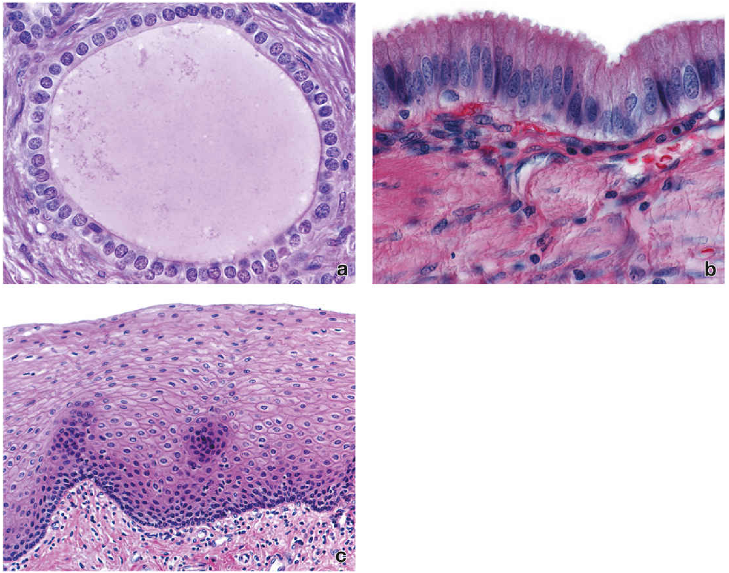
Source: Histology: A Text and Atlas with Correlated Cell and Molecular Biology
- Panel (a) - Pancreatic duct - Single layer of cuboidal cells around a lumen
- Panel (b) - Gallbladder - Tall columnar cells with elongated dark nuclei
- Panel (c) - Esophagus wall - Multiple layers of squamous cells
✏️ QUESTION: "What is epithelium? Describe its types with diagrams." (5 Marks)
📝 MODEL ANSWER — Read Once, Draw & Write Directly in Exam
DEFINITION (1 mark)
Epithelium is a tissue composed of closely packed cells with minimal intercellular substance, resting on a basement membrane (basal lamina), always covering a free surface of the body. It is avascular (no blood vessels; nutrients diffuse from underlying connective tissue).
CLASSIFICATION (1 mark)
Epithelium is classified based on:
- Number of layers - Simple (one layer) or Stratified (multiple layers)
- Shape of cells - Squamous (flat), Cuboidal, Columnar
TYPES WITH DIAGRAMS (2 marks)
1. Simple Squamous Epithelium
Draw: Flat, scale-like cells, single layer, nuclei appear like bulging bumps
 ___________________________________________
|   flat  |  flat  |  flat  |  flat  |     |
|___nucleus___|___|_____|_____|_____|________|
|=============== BASEMENT MEMBRANE ========|
- Location: Lining of blood vessels (endothelium), body cavities (mesothelium), alveoli of lung
- Function: Diffusion, filtration, secretion of serous fluid
2. Simple Cuboidal Epithelium ← Panel (a) in the image above
Draw: Square/cube-shaped cells, central round nuclei, single layer
 _____  _____  _____  _____
|  O  ||  O  ||  O  ||  O  |   (O = round central nucleus)
|_____||_____||_____||_____|
|======= BASEMENT MEMBRANE =|
- Location: Kidney tubules, thyroid follicles, pancreatic ducts, ovarian surface
- Function: Secretion and absorption
3. Simple Columnar Epithelium ← Panel (b) in the image above
Draw: Tall rectangular cells, oval nuclei at BASE, single layer
 _  _  _  _  _  _
| || || || || || |   (nuclei near base = oval)
|O||O||O||O||O||O|
|_||_||_||_||_||_|
|== BASEMENT MEMBRANE ==|
- Location: Gallbladder, stomach, intestine (with goblet cells), uterine tube
- Function: Secretion, absorption; may bear cilia or microvilli
4. Stratified Squamous Epithelium ← Panel (c) in the image above
Draw: Multiple layers - bottom layer = columnar/cuboidal (basal), middle = polygonal, top = flat squamous
 ___________________________________
 flat flat flat flat flat flat flat   <- TOP (squamous)
  poly  poly  poly  poly  poly        <- MIDDLE
 [col][col][col][col][col][col]       <- BASAL LAYER (cuboidal/columnar)
 |===== BASEMENT MEMBRANE ===========|
- Non-keratinized: Mouth, esophagus, vagina, cornea
- Keratinized: Skin (epidermis)
- Function: Protection against abrasion and mechanical stress
5. Pseudostratified Columnar Epithelium (Bonus type)
Draw: All cells touch basement membrane but nuclei at DIFFERENT levels - LOOKS stratified but is NOT
 cilia cilia cilia cilia
 _  _  _  _  _  _  _
| || || | | || || ||
|O|||O || |O|  ||O||    <- nuclei at different heights
||  | ||O||  |O|  ||
|_||_||_||_||_||_||_|
|===== BASEMENT MEMBRANE ===|
- Location: Respiratory tract (trachea, bronchi), epididymis
- Function: Mucociliary clearance; secretion
SPECIAL FEATURES OF ALL EPITHELIA (1 mark)
| Feature | Detail |
|---|---|
| Avascular | No blood vessels; nourished by diffusion |
| Basement membrane | All epithelium rests on it; made of basal lamina + reticular lamina |
| Free surface modifications | Microvilli (absorption), Cilia (movement), Stereocilia (sensory) |
| Cell junctions | Tight junctions (barrier), Desmosomes (adhesion), Gap junctions (communication) |
| Regeneration | High mitotic activity - rapid renewal |
QUICK MEMORY TRICK
"Simple = Single, Shape = S/C/Col, Site tells the function"
- Squamous = Surface lining (diffusion)
- Cuboidal = Collecting ducts (kidney)
- Columnar = Content secretion (gut, gallbladder)
- Stratified = Strength/protection (skin, esophagus)
Key point to write in exam: "Epithelium is always avascular, always rests on a basement membrane, and is classified by the number of layers and cell shape." - (Histology: A Text and Atlas, p. 298-300)
This is a shared conversation. Sign in to Orris to start your own chat.