
Ild
"interstitial lung disease"[MeSH Terms] AND diagnosis
interstitial lung disease HRCT pattern honeycombing
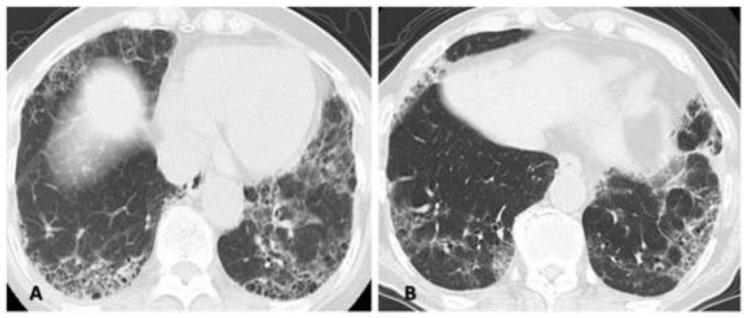
Two high-resolution computed tomography (HRCT) axial lung window images (A and B) demonstrating interstitial lung disease (ILD) patterns with overlapping features. Case A (left) shows a combination of diffuse ground-glass opacities (GGO), particularly in the right lung, and subpleural reticulation. Peripheral honeycombing is visible in the posterior lower lung fields, creating a mixed picture of nonspecific interstitial pneumonia (NSIP) and usual interstitial pneumonia (UIP). Case B (right) reveals predominant peripheral and subpleural reticular opacities and honeycombing in the bilateral lower lobes, more pronounced on the left side, which are classic hallmarks of a UIP pattern. These images illustrate the diagnostic challenge of 'unclassifiable' or 'mixed' patterns in rheumatology-associated ILD, where inflammatory (NSIP-like) and fibrotic (UIP-like) components coexist. Key educational features include the distinction between reversible GGO and irreversible honeycombing/traction bronchiectasis in the context of progressive fibrosing interstitial lung disease (PF-ILD).
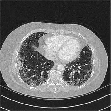
High-resolution computed tomography (HRCT) axial section of the chest demonstrating advanced interstitial lung disease. The image shows extensive reticular opacities with a predominant peripheral and subpleural distribution. There is evidence of architectural distortion characterized by early honeycombing, visible as small, clustered cystic spaces in the subpleural regions of both lower lobes. These findings are consistent with a Usual Interstitial Pneumonia (UIP) pattern, often associated with Idiopathic Pulmonary Fibrosis (IPF). The central lung parenchyma remains relatively spared compared to the dense fibrotic changes at the periphery. The imaging highlights the characteristic superior-to-inferior gradient of UIP, where fibrotic destruction is most pronounced in the lung bases. This diagnostic image serves as an educational tool for identifying the hallmarks of chronic fibrosing interstitial pneumonia, including subpleural reticulation, traction bronchiectasis, and honeycombing.
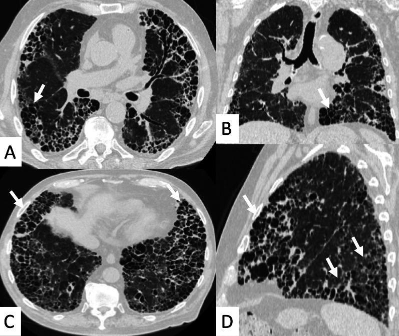
High-resolution computed tomography (HRCT) of the chest in axial (A, C), coronal (B), and sagittal (D) planes demonstrating advanced interstitial lung disease. The images show diffuse, extensive honeycombing involving more than 50% of the lung parenchyma. This finding is characterized by the 'exuberant honeycombing sign,' featuring hypertrophic, well-defined macrocystic spaces (white arrows) that are larger than those seen in typical honeycomb patterns. The cystic changes are most prominent in the peripheral and subpleural regions of both lungs, with a notable basal predominance. These findings represent a Usual Interstitial Pneumonia (UIP) pattern in the context of Rheumatoid Arthritis-associated Interstitial Lung Disease (RA-ILD). Associated features include marked architectural distortion and the absence of significant ground-glass opacities, indicating end-stage fibrotic disease.
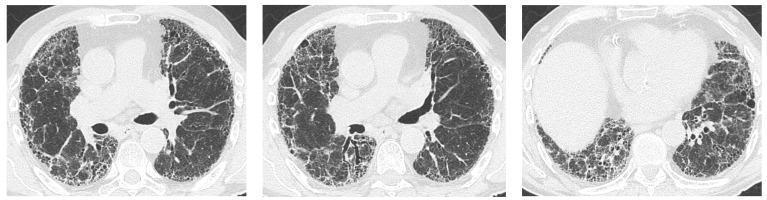
High-resolution computed tomography (HRCT) of the chest in three axial views demonstrating a classic Usual Interstitial Pneumonia (UIP) pattern. The images show bilateral, predominantly peripheral, and subpleural lung disease. Key diagnostic features include honeycombing, characterized by clusters of cystic airspaces with thick, well-defined walls, and significant traction bronchiectasis, where the fibrotic lung parenchyma pulls on and dilates the airways. These findings result in extensive architectural distortion of the lung fields, particularly in the lower lobes and subpleural regions. This diagnostic imaging is characteristic of advanced interstitial lung disease (ILD), often seen in conditions such as primary Sjögren's syndrome (pSS) or idiopathic pulmonary fibrosis. The visualization is essential for distinguishing UIP from other interstitial patterns like non-specific interstitial pneumonia (NSIP) or organizing pneumonia (OP) in clinical practice.
Interstitial Lung Disease (ILD)
Definition
Classification
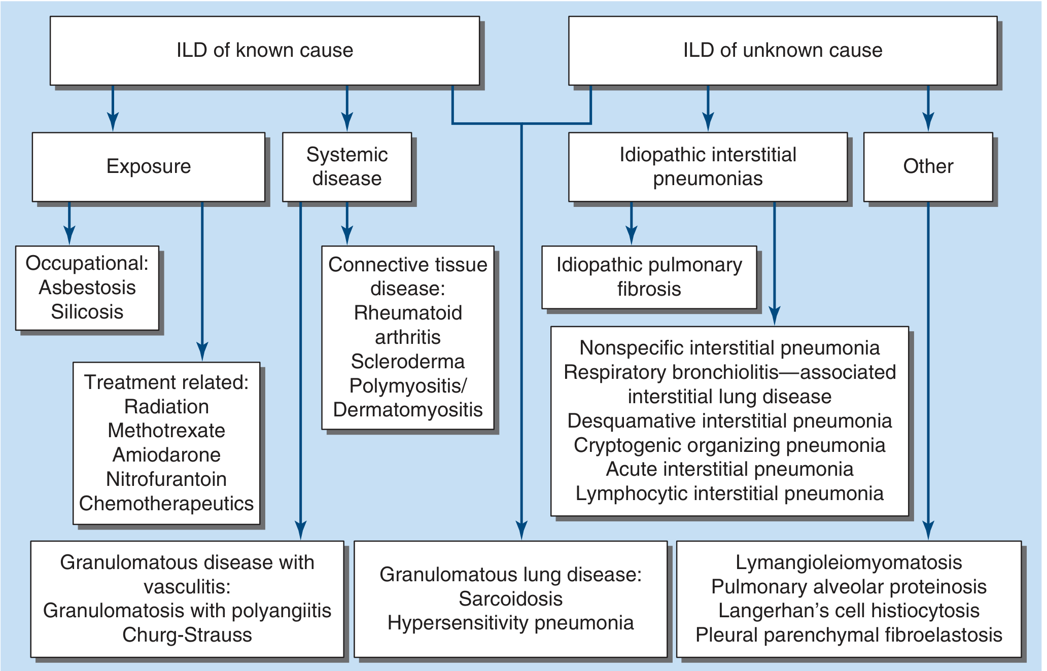
ILD of Known Cause
| Category | Examples |
|---|---|
| Occupational/Environmental | Asbestosis, Silicosis, Coal worker's pneumoconiosis |
| Drug/Treatment-related | Methotrexate, Amiodarone, Nitrofurantoin, Radiation, Chemotherapeutics |
| Connective Tissue Disease (CTD) | Rheumatoid arthritis (RA), Scleroderma (SSc), Polymyositis/Dermatomyositis, Sjogren's, SLE |
| Granulomatous with vasculitis | Granulomatosis with Polyangiitis (GPA), Eosinophilic granulomatosis (Churg-Strauss) |
| Granulomatous lung disease | Sarcoidosis, Hypersensitivity Pneumonitis (HP) |
ILD of Unknown Cause (Idiopathic Interstitial Pneumonias - IIPs)
| Entity | Key Feature |
|---|---|
| Idiopathic Pulmonary Fibrosis (IPF) | Most common IIP; UIP pattern; poor prognosis |
| Nonspecific Interstitial Pneumonia (NSIP) | NSIP pattern; often CTD-associated |
| Cryptogenic Organizing Pneumonia (COP) | Subacute; responds to steroids |
| Desquamative Interstitial Pneumonia (DIP) | Smoking-related |
| Respiratory Bronchiolitis-ILD (RB-ILD) | Smoking-related |
| Acute Interstitial Pneumonia (AIP) | Acute/fulminant; DAD pattern |
| Lymphocytic Interstitial Pneumonia (LIP) | Lymphocytic infiltrate; associated with Sjogren's |
Other ILDs
- Lymphangioleiomyomatosis (LAM) - young women
- Pulmonary Langerhans Cell Histiocytosis (PLCH) - smokers
- Pulmonary Alveolar Proteinosis (PAP)
- Pleural Parenchymal Fibroelastosis (PPFE)
Clinical Presentation
- Progressive exertional dyspnea - most common complaint
- Dry cough - especially prominent in IPF
- Fatigue - common across all ILDs
- Rare: hemoptysis (suggests DAH, GPA, LAM), chest pain (suggests sarcoidosis)
- Acute presentations (days-weeks): eosinophilic pneumonia, AIP, HP, acute exacerbation of IPF
- Subacute (weeks-months): sarcoidosis, CTD-ILD, drug-induced ILD, COP
- Chronic/indolent (months-years): IPF - Harrison's, p. 2304
- Bibasal fine ("Velcro") crackles
- Digital clubbing (especially IPF)
- Signs of underlying CTD (skin thickening in SSc, joint changes in RA)
Diagnosis
Pulmonary Function Tests (PFTs)
- Restrictive pattern: reduced TLC, FVC, normal or elevated FEV1/FVC ratio
- Reduced DLCO (diffusing capacity)
HRCT - The Key Imaging Tool
| Pattern | Key HRCT Features | Associated Disease |
|---|---|---|
| UIP | Subpleural, bibasal reticulation; honeycombing; traction bronchiectasis | IPF, RA-ILD, asbestosis |
| NSIP | Bilateral GGO; subpleural sparing; lower lobe predominance; no/minimal honeycombing | CTD-ILD, drug-induced |
| COP | Peripheral/peribronchial consolidation; migratory; upper/mid lobe | Cryptogenic OP, infection |
| DIP/RB-ILD | Diffuse GGO (DIP), centrilobular nodules (RB-ILD) | Smoking-related |
| HP | Upper/mid lobe involvement; mosaic attenuation; GGO; poorly defined centrilobular nodules | Hypersensitivity pneumonitis |
Serologies
- ANA, anti-dsDNA, anti-Scl-70, anti-Jo-1, RF, anti-CCP, anti-MDA5 - screen for CTD
- Precipitating antibodies for HP
Bronchoalveolar Lavage (BAL)
- Lymphocytosis suggests HP, sarcoidosis, COP, NSIP
- Eosinophilia suggests eosinophilic pneumonia
- Hemosiderin-laden macrophages suggest DAH
- Used on a case-by-case basis after HRCT pattern assessment - Murray & Nadel's, block21
Surgical Lung Biopsy
- VATS approach preferred over open thoracotomy
- Short-term mortality ~5% (higher in IPF, acute presentations)
- Most useful when HRCT is non-diagnostic - Harrison's, p. 2305
Multidisciplinary Discussion (MDD)
- Standard of care: Pulmonologist + Radiologist + Pathologist (+ Rheumatologist if CTD suspected) together reach the most accurate diagnosis.
Key Individual Diseases
Idiopathic Pulmonary Fibrosis (IPF)
- Most common IIP; prevalence 50-200/100,000; increases with age
- Men > women; associated with smoking; typically >60 years
- 50% 3-5 year survival - worst prognosis among IIPs
- HRCT: UIP pattern (subpleural, basal, reticulation + honeycombing)
- Histology: UIP - temporal and spatial heterogeneity, fibroblast foci, honeycombing
- Treatment: Antifibrotics (pirfenidone, nintedanib) slow FVC decline; lung transplant for advanced disease - Harrison's, p. 2306
NSIP
- Often secondary to CTD (especially SSc), drugs, HP
- Ground-glass opacities with subpleural sparing on HRCT
- Responds better to immunosuppression than IPF; better prognosis
Hypersensitivity Pneumonitis (HP)
- Due to inhaled organic antigens (bird proteins, fungal spores, molds)
- Acute, subacute, or chronic
- BAL: lymphocytosis; HRCT: upper lobe GGO, mosaic attenuation
- Key treatment: antigen avoidance
Sarcoidosis
- Granulomatous; bilateral hilar lymphadenopathy + pulmonary infiltrates
- Serum ACE elevated; BAL lymphocytosis with CD4:CD8 ratio >3.5
- Corticosteroids for symptomatic/progressive disease
CTD-ILD
- All CTDs can cause ILD; SSc has highest prevalence (~75%)
- RA-ILD: more common in men; UIP pattern associated with worse prognosis
- Anti-Jo-1 positive myositis: NSIP most common; "mechanic's hands," ILD, fever triad
Treatment
Antifibrotics (for IPF)
| Drug | Mechanism | Key Trial | Side Effects |
|---|---|---|---|
| Nintedanib | Tyrosine kinase inhibitor (FGF-R, PDGF-R, VEGF-R) | TOMORROW, INPULSIS-1/2 | Diarrhea (>60%) |
| Pirfenidone | Inhibits TGF-β-stimulated fibroblast proliferation & collagen production | CAPACITY, ASCEND | Photosensitivity, nausea, fatigue |
Immunosuppression (for non-IPF ILDs)
- Corticosteroids: first-line for COP, acute HP, CTD-ILD, sarcoidosis
- Caution: Steroids + azathioprine + N-acetylcysteine combination is harmful in IPF (PANTHER trial)
- Steroid-sparing agents: azathioprine, mycophenolate, cyclophosphamide (in CTD-ILD)
- Rituximab: for refractory myositis-associated ILD
Lung Transplantation
- Standard of care for advanced, rapidly progressive ILD
- Single or bilateral lung transplant
Supportive Management
- Oxygen supplementation for hypoxemia
- Pulmonary rehabilitation
- Treating pulmonary hypertension (complication of advanced ILD)
- Antacids/proton pump inhibitors (gastroesophageal reflux is common in IPF)
- Vaccinations (influenza, pneumococcal)
Prognostic Indicators (worse prognosis)
- UIP pattern on HRCT/histology
- Greater extent of fibrosis on HRCT
- Low baseline FVC and DLCO
- Rapid decline in FVC (>10% in 6-12 months)
- Acute exacerbations
- Pulmonary hypertension as complication
- IPF > RA-ILD > SSc-ILD > NSIP (in terms of mortality)
- Harrison's Principles of Internal Medicine 22E, Chapter 304 (ILD), pp. 2303-2308
- Murray & Nadel's Textbook of Respiratory Medicine, Chapters on ILD and CTD-ILD
- Fishman's Pulmonary Diseases and Disorders, Chapter on IPF management