
Ligament of uterus
Reading File
Now let me search the medical library for ligaments of the uterus.
Finding Sources
Finding Sources
Good results. Let me now read the most relevant sections in parallel - the Berek & Novak's Gynecology section and the Color Atlas section, plus the Gray's Anatomy section.
Reading File
Reading File
Reading File
Now let me get more detail from Campbell Walsh Urology on the ligaments, and fetch the anatomical image from Netter's.
Reading File
Reading File
Excellent! I have comprehensive information. Let me fetch the most relevant anatomical images from Netter's to include in the response.
Loading Image
Loading Image
I now have comprehensive information from multiple authoritative sources. Here is a detailed answer on the ligaments of the uterus.
Ligaments of the Uterus
The uterus is held in position within the pelvis by a combination of peritoneal folds, condensations of pelvic fascia, and fibromuscular bands. The primary ligaments are described below.
Anatomical Overview
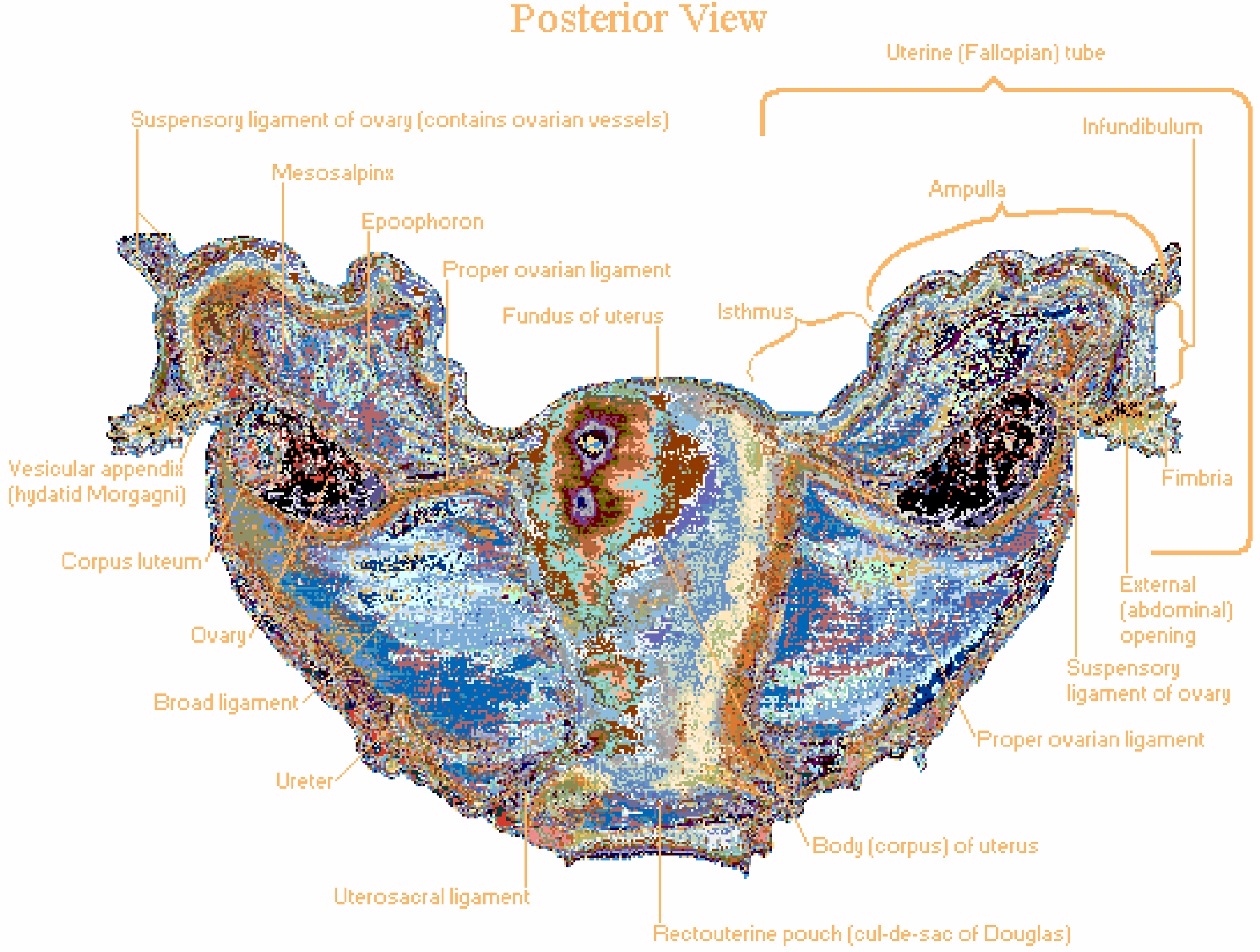
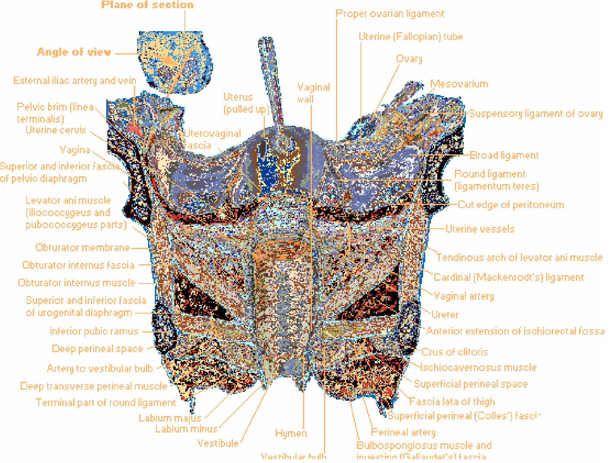
1. Broad Ligament
- A peritoneal fold that extends from the anterior and posterior surfaces of the uterus to the lateral pelvic wall.
- It is not a true ligament but a double layer of peritoneum that carries the neurovascular structures.
- Contents: uterine artery, vein, nerves, fallopian tube (in the mesosalpinx), ovary (in the mesovarium), round ligament, ovarian ligament, lymphatics, connective tissue, and adipose tissue.
- Subdivisions:
- Mesometrium - largest part, contains uterine vessels
- Mesosalpinx - between fallopian tube and ovary; also houses a vascular network
- Mesovarium - suspends the ovary
- Superiorly, it continues as the infundibulopelvic (suspensory) ligament of the ovary.
- Inferiorly, it is in contact with the pararectal space, obturator fossa, and uterosacral ligament.
(Berek & Novak's Gynecology; Color Atlas of Human Anatomy Vol. 2)
2. Round Ligament (Ligamentum Teres Uteri)
- A cord-like fibromuscular structure that arises near the uterine horns (just in front of and below the fallopian tube attachment).
- Runs anterolaterally within the broad ligament, crosses the external iliac artery, enters the deep inguinal ring, passes through the inguinal canal, exits at the superficial inguinal ring, and ends as fibrous strands in the connective tissue of the labia majora.
- Composed of smooth muscle cells.
- Embryological origin: it is the long distal part of the original gubernaculum in the fetus; the proximal part of the gubernaculum becomes the ligament of the ovary (proper ovarian ligament). Male homolog = gubernaculum testis.
- As it traverses the inguinal canal, it acquires the same fascial coverings as the spermatic cord in males.
- Function: maintains the anteverted position of the uterus; does NOT provide significant structural support.
(Gray's Anatomy for Students; Color Atlas of Human Anatomy Vol. 2; Campbell-Walsh-Wein Urology)
3. Cardinal Ligament (Mackenrodt's Ligament / Transverse Cervical Ligament)
- A condensation of endopelvic fascia at the base of the broad ligament.
- Originates from S2-S4 and inserts into the posterolateral pericervical fascia and lateral vaginal wall.
- Provides primary support to the cervix and upper vagina.
- Fuses posteriorly with the uterosacral ligaments.
- Contains major blood vessels from the internal iliac artery, autonomic nerves, and connective tissue.
- The uterine artery travels within the cardinal ligament - the ureter passes beneath the uterine artery here ("water under the bridge" - ureter lies ~2-3 cm lateral to the cervix).
- The vesicopelvic fascia is continuous with the cardinal ligament and contributes to bladder support.
(Berek & Novak's Gynecology; Sabiston Textbook of Surgery; Campbell-Walsh-Wein Urology)
4. Uterosacral Ligament (Sacrouterine / Rectouterine Ligament)
- Fibromuscular bands of dense subperitoneal connective tissue (and possibly smooth muscle - the rectouterinus muscle - though debated).
- Origin: greater sciatic foramen / posterolateral pelvic wall (roughly sacral levels S2-S4).
- Insertion: lateral aspect of the fascia encircling the cervix, isthmus of uterus, and vaginal wall.
- Form the lateral boundaries of the rectouterine pouch (pouch of Douglas).
- Fuse anteriorly with the cardinal ligaments near the cervix.
- Function: primary suspensory support for the cervix and upper vagina; commonly used as anchoring structures in apical suspension procedures (vaginal vault prolapse repair).
- Important relation: the ureter lies lateral to the anterior portion of the uterosacral ligament (closest proximity near the cervix - warrants caution during uterosacral suspension). Sacral nerves (S1-S4) pass dorsally and can be entrapped during suspension.
(Berek & Novak's Gynecology; Campbell-Walsh-Wein Urology; Color Atlas of Human Anatomy Vol. 2)
5. Pubocervical Ligament
- Condensation of fascia running from the pubic symphysis to the cervix, passing anteriorly.
- Contributes to anterior support of the uterus and bladder base.
6. Proper Ovarian Ligament (Ligament of the Ovary)
- A fibromuscular cord running from the medial pole of the ovary to the lateral uterine wall, just below the fallopian tube attachment.
- Lies within the broad ligament.
- Embryological origin: proximal part of the gubernaculum (homologous to the proximal gubernaculum of the testis).
7. Infundibulopelvic Ligament (Suspensory Ligament of the Ovary)
- Not a ligament of the uterus per se, but lies behind the broad ligament and connects the ovary to the lateral pelvic wall.
- Contains the ovarian vessels (ovarian artery and vein) and lymphatics.
- Clinically important: the ureter crosses beneath the ovarian artery as it runs under the infundibulopelvic ligament.
Summary Table
| Ligament | Type | Attachment | Key Function |
|---|---|---|---|
| Broad ligament | Peritoneal fold | Uterus → lateral pelvic wall | Carries neurovascular structures |
| Round ligament | Fibromuscular cord | Uterine horn → labia majora (via inguinal canal) | Maintains anteversion |
| Cardinal (Mackenrodt's) | Fascial condensation | Cervix → lateral pelvic wall | Main cervical/upper vaginal support |
| Uterosacral | Fibromuscular band | Cervix → sacrum (S2-S4) | Main cervical/upper vaginal support |
| Pubocervical | Fascial band | Cervix → pubic symphysis | Anterior uterine support |
| Ovarian ligament | Fibromuscular cord | Ovary → lateral uterine wall | Ovarian tethering |
| Infundibulopelvic | Peritoneal fold | Ovary → lateral pelvic wall | Carries ovarian vessels |
Clinical Notes
- True support of the uterus comes primarily from the pelvic floor muscles (levator ani complex), not the ligaments. The cardinal and uterosacral ligaments provide the most important ligamentous support at the level of the cervix.
- Damage to cardinal and uterosacral ligaments leads to uterovaginal prolapse.
- The ureter is most at risk during hysterectomy where it passes: (1) under the ovarian vessels at the infundibulopelvic ligament, (2) through the cardinal ligament beneath the uterine artery (2-3 cm lateral to the cervix), and (3) near the insertion of the uterosacral ligament.
- The round ligament is used as a surgical landmark to identify the external iliac artery and is ligated early in total abdominal hysterectomy.
- The broad ligament does NOT provide significant pelvic organ support (confirmed in Campbell-Walsh-Wein Urology citing Barber, 2005).
(Berek & Novak's Gynecology, p.207; Netter's Atlas of Human Anatomy, p.623-625; Color Atlas of Human Anatomy Vol. 2, p.461; Gray's Anatomy for Students, p.355; Campbell-Walsh-Wein Urology; Sabiston Textbook of Surgery)
Perineum
Finding Sources
Finding Sources
Reading File
Reading File
Reading File
Reading File
Reading File
Now let me fetch the key anatomical diagrams and read the Gray's section on deep perineal pouch and muscles.
Loading Image
Loading Image
Reading File
Reading File
Reading File
Reading File
I now have comprehensive information from multiple sources. Let me compile the full answer.
Perineum
Definition and Overview
The perineum is a diamond-shaped region at the inferior end of the trunk, lying inferior to the pelvic floor and between the thighs. Its ceiling is formed by the pelvic diaphragm (levator ani and coccygeus muscles), which separates the pelvic cavity above from the perineum below.
The major nerve and artery of the entire perineum are the pudendal nerve (S2-S4) and the internal pudendal artery.
(Gray's Anatomy for Students)
Boundaries (Pelvic Outlet)
The peripheral boundary of the perineum corresponds to the pelvic outlet:
| Point | Landmark |
|---|---|
| Anterior | Inferior border of pubic symphysis |
| Posterior | Tip of coccyx |
| Lateral (x2) | Ischial tuberosities |
| Anterolateral margins | Ischiopubic rami |
| Posterolateral margins | Sacrotuberous ligaments |
Division into Two Triangles
An imaginary transverse line joining the two ischial tuberosities (at the level of the perineal body) divides the diamond-shaped perineum into two triangular regions:
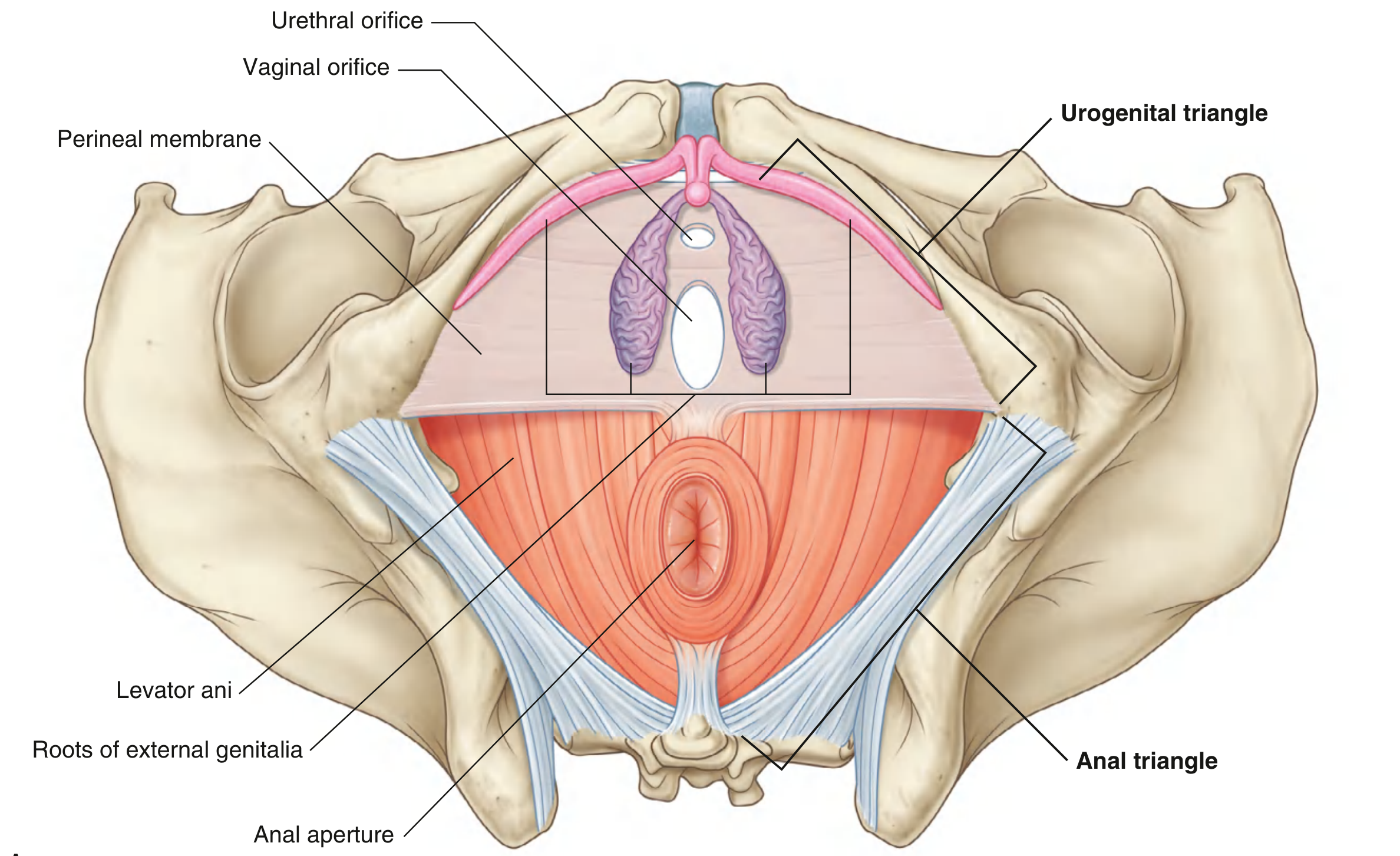
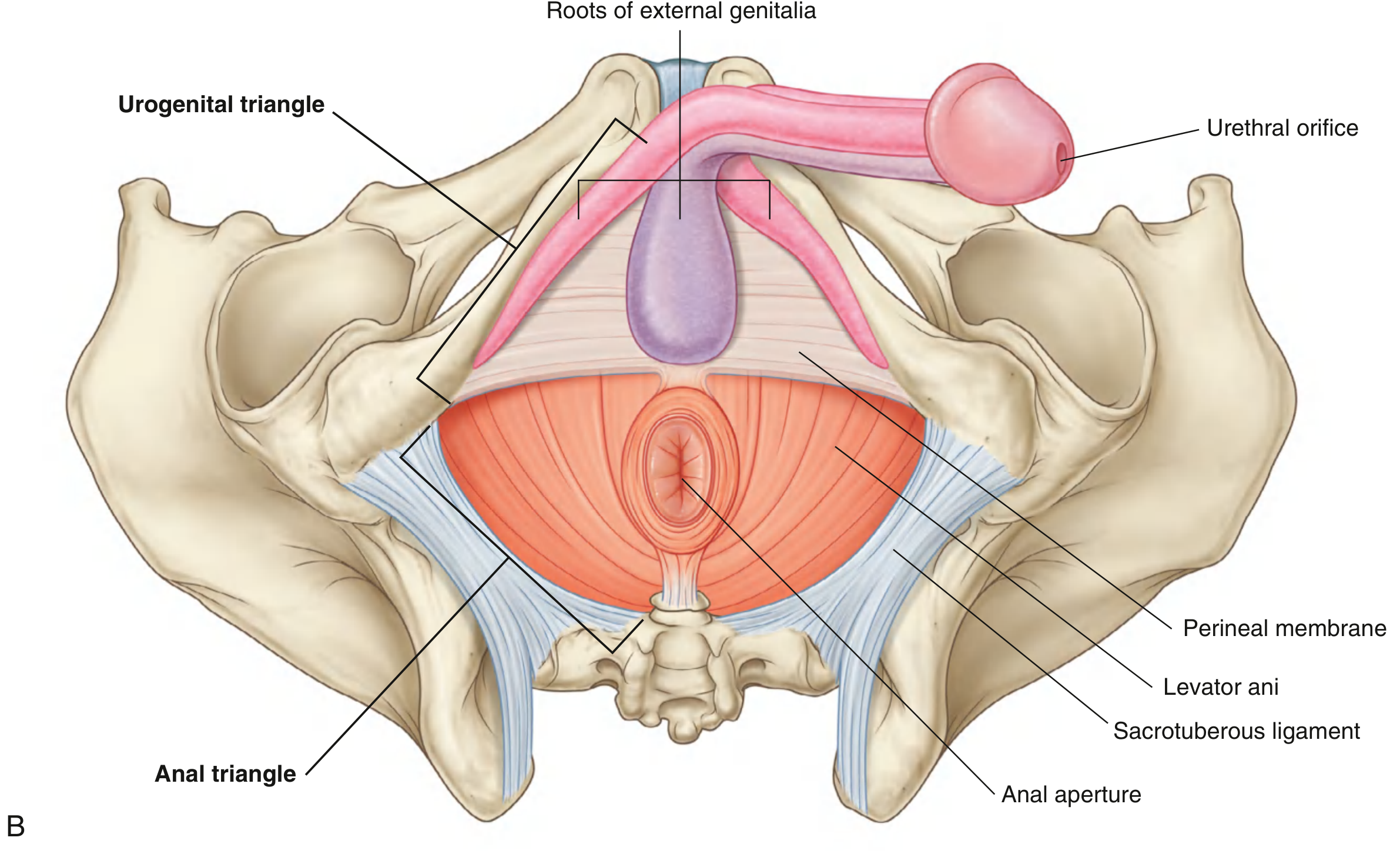
Note: The two triangles are not in the same plane. In the anatomical position, the urogenital triangle is oriented roughly horizontally, while the anal triangle is tilted upward so it faces more posteriorly.
1. Urogenital Triangle (Anterior)
Boundaries: pubic symphysis, pubic rami, ischial rami, and the deep transverse perineal muscles spanning between the ischial tuberosities.
Contains the roots of the external genitalia and the openings of the urinary/reproductive tracts.
Perineal Membrane (formerly Urogenital Diaphragm)
- A thick fibrous sheet filling the urogenital triangle.
- Attached laterally to the pubic arch; free posterior border anchored in the midline to the perineal body.
- The urethra and vagina (in women) / urethra (in men) pass through it via the urogenital hiatus.
- Divides the urogenital triangle into a deep perineal pouch above and a superficial perineal pouch below.
- Provides support for the external genitalia (attached to its inferior surface) and for the pelvic viscera above.
Deep Perineal Pouch (Space)
Located immediately superior to the perineal membrane. Contains:
| Structure | Notes |
|---|---|
| External urethral sphincter | Skeletal muscle; primary sphincter |
| Urethrovaginalis (= female external sphincter) | Also called urethrovaginalis |
| Compressor urethrae | Compresses urethra |
| Deep transverse perineal muscles | Stabilize perineal body |
| Neurovascular tissues | Branches of pudendal nerve and vessels |
(Campbell-Walsh-Wein Urology; Gray's Anatomy for Students)
Superficial Perineal Pouch (Space)
Located inferior to the perineal membrane. Roofed by Colles fascia (superficial perineal fascia).
Colles fascia attachments:
- Laterally: fused to fascia lata of thigh at ischiopubic ramus (defines the perineal-thigh crease)
- Posteriorly: fuses with inferior fascia of urogenital diaphragm at the perineal body
- Anteriorly: continues as dartos fascia over the scrotum/labia majora
Contents of the superficial perineal pouch:
Muscles (all innervated by pudendal nerve S2-S4):
| Muscle | Origin | Insertion | Function |
|---|---|---|---|
| Ischiocavernosus | Ischial tuberosity and ramus | Crus of penis/clitoris | Forces blood from crura into body of penis/clitoris (maintains erection) |
| Bulbospongiosus | In women: perineal body; in men: perineal body + midline raphe | Perineal membrane, bulb/vestibule, body of clitoris/corpus cavernosum | Forces blood into glans; in men: expels residual urine, pulsatile semen emission |
| Superficial transverse perineal | Ischial tuberosity and ramus | Perineal body | Stabilizes the perineal body |
Erectile Tissues:
- Corpora cavernosa (crura): paired, anchored to ischiopubic rami; unite to form body of clitoris/penis
- Bulbs of vestibule (women) / Corpus spongiosum (men): surround the urogenital openings; anchored to perineal membrane
- Glans clitoris / Glans penis: at the distal end
Glands:
- Greater vestibular glands (Bartholin glands) - in women; lie at the posterior end of each vestibular bulb; site of Bartholin cysts/abscesses.
2. Anal Triangle (Posterior)
Boundaries: laterally by sacrotuberous ligaments, anteriorly by the transverse line between ischial tuberosities, posteriorly by the coccyx.
Ceiling: pelvic diaphragm (levator ani + coccygeus).
Central content: anal aperture, flanked on each side by the ischio-anal fossa.
External Anal Sphincter
A skeletal muscle with three parts (from distal to proximal):
| Part | Position |
|---|---|
| Subcutaneous | Encircles distal anal canal |
| Superficial | Anchored to perineal body anteriorly and anococcygeal body posteriorly |
| Deep | Adjacent to deep transverse perineal muscles and levator ani |
- Innervated by the inferior rectal nerve and branches of the pudendal nerve (S2-S4).
- The internal anal sphincter is circular smooth muscle; receives autonomic innervation from the iliohypogastric plexus.
Ischio-anal (Ischiorectal) Fossa
- Inverted wedge-shaped fat-filled space on each side of the anal aperture.
- Lateral wall: ischium, obturator internus muscle, sacrotuberous ligament.
- Medial wall: levator ani muscle.
- Contents: fat, Colles fascia, internal pudendal vessels and pudendal nerve running in Alcock's canal (pudendal canal - made up of fascia of obturator internus).
- Extends anteriorly as recesses into the urogenital triangle above the deep perineal pouch.
- Function: allows movement of pelvic diaphragm and expansion of anal canal during defecation.
- Clinically: site of ischio-anal abscesses and fistula-in-ano.
Perineal Body (Central Tendon of Perineum)
- Located at the central point of the perineum, posterior to the vestibule/vaginal opening and anterior to the anal canal.
- A fibromuscular mass of collagen and elastic fibres.
- Acts as the convergence point for:
- Bulbospongiosus
- External anal sphincter
- Superficial and deep transverse perineal muscles
- Attaches to the posterior border of the perineal membrane.
- Clinical significance: damage during parturition can injure the external anal sphincter. Episiotomy is angled laterally (mediolateral) to avoid these fibres.
(Campbell-Walsh-Wein Urology)
Neurovascular Supply
| Structure | Details |
|---|---|
| Nerve | Pudendal nerve (S2-S4) - main nerve of the entire perineum |
| Artery | Internal pudendal artery (branch of internal iliac) |
| Vein | Internal pudendal vein |
| Lymphatics | Superficial inguinal nodes |
The internal pudendal vessels and pudendal nerve travel in Alcock's canal along the lateral wall of the ischio-anal fossa before branching into the perineum.
Sex Differences Summary
| Feature | Female | Male |
|---|---|---|
| UG triangle openings | Urethra + vagina | Urethra only (within penis) |
| Erectile tissue type | Bulbs of vestibule (paired) + corpora cavernosa | Corpus spongiosum (single, fused) + corpora cavernosa |
| Bulbospongiosus | Splits to surround introitus; attached to clitoris | Surrounds bulb of penis; fused midline raphe |
| Vestibular glands | Bartholin glands (greater vestibular) | Bulbourethral (Cowper's) glands |
Clinical Relevance
- Pudendal nerve block: administered at the ischial spine - anaesthetises the entire perineum; used in obstetric procedures.
- Episiotomy: incision of the perineal body to facilitate delivery; mediolateral direction avoids the external anal sphincter.
- Perineal tears (obstetric): classified by depth of involvement:
- 1st degree: skin only
- 2nd degree: skin + superficial perineal muscles
- 3rd degree: involves external anal sphincter
- 4th degree: involves anal mucosa
- Bartholin gland abscess: in the superficial perineal pouch; requires incision and drainage or marsupialisation.
- Ischio-anal abscess: infection spreads from anal crypts into the fat of the ischio-anal fossa; can form horseshoe abscess communicating across the midline.
- Hemorrhoids: engorgement of the internal or external rectal venous plexus.
(Gray's Anatomy for Students, p.580-587; Berek & Novak's Gynecology, p.194; Campbell-Walsh-Wein Urology, p.3218)
Perineal body
Finding Sources
Finding Sources
Reading File
Reading File
Reading File
Reading File
Reading File
Reading File
Now let me fetch the perineal body diagram and one more for sex differences.
Loading Image
Reading File
I now have comprehensive information from four major textbooks. Here is the full answer.
Perineal Body (Central Tendon of the Perineum)
Definition
The perineal body (also called the central perineal tendon or central tendon of the perineum) is a poorly defined but anatomically and functionally critical fibromuscular node located at the geometric centre of the perineum.
It is described variously as:
- A pyramidal / triangular block of fibromuscular connective tissue (Campbell-Walsh-Wein)
- An ill-defined fibromuscular node into which muscles of the pelvic floor and perineum converge (Gray's Anatomy for Students)
- The central connection between the two layers of pelvic floor support - the pelvic diaphragm and the urogenital diaphragm (Berek & Novak's)
Location
- Lies in the midline, along the posterior border of the perineal membrane.
- Situated anterior to the anus and posterior to the vaginal opening (in women) / posterior to the bulb of the penis (in men).
- At the midpoint of an imaginary line joining the two ischial tuberosities - the same line that divides the perineum into the urogenital and anal triangles.
- Separates the distal portions of the anal and vaginal canals.
Diagram
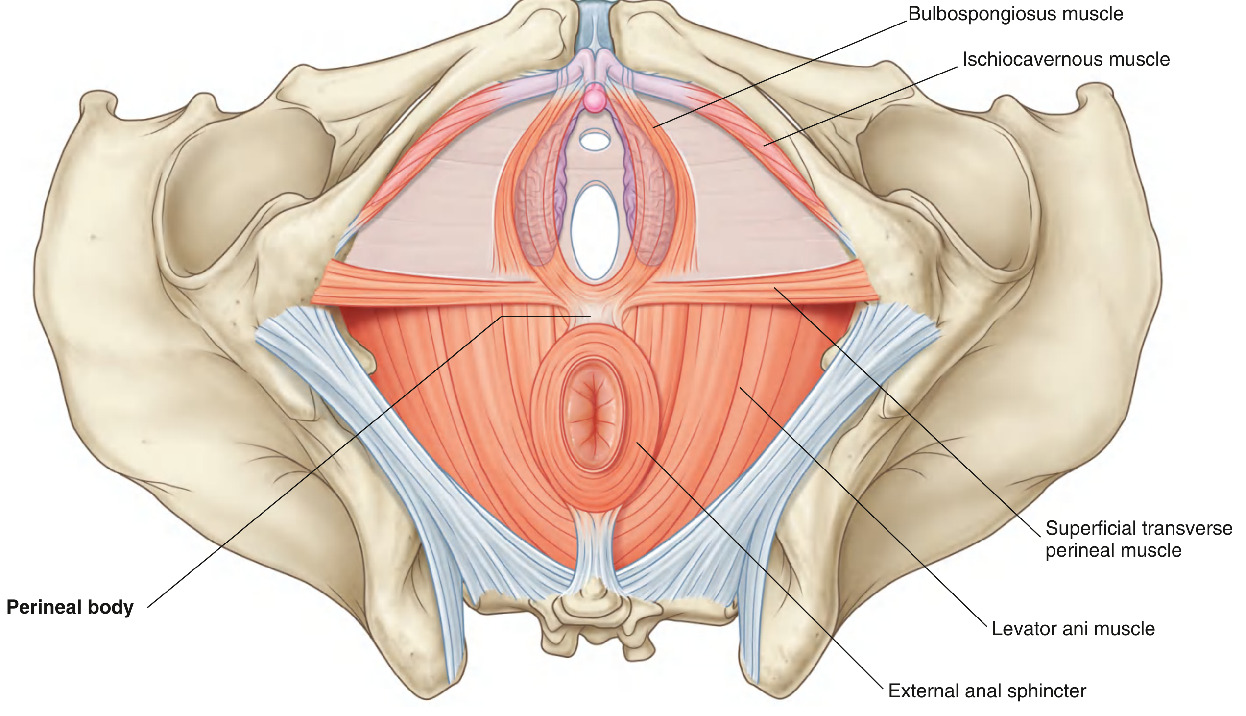
Muscles Converging at the Perineal Body
According to Campbell-Walsh-Wein Urology, 8 muscles converge at the perineal body:
| Muscle | Layer | Notes |
|---|---|---|
| Bulbospongiosus | Superficial perineal pouch | Fixed to perineal body by its most posterior fibres |
| Superficial transverse perineal (x2) | Superficial perineal pouch | Arise from ischial tuberosities; insert centrally - major stabilisers |
| Deep transverse perineal (x2) | Deep perineal pouch | Attach to the deep aspect |
| External anal sphincter | Anal triangle | Anterior portion inserts into perineal body (especially in women) |
| Sphincter urethrovaginalis | Deep perineal pouch | In women only; also attaches to perineal body |
| Levator ani (pubococcygeus) | Pelvic diaphragm | Anterior fibres attach to the deep aspect |
(Campbell-Walsh-Wein Urology; Gray's Anatomy for Students; Berek & Novak's Gynecology)
Structural Composition
- Collagen and elastic fibres embedded in a fibromuscular matrix.
- Receives tendinous attachments from all the above muscles.
- Its superior border is the point of insertion of the rectovaginal fascia (Denonvilliers' fascia), which extends superiorly to the underside of the peritoneum covering the rectouterine pouch (pouch of Douglas), effectively separating the anorectal compartment from the urogenital compartment.
- Connected posteriorly to the anococcygeal raphe (via the external anal sphincter and levator ani).
Functions
- Structural anchor: Acts as the central anchoring point for all perineal muscles - stability of the entire perineal floor depends on its integrity.
- Pelvic organ support: In women, it provides posterior support for the lower anterior vaginal wall and the pelvic organs above (uterus, bladder, vagina). If damaged, prolapse of these organs can occur.
- Bi-level pelvic floor connection: Connects the pelvic diaphragm (levator ani, above) with the urogenital diaphragm (perineal membrane, below) - it is the central linkage between these two support layers.
- Separates compartments: Separates the anorectal compartment from the urogenital compartment.
- Elastic reserve: Although small at rest, it has the capacity to stretch significantly without tearing during childbirth to permit passage of the fetal head.
Sex Differences
| Feature | Female | Male |
|---|---|---|
| External anal sphincter relationship | Inferior fibres fuse with bulbospongiosus and transverse perineal muscles at the perineal body | Sphincter does NOT fuse with perineal body - a fascial plane exists between them |
| Sphincter urethrovaginalis | Attaches to perineal body | Absent |
| Clinical importance | Much greater - major support structure for pelvic organs | Less significant structurally |
Obstetric & Clinical Significance
- Episiotomy: An incision deliberately made through the perineum during delivery. The mediolateral direction is preferred to avoid the perineal body and external anal sphincter. A midline episiotomy, though technically easier, carries greater risk of extending into the sphincter.
- Obstetric perineal tears (classified by involvement of the perineal body):
- 1st degree: Skin only
- 2nd degree: Skin + perineal body muscles (superficial transverse perineal, bulbospongiosus)
- 3rd degree: Extends into external anal sphincter
- 4th degree: Extends through sphincter into anal mucosa
- Pelvic organ prolapse: Damage to the perineal body during parturition leads to deficient posterior vaginal support, contributing to rectocele, uterine prolapse, and cystocele.
- Perineorrhaphy: Surgical repair of the perineal body to restore its integrity and pelvic floor support.
- Sacral colpoperineopexy: In prolapse surgery, a continuous graft may be placed from the sacral promontory all the way down to the perineal body to restore apical and posterior support.
- Rectovaginal fistula repair: The perineal body is a key surgical landmark; low fistulae (between lower rectum and lower vagina) involve the space immediately around the perineal body.
- After being stretched during delivery, the muscles attached to the perineal body regain most of their tone over the following months.
(Gray's Anatomy for Students, p.514, 534, 580; Berek & Novak's Gynecology, p.203; Campbell-Walsh-Wein Urology, p.3218; Campbell-Walsh-Wein Urology, Female Anatomy block)
This is a shared conversation. Sign in to Orris to start your own chat.