
Clinical feature of ckd
chronic kidney disease clinical features manifestations
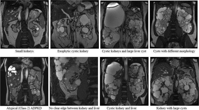
This composite diagnostic image features eight coronal MRI slices (T2-weighted) demonstrating the phenotypic spectrum of Autosomal Dominant Polycystic Kidney Disease (ADPKD). The images illustrate varied morphological presentations used for testing Total Kidney Volume (TKV) segmentation tools. Key clinical findings include progressive renal enlargement (nephromegaly) due to bilateral, multifocal cysts of varying sizes and distributions. Specific panels highlight ‘small kidneys’ with early-stage disease, ‘exophytic cystic kidneys’ where cysts protrude from the cortex, and kidneys with ‘large cysts’ causing significant architectural distortion. The series also depicts extrarenal manifestations, specifically polycystic liver disease (PLD), ranging from small focal lesions to large, hypointense hepatic cysts that obscure the renal-hepatic interface. One panel identifies Atypical (Class 2) ADPKD, characterized by asymmetric or focal cyst distribution. This collection serves as an educational resource for identifying Mayo Clinic Imaging Classification variations and understanding the challenges of automated renal segmentation in chronic kidney disease.
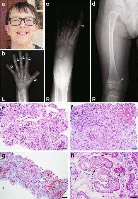
Educational panel illustrating skeletal and renal manifestations of a ciliopathy, likely Mainzer-Saldino syndrome.
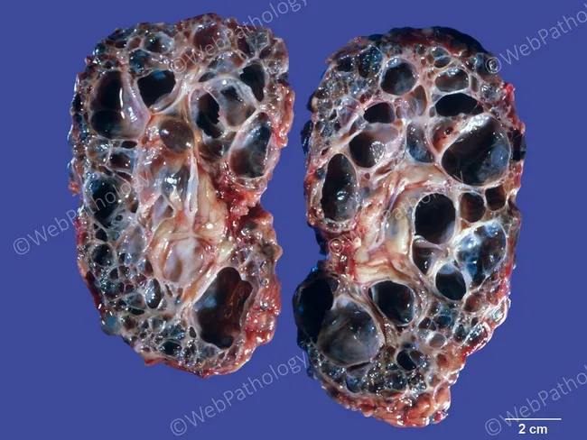
Gross pathology photograph of bilateral kidneys displaying classic autosomal dominant polycystic kidney disease (ADPKD) morphology. The organs are enlarged, yet retain their reniform shape. Longitudinally sectioned cut surfaces reveal numerous variably sized cysts that dissect the renal parenchyma and are separated by only a scant amount of residual cortex and medulla. Cysts range from tiny, sub-centimeter ducts to large, fluid-filled compartments; walls appear thin and translucent, and the cysts are often filled with clear or straw-colored fluid. The remaining parenchyma is markedly thinned, with loss of normal cortical architecture and parenchymal compression by the extensive cystic burden. This macroscopic pattern is characteristic of ADPKD, reflecting underlying genetic mutations (PKD1 or PKD2) and progressive replacement of functional nephrons by cysts. Diagnostic significance lies in correlating with bilateral nephromegaly, diffuse cortical-medullary cystic disease, and potential progression to chronic kidney disease and hypertension. Clinical relevance includes anticipation of renal insufficiency, risk of berry aneurysms in some patients, and the need for surveillance for extrarenal manifestations. Potential educational use includes teaching gross morphologic criteria for ADPKD, differential diagnosis versus ARPKD, and correlation with imaging modalities such as ultrasound, CT, and MRI. These features underscore ADPKD pathophysiology.

This clinical photograph displays the fingernails of a patient's hand, demonstrating the characteristic appearance of Lindsay's nails, also known as half-and-half nails. The visual presentation features a distinct transverse bicolor distribution across the nail plates. The proximal portion (closest to the cuticle) appears dull, opaque, or whitish, while the distal portion (occupying approximately 20% to 60% of the nail bed) exhibits a dark brown or reddish-brown discoloration. The boundary between these two zones is sharply demarcated. This finding is a specific dermatological sign highly associated with chronic kidney disease (CKD) and uremia, specifically end-stage renal disease (ESRD). The photograph serves as a diagnostic educational tool for identifying systemic manifestations of renal failure through nail bed examination. The nail texture remains largely smooth without significant ridging or pitting, focusing the clinical significance on the vascular bed discoloration.
Clinical Features of Chronic Kidney Disease (CKD)
Definition & Staging
| Stage | eGFR | Description |
|---|---|---|
| G1 | ≥90 | Normal/high, with kidney damage markers |
| G2 | 60-89 | Mildly decreased |
| G3a | 45-59 | Mild-moderate decrease |
| G3b | 30-44 | Moderate-severe decrease |
| G4 | 15-29 | Severely decreased |
| G5 | <15 | Kidney failure (ESRD/uremia) |
General / Nonspecific Symptoms (Early to Mid CKD)
- Fatigue and weakness - one of the most common early complaints
- Anorexia (loss of appetite) and altered taste sensation
- Weight loss and wasting
- Mood changes - depression is common
- Cognitive changes - decline in executive function, memory, and calculation ability
- Nocturia - loss of urinary concentrating ability forces ~2 L obligate urine output, bladder fills at night (importantly, loss of nocturia in late CKD may signal worsening oliguria)
The Full Uremic Syndrome (Advanced CKD, eGFR <15 mL/min)
- Severe hypertension
- Encephalopathic manifestations (confusion, cognitive decline, seizures)
- Gastrointestinal bleeding
- Uremic pericarditis (fibrinous pericarditis - a classic feature of severe uremia)
- Severe electrolyte disturbances, especially hyperkalemia
- Secondary hyperparathyroidism
Harrison's Principles of Internal Medicine 22E, p. 2404
System-by-System Manifestations
1. Cardiovascular
- Hypertension - nearly universal; due to salt/water retention and RAAS activation
- Accelerated atherosclerosis and increased cardiovascular mortality
- Pericarditis (uremic) - friction rub on auscultation; can progress to cardiac tamponade
- Cardiomyopathy and left ventricular hypertrophy
- Arrhythmias (from hyperkalemia/hypocalcemia)
2. Hematological
- Normocytic normochromic anemia - the most common hematologic finding; primarily from decreased erythropoietin production; target Hb 10-12 g/dL with ESAs
- Uremic bleeding tendency - platelet dysfunction (abnormal platelet-vessel wall interaction)
- Note: Some patients (PCKD, renal vascular disease, renal cell carcinoma) may actually have erythrocytosis
3. Neurological
- Peripheral neuropathy - "glove and stocking" distribution; sensory > motor
- Autonomic neuropathy
- Uremic encephalopathy - confusion, asterixis (metabolic flap), myoclonus
- Restless legs syndrome - common, particularly at night
- Sleep disorders
4. Gastrointestinal
- Nausea and vomiting (uremic gastritis)
- Anorexia, metallic taste in mouth (uremic fetor)
- Gastrointestinal bleeding (due to platelet dysfunction, uremic erosions)
- Constipation
5. Musculoskeletal / Bone Disease (CKD-MBD)
- Renal osteodystrophy - from low calcitriol → secondary hyperparathyroidism → high PTH → bone resorption
- Osteitis fibrosa cystica (high-turnover bone disease from hyperPTH)
- Osteomalacia (low-turnover, vitamin D-related)
- Vascular calcification (soft tissue calcification from high Ca × Phosphate product)
- Fractures, bone pain, myopathy
6. Fluid and Electrolytes
- Hyperkalemia - can be life-threatening; from decreased renal K⁺ excretion
- Metabolic acidosis - high anion gap in advanced CKD (uremic acids accumulate)
- Hyponatremia (dilutional) in fluid-overloaded states
- Hyperphosphatemia with hypocalcemia
- Edema / fluid overload
7. Endocrine / Metabolic
- Secondary hyperparathyroidism - from low calcitriol and phosphate retention
- Insulin resistance and altered glucose metabolism
- Dyslipidemia (elevated triglycerides, reduced HDL)
- Growth retardation in children
- Amenorrhea / infertility in women; sexual dysfunction in men
8. Dermatological
- Pruritus - extremely common and distressing; from uremic toxins, secondary HPT, xerosis
- Uremic frost - white crystalline urea deposits on skin (very advanced uremia, rare today)
- Pallor (anemia)
- Half-and-half nails (Lindsay's nails) - proximal whitish + distal brown/red bands; highly specific for CKD/uremia
9. Immunological
- Immune dysfunction - increased susceptibility to infections (leading cause of death after cardiovascular disease)
- Impaired T-cell and neutrophil function
- Reduced vaccine efficacy
10. Pulmonary
- Uremic pleuritis (pleuritic chest pain, pleural rub/effusion)
- Pulmonary edema (from fluid overload and cardiac dysfunction)
- "Butterfly" pattern on CXR in severe fluid overload
Key Laboratory Findings
| Test | Finding |
|---|---|
| Serum creatinine | Elevated, rising |
| eGFR | Reduced |
| BUN | Elevated |
| Serum potassium | Elevated (hyperkalemia) |
| Bicarbonate | Low (metabolic acidosis) |
| Phosphate | High |
| Calcium | Low (secondary to low calcitriol) |
| PTH | High (secondary hyperparathyroidism) |
| Hemoglobin | Low (normocytic anemia) |
| Urinalysis | Proteinuria, hematuria, casts |
Imaging Clue to Chronicity
- Harrison's Principles of Internal Medicine 22E (2025), Chapter on Chronic Kidney Disease and the Uremic Syndrome, p. 2404
- Brenner and Rector's The Kidney, 2-Volume Set
- Comprehensive Clinical Nephrology, 7th Edition