
Signa and Symptoms of Acute Heart Failure
acute heart failure signs symptoms clinical presentation
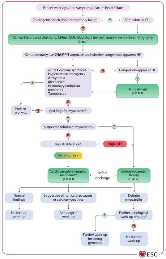
Summary : This flowchart outlines the diagnostic and management pathway for patients presenting with signs and symptoms of acute heart failure, with a focus on identifying myocarditis and guiding further work-up based on risk stratification and findings.
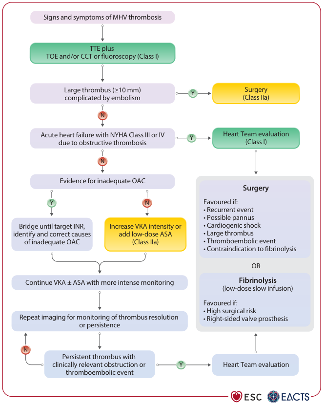
Summary : This flowchart outlines the diagnostic and management pathway for patients presenting with signs and symptoms of mechanical heart valve (MHV) thrombosis, according to ESC/EACTS guidelines. It details the decision-making process based on imaging findings, clinical status, and adequacy of oral anticoagulation (OAC), leading to options such as surgery, fibrinolysis, or adjustment of anticoagulation therapy.
"acute heart failure" AND "signs symptoms" AND "clinical presentation"
acute heart failure pulmonary edema dyspnea orthopnea
Signs and Symptoms of Acute Heart Failure (AHF)
Symptoms
Respiratory (Left-Sided Congestion)
| Symptom | Description |
|---|---|
| Dyspnea on exertion | The earliest and most sensitive symptom (~84% sensitivity); caused by rising pulmonary venous pressure reducing lung compliance |
| Orthopnea | Dyspnea in the recumbent position; occurs because lying flat increases venous return from the lower limbs and elevates the diaphragm; relieved by sitting up; quantified by the number of pillows needed |
| Paroxysmal nocturnal dyspnea (PND) | Dramatic breathlessness awakening the patient from sleep with extreme dyspnea bordering on suffocation; one of the most specific symptoms (76-84% specificity) |
| Cough | Consequence of fluid transudation into airspaces; may be productive of frothy or blood-tinged sputum in pulmonary edema |
| Acute pulmonary edema | Sudden, severe breathlessness at rest; the most dramatic presentation of AHF |
Systemic / Low Output Symptoms
| Symptom | Description |
|---|---|
| Fatigue / Lethargy | Reduced cardiac output leads to poor skeletal muscle perfusion |
| Reduced exercise tolerance | Progressive limitation of physical activity |
| Weight gain | Fluid retention from neurohormonal activation (RAAS + SNS) |
| Ankle/leg swelling (edema) | Peripheral fluid accumulation; more common in right-sided or biventricular failure |
| Nausea, early satiety, abdominal discomfort | Visceral/splanchnic congestion and gut edema |
| Oliguria | Reduced renal perfusion; cardiorenal syndrome develops in 20-40% of patients |
| Confusion / delirium | Cerebral hypoperfusion in severe low-output states |
Signs
Vital Signs
- Tachycardia - compensatory response to low stroke volume
- Tachypnea - elevated respiratory rate, often >20 breaths/min
- Hypertension - common in acute hypertensive AHF
- Hypotension - in cardiogenic shock (cold-wet profile); a sign of severely reduced output
- Low oxygen saturation (SpO2) - pulmonary edema impairs gas exchange
Respiratory Signs
- Fine bibasal crepitations (rales) - caused by opening of edematous pulmonary alveoli during inspiration; heard at lung bases initially, spreading upward with increasing severity
- Wheeze ("cardiac asthma") - bronchospasm from mucosal edema
- Dullness to percussion at bases - pleural effusion (transudative, from elevated hydrostatic pressure)
Cardiovascular Signs
- S3 gallop - the single most specific physical sign; positive LR = 4.0; represents rapid passive ventricular filling in a dilated, poorly compliant ventricle
- S4 gallop - atrial contraction against a stiff ventricle; suggests diastolic dysfunction
- Displaced apex beat - laterally and inferiorly displaced due to left ventricular enlargement (cardiomegaly)
- Pulsus alternans - alternating strong and weak pulses; sign of severe LV dysfunction
- Mitral regurgitation murmur - from papillary muscle displacement due to ventricular dilation
- Elevated Jugular Venous Pressure (JVP) - key sign of venous congestion; reflects raised right atrial pressure; look for hepatojugular reflux
- Right ventricular heave - in right-sided or biventricular failure
- Cool extremities / cyanosis - peripheral vasoconstriction and poor perfusion (cold profile)
Abdominal / Peripheral Signs
- Pitting pedal/ankle edema - bilateral in most cases
- Hepatomegaly - tender, pulsatile liver from hepatic venous congestion
- Ascites - in severe right-sided or chronic failure
- Hepatojugular reflux - JVP rises on abdominal pressure; sign of elevated venous pressure
Hemodynamic Profiles (Clinical Classification)
| Profile | Perfusion | Congestion | Typical Presentation |
|---|---|---|---|
| Warm & Wet | Adequate | Yes | Most common; responds to diuretics/vasodilators |
| Cold & Wet | Impaired | Yes | Cardiogenic shock with congestion; worst prognosis |
| Cold & Dry | Impaired | No | Hypovolemia + low output; requires fluid challenge |
| Warm & Dry | Adequate | No | Compensated; consider other diagnoses |
Chest Radiograph Findings
- Cardiomegaly (cardiothoracic ratio >0.5)
- Pulmonary venous congestion ("upper lobe diversion")
- Kerley B lines (interstitial edema)
- Perihilar "bat wing" opacification (alveolar edema)
- Pleural effusions (blunting of costophrenic angles)
Note: Up to 20% of patients subsequently diagnosed with AHF have chest X-rays without signs of congestion at the initial ED evaluation. (Tintinalli's Emergency Medicine, A Comprehensive Study)
ESC Diagnostic and Management Pathway
Key Diagnostic Pearls
- Dyspnea on exertion has the highest sensitivity (~84%) but is non-specific
- PND, orthopnea, and edema are most specific (76-84%)
- S3 gallop is the most specific physical sign (positive LR 4.0), but has poor interrater reliability
- BNP/NT-proBNP should be measured when the diagnosis is uncertain; elevated levels corroborate the clinical picture
- In chronic left-sided HF, the lungs may be clear despite elevated filling pressures (lymphatic compensation)
- Braunwald's Heart Disease, 2-Vol Set - A Textbook of Cardiovascular Medicine (Chapter 49)
- Tintinalli's Emergency Medicine: A Comprehensive Study (Chapter 53)
- Fuster and Hurst's The Heart, 15th Edition (Chapter on AHF)
- Robbins & Kumar Basic Pathology (Chapter 9 - Heart Failure morphology and clinical features)
- Frameworks for Internal Medicine (Heart Failure Key Points)