
Management of osteoarthritis in female of 53 yr old
"osteoarthritis" AND "management"
knee osteoarthritis radiograph joint space narrowing osteophytes
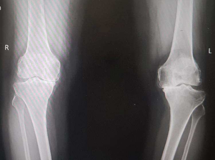
This diagnostic image is an anteroposterior (AP) standing radiograph of bilateral knee joints, demonstrating findings consistent with advanced primary osteoarthritis. The right knee shows significant medial compartment joint space narrowing, subchondral sclerosis, and marginal osteophyte formation at the femoral and tibial condyles. The left knee exhibits even more severe degenerative changes, characterized by near-total loss of medial joint space (bone-on-bone contact), pronounced subchondral sclerosis, large osteophytes, and visible bony irregularity indicating advanced articular cartilage destruction. There is evidence of varus malalignment, particularly on the left. This radiograph serves as a classic educational example of end-stage degenerative joint disease requiring clinical correlation for surgical planning, such as total knee arthroplasty. The image highlights key radiographic hallmarks of osteoarthritis: joint space narrowing, osteophytosis, subchondral sclerosis, and subchondral cyst formation.
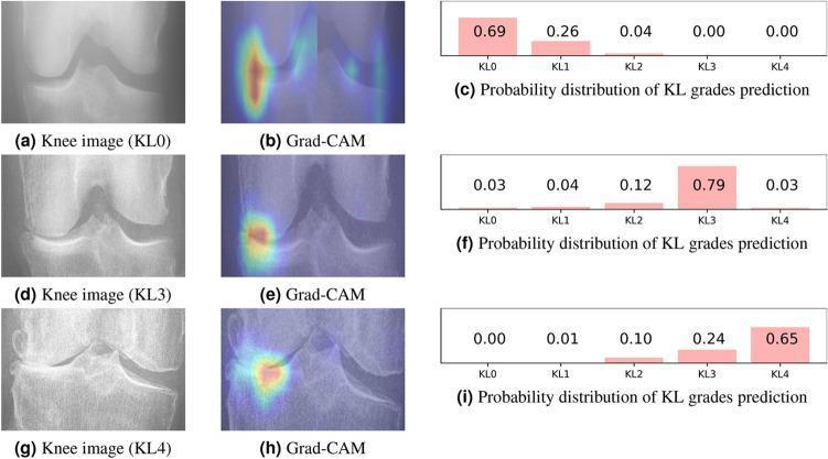
A comparative diagnostic visualization showcasing different stages of knee osteoarthritis using the Kellgren-Lawrence (KL) grading system, integrated with AI explainability heatmaps. The figure is organized into three rows corresponding to KL0 (no OA), KL3 (moderate OA), and KL4 (severe OA). Each row contains a frontal knee radiograph, a Grad-CAM (Gradient-weighted Class Activation Mapping) visualization, and a bar chart of prediction probabilities. Radiographically, KL0 shows a normal joint space and healthy bone contours. KL3 displays definitive joint space narrowing and visible osteophyte formation. KL4 demonstrates severe joint space narrowing with subchondral sclerosis and large, prominent osteophytes. The Grad-CAM overlays indicate the neural network's focus, which shifts from diffuse areas in KL0 to highly localized, intense heatmaps concentrated on the medial joint compartment and marginal osteophytes in the pathological KL3 and KL4 cases. This clinical imaging tool illustrates how computer-aided diagnosis identifies disease markers like joint space loss and bone remodeling to classify osteoarthritis severity.
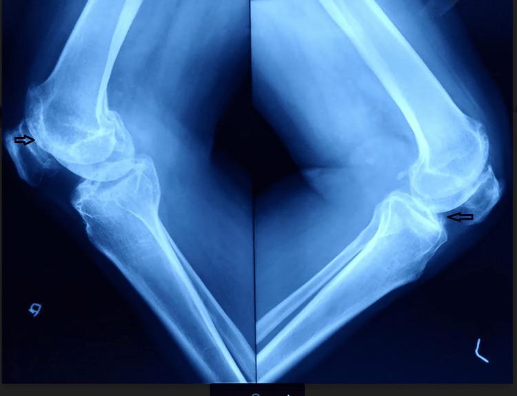
This diagnostic image is a lateral-view plain radiograph of both knee joints showing advanced degenerative changes consistent with Kellgren-Lawrence grade IV osteoarthritis. The imaging demonstrates significant joint space narrowing in the tibiofemoral compartments and evidence of patellofemoral osteoarthritis. Key radiological features include subchondral sclerosis, which appears as increased bone density at the joint margins, and prominent osteophyte formation. Black arrows specifically highlight osteophytes at the superior and inferior poles of the patella and the anterior tibial margins. There is visible narrowing of the patellofemoral joint space. While lateral views typically focus on sagittal alignment, the severe degeneration shown is associated with clinical varus deformity as noted in the patient's history. These findings are representative of end-stage joint disease, which in this clinical context was secondary to ochronotic arthropathy, and indicate the need for total knee arthroplasty (TKA).
Heberden and Bouchard nodes hand osteoarthritis
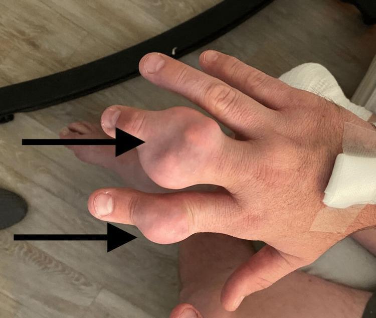
Clinical photograph of the dorsal aspect of a right hand demonstrating chronic tophaceous gout. The image prominently features two large, nodular, firm swellings (tophi) located at the distal interphalangeal (DIP) and proximal interphalangeal (PIP) joint regions of the second (index) and third (middle) digits, highlighted by black arrows. The larger mass on the index finger appears bulbous and irregular, with the overlying skin appearing thin, taut, and slightly erythematous or translucent, characteristic of underlying monosodium urate crystal deposition. Proximal to the metacarpophalangeal joints, a white medical dressing is visible on the wrist area, suggesting recent clinical intervention or intravenous access. The photograph illustrates advanced manifestations of gouty arthritis, emphasizing the typical presentation of tophi in the small joints of the hand. The clinical significance lies in identifying chronic hyperuricemia complications and distinguishing these masses from other rheumatological conditions like rheumatoid nodules or osteoarthritis-related Heberden's and Bouchard's nodes.
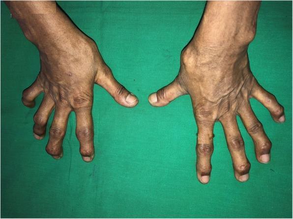
Clinical photograph showing the dorsal view of both hands against a green background, demonstrating advanced articular deformities. The image highlights significant bony enlargement at the distal interphalangeal (DIP) and proximal interphalangeal (PIP) joints, consistent with Heberden's and Bouchard's nodes typical of severe nodal osteoarthritis. There is visible malalignment and deviation of multiple digits, giving the fingers a gnarled appearance. A prominent bony swelling is also noted on the dorsal aspect of the right wrist. The overlying skin appears thin, dry, and wrinkled with prominent creases over the joints, but notably lacks the taut, shiny appearance characteristic of sclerodactyly. The medical context suggests these musculoskeletal changes are comorbid with a systemic condition like scleredema, although the hand deformities themselves are primarily osteoarthritic in nature. This image serves as an educational tool for distinguishing chronic degenerative joint disease from the skin tightening seen in systemic sclerosis.
Osteoarthritis Management — 53-year-old Woman
1. Core / first-line management (all patients, before or alongside drugs)
- Patient education and self-management programs — explain the chronic, fluctuating nature of OA; avoid fear-inducing language (Firestein & Kelley's Rheumatology).
- Weight management — if BMI ≥25 kg/m², aim for 5-10% body weight loss; benefit is dose-dependent (greater loss = greater pain/function benefit) (Goldman-Cecil Medicine, Table 241-2).
- Exercise / physical activity — supervised or unsupervised strengthening (especially quadriceps for knee OA), aerobic conditioning, aquatic exercise; ~150 min/week moderate activity. Evidence shows benefit comparable to drugs without the adverse-effect burden, and it's strongly recommended by AAOS and ACR guidelines.
- Physical/occupational therapy referral — gait aids, bracing if varus malalignment, footwear/orthotic advice, joint-protection techniques, assistive devices (canes, jar openers, dressing sticks) for hand/hip/knee OA.
- Screen for and address mood disorders, sleep problems, and fall risk, which worsen pain perception and function.
2. Pharmacologic therapy (added if pain persists despite core measures)
| Line | Options | Notes |
|---|---|---|
| First-line | Topical NSAIDs (esp. for knee/hand OA) | Preferred initially — good efficacy, lower systemic risk (AAOS strong recommendation) |
| First-line | Oral NSAIDs or oral acetaminophen | Both strongly recommended by AAOS; NSAIDs generally more effective but weigh GI, renal, and cardiovascular risk — relevant if she has hypertension, cardiac disease, or is on antiplatelet/anticoagulant therapy |
| Adjunct | Topical capsaicin | Option for localized joint pain |
| Second-line | Intra-articular corticosteroid injection | Recommended for short-term flare relief (moderate-strength evidence); avoid frequent repeated injections |
| Situational | Intra-articular hyaluronic acid | Evidence is mixed/conflicting - AAOS lists as "not recommended," though some guidelines and patients report benefit |
| Not recommended | Oral opioids/tramadol, lateral wedge insoles, routine glucosamine/chondroitin | AAOS strong evidence against benefit |
| Adjunct for centrally-mediated pain | Duloxetine | Considered for patients with widespread pain sensitization or coexisting depression |
3. Referral / surgical management
- Orthopedic referral for joint locking, instability, or advanced radiographic disease.
- Arthroscopic partial meniscectomy — only if a concomitant meniscal tear with mechanical symptoms in mild-moderate OA; not for OA alone.
- High tibial osteotomy — selected younger patients with unicompartmental knee OA and malalignment.
- Total joint arthroplasty (knee/hip replacement) — definitive option once conservative measures fail and pain/disability is significant; particularly reasonable to discuss at this age if disease is advanced, since implants now have good longevity.
Practical approach for this patient
- Confirm site(s) involved (knee, hip, hand are most common at this age) and rule out inflammatory arthritis/red flags.
- Start education + weight loss (if overweight) + structured exercise/physiotherapy immediately — this is the foundation, not an afterthought.
- Add topical NSAID first; step up to oral NSAID (checking GI/CV/renal risk) or acetaminophen if inadequate.
- Consider intra-articular corticosteroid for a painful flare while other measures take effect.
- Reassess in 6-12 weeks; refer to orthopedics if function remains significantly impaired or imaging shows advanced disease.
- Address bone health and cardiovascular risk given her menopausal transition, since these often coexist and influence drug choice (e.g., NSAID caution) and overall care.
- Textbook of Family Medicine 9e, p. 875
- Goldman-Cecil Medicine, Table 241-2 & OSTEOARTHRITIS section
- Firestein & Kelley's Textbook of Rheumatology, "Approach to Management of Osteoarthritis"
- Miller's Review of Orthopaedics 9th Edition, Table 5.7 (AAOS Guideline)
Pt management
By 'Pt management' do you mean the physiotherapy (PT) treatment plan specifically, or a general patient management summary?
Osteoarthritis Management — 53-year-old Woman
1. Core / first-line management (all patients, before or alongside drugs)
- Patient education and self-management programs — explain the chronic, fluctuating nature of OA; avoid fear-inducing language (Firestein & Kelley's Rheumatology).
- Weight management — if BMI ≥25 kg/m², aim for 5-10% body weight loss; benefit is dose-dependent (greater loss = greater pain/function benefit) (Goldman-Cecil Medicine, Table 241-2).
- Exercise / physical activity — supervised or unsupervised strengthening (especially quadriceps for knee OA), aerobic conditioning, aquatic exercise; ~150 min/week moderate activity. Evidence shows benefit comparable to drugs without the adverse-effect burden, and it's strongly recommended by AAOS and ACR guidelines.
- Physical/occupational therapy referral — gait aids, bracing if varus malalignment, footwear/orthotic advice, joint-protection techniques, assistive devices (canes, jar openers, dressing sticks) for hand/hip/knee OA.
- Screen for and address mood disorders, sleep problems, and fall risk, which worsen pain perception and function.
2. Pharmacologic therapy (added if pain persists despite core measures)
| Line | Options | Notes |
|---|---|---|
| First-line | Topical NSAIDs (esp. for knee/hand OA) | Preferred initially — good efficacy, lower systemic risk (AAOS strong recommendation) |
| First-line | Oral NSAIDs or oral acetaminophen | Both strongly recommended by AAOS; NSAIDs generally more effective but weigh GI, renal, and cardiovascular risk — relevant if she has hypertension, cardiac disease, or is on antiplatelet/anticoagulant therapy |
| Adjunct | Topical capsaicin | Option for localized joint pain |
| Second-line | Intra-articular corticosteroid injection | Recommended for short-term flare relief (moderate-strength evidence); avoid frequent repeated injections |
| Situational | Intra-articular hyaluronic acid | Evidence is mixed/conflicting - AAOS lists as "not recommended," though some guidelines and patients report benefit |
| Not recommended | Oral opioids/tramadol, lateral wedge insoles, routine glucosamine/chondroitin | AAOS strong evidence against benefit |
| Adjunct for centrally-mediated pain | Duloxetine | Considered for patients with widespread pain sensitization or coexisting depression |
3. Referral / surgical management
- Orthopedic referral for joint locking, instability, or advanced radiographic disease.
- Arthroscopic partial meniscectomy — only if a concomitant meniscal tear with mechanical symptoms in mild-moderate OA; not for OA alone.
- High tibial osteotomy — selected younger patients with unicompartmental knee OA and malalignment.
- Total joint arthroplasty (knee/hip replacement) — definitive option once conservative measures fail and pain/disability is significant; particularly reasonable to discuss at this age if disease is advanced, since implants now have good longevity.
Practical approach for this patient
- Confirm site(s) involved (knee, hip, hand are most common at this age) and rule out inflammatory arthritis/red flags.
- Start education + weight loss (if overweight) + structured exercise/physiotherapy immediately — this is the foundation, not an afterthought.
- Add topical NSAID first; step up to oral NSAID (checking GI/CV/renal risk) or acetaminophen if inadequate.
- Consider intra-articular corticosteroid for a painful flare while other measures take effect.
- Reassess in 6-12 weeks; refer to orthopedics if function remains significantly impaired or imaging shows advanced disease.
- Address bone health and cardiovascular risk given her menopausal transition, since these often coexist and influence drug choice (e.g., NSAID caution) and overall care.
- Textbook of Family Medicine 9e, p. 875
- Goldman-Cecil Medicine, Table 241-2 & OSTEOARTHRITIS section
- Firestein & Kelley's Textbook of Rheumatology, "Approach to Management of Osteoarthritis"
- Miller's Review of Orthopaedics 9th Edition, Table 5.7 (AAOS Guideline)
Physiotherapy Management of Osteoarthritis
1. Assessment before starting
- Joint(s) involved, pain pattern, ROM, quadriceps/hip muscle strength, gait, alignment (varus/valgus thrust), balance, and fall history (Goldman-Cecil Medicine, Table 241-2).
- Functional ability (walking, stairs, sit-to-stand) and footwear.
2. Therapeutic exercise (mainstay)
| Type | Purpose | Example |
|---|---|---|
| Strengthening | Quadriceps/hip abductor weakening precedes and worsens knee OA | Straight-leg raises, isometric quad sets, resisted leg press, mini-squats |
| Aerobic conditioning | Improves function, weight control, cardiovascular health | Walking, cycling, elliptical |
| Aquatic exercise | Low joint-loading, good for painful flares or obese patients | Pool-based strengthening/aerobic programs |
| Range-of-motion/flexibility | Maintains joint mobility, reduces stiffness | Gentle stretching, tai chi |
| Neuromuscular training | Improves proprioception and joint stability | Balance/agility drills, combined with strengthening (AAOS moderate recommendation) |
3. Manual therapy and gait correction
- Joint mobilization, soft-tissue techniques as an adjunct to exercise (AAOS: limited-evidence recommendation, "may be used").
- Gait retraining and correction of varus thrust if present.
- Assistive devices: cane (recommended, contralateral hand to affected knee/hip), walker if balance-impaired.
- Bracing: unloader/valgus brace for medial compartment knee OA with malalignment (AAOS moderate recommendation).
4. Physical modalities
- Heat/cold therapy: symptomatic relief, low cost, no strong evidence but commonly used adjunct.
- TENS, acupuncture, laser, extracorporeal shockwave, PRP: AAOS classifies these as limited-evidence ("may improve pain/function") — reasonable adjuncts but not primary treatment.
- Massage: adjunct to usual care only, limited evidence.
5. Weight and lifestyle counseling (often delivered via PT/dietitian collaboration)
- Target 5-10% body weight loss if BMI ≥25 kg/m² — greater loss gives greater benefit for both pain and function.
- Activity pacing and joint-protection education for daily tasks (especially relevant for hand OA if DIP/PIP joints involved).
6. Patient education and self-management
- Explain that OA is a manageable chronic condition, not a progressively disabling one for most patients.
- Teach a home exercise program with periodic PT follow-up/refreshers, since benefits fade if exercise is discontinued.
- Use of activity trackers/apps to encourage consistent movement.
Practical PT prescription for this patient
- Referral to physiotherapist for a formal assessment and individualized program (strength + aerobic + neuromuscular components).
- Start low-impact aerobic activity (walking/cycling/swimming) 3-5x/week, 30-60 min sessions, building toward ~150 min/week.
- Quadriceps and hip strengthening exercises 2-3x/week if knee is the primary joint.
- Consider unloader brace or cane if malalignment/instability present.
- Reassess at 6-12 weeks; escalate to pharmacologic or intra-articular therapy (already discussed) if pain limits participation in the exercise program, since pain control and PT work best together, not sequentially.
- Goldman-Cecil Medicine, Table 241-2 (Comprehensive Assessment of Knee OA)
- Firestein & Kelley's Textbook of Rheumatology, "Education and Physical Interventions"
- Miller's Review of Orthopaedics 9th Edition, Table 5.7 (AAOS Guideline for Knee OA)