
Fetal circulation
Reading File
Now let me search the medical library for fetal circulation content in parallel.
Finding Sources
Finding Sources
Excellent results. Now let me read the key sections in parallel - from The Developing Human (embryology), Ganong's (physiology), and Langman's (embryology), plus grab the diagram image.
Reading File
Reading File
Reading File
Loading Image
Now let me get the Ganong's detailed physiology content and the neonatal circulation changes content:
Reading File
Reading File
Loading Image
Now let me get the neonatal changes at birth section and the fetal hemoglobin content:
Reading File
Reading File
I now have comprehensive content from multiple authoritative textbooks. Let me compile the full answer.
Fetal Circulation
Fetal circulation is a specialized arrangement designed to deliver oxygenated, nutrient-rich blood from the placenta to fetal tissues while bypassing two non-functional organs - the liver (partially) and the lungs. It relies on three key shunts that close at birth. The system operates with the two ventricles working in parallel rather than in series as in adults.
The Placenta as the "Fetal Lung"
The placenta acts as the site of gas exchange in fetal life. Oxygenated blood leaves the placenta at approximately 80% O2 saturation in the umbilical vein - compared with 98% in adult arterial blood. This lower saturation is compensated by fetal hemoglobin (HbF), which has a higher O2 affinity (left-shifted dissociation curve) than adult hemoglobin, enabling adequate oxygen delivery to fetal tissues.
- Ganong's Review of Medical Physiology, 26th ed.
The Three Fetal Shunts
1. Ductus Venosus
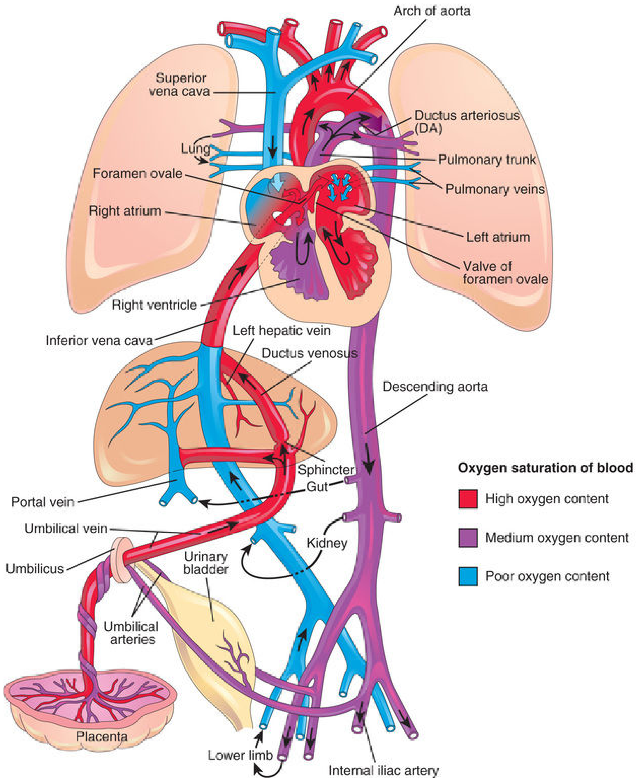
On approaching the liver, approximately 50% of umbilical venous blood passes directly through the ductus venosus - a fetal vessel connecting the umbilical vein to the inferior vena cava (IVC) - bypassing the hepatic sinusoids. The remaining 50% enters the portal system and liver sinusoids before joining the IVC via hepatic veins. A physiologic sphincter near the umbilical vein regulates this split; when IVC pressure is high (e.g., during uterine contractions), more blood is diverted to the portal vein to prevent cardiac overloading.
- The Developing Human: Clinically Oriented Embryology
2. Foramen Ovale
Blood entering the right atrium via the IVC is directed by the crista dividens (inferior border of the septum secundum) preferentially through the foramen ovale into the left atrium. This is the most oxygen-rich blood in the fetal heart. It mixes with a small volume of (low-oxygen) pulmonary venous return, then passes to the left ventricle and out the ascending aorta. Critically, the coronary arteries and carotid arteries are the first branches of the ascending aorta - so the heart muscle and brain receive the best-oxygenated blood in the fetal circulation.
A small amount of IVC blood is blocked by the crista dividens and remains in the right atrium, where it mixes with desaturated blood from the superior vena cava (SVC) draining the head and upper limbs.
- Langman's Medical Embryology; The Developing Human
3. Ductus Arteriosus
During fetal life, pulmonary vascular resistance is high (collapsed lungs), and pulmonary artery pressure is actually higher than aortic pressure. As a result, blood expelled by the right ventricle into the pulmonary trunk preferentially flows through the ductus arteriosus into the descending aorta, bypassing the lungs. Only about 10% of combined cardiac output reaches the lungs in fetal life.
The ductus arteriosus delivers relatively deoxygenated blood to the lower body, trunk, and umbilical arteries. Approximately 65% of descending aortic blood returns to the placenta for reoxygenation via the paired umbilical arteries (O2 saturation ~58-60%). The remaining 35% supplies abdominal viscera and the lower body.
- Ganong's; The Developing Human
Blood Flow Diagram (Medical Physiology)
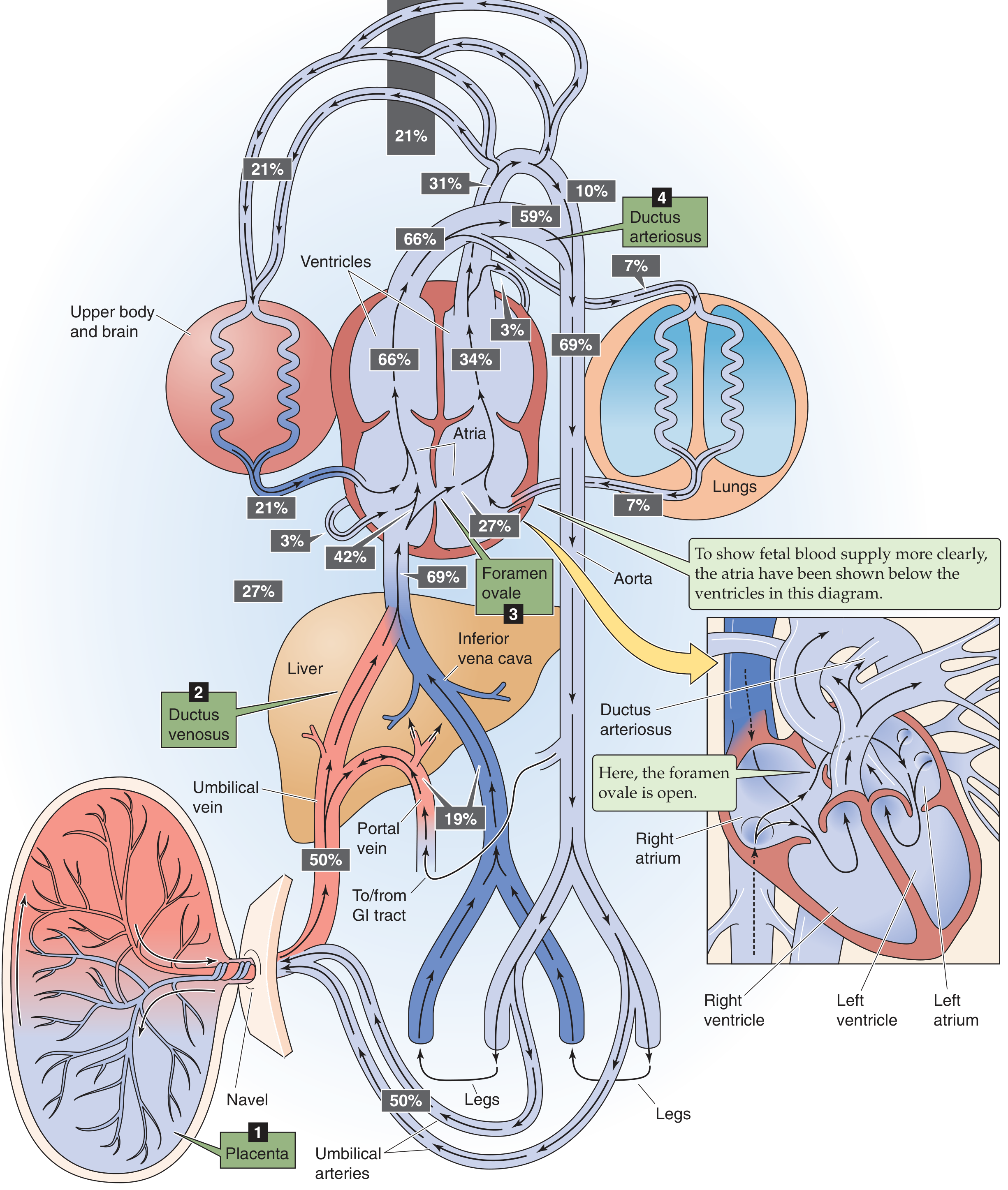
Complete Circuit - Step by Step
| Step | Vessel/Structure | O2 Status | Notes |
|---|---|---|---|
| 1 | Umbilical vein | ~80% sat. | From placenta to fetus |
| 2 | Ductus venosus | ~80% | Bypasses liver → IVC |
| 3 | IVC | ~67% | Mixed with deoxygenated lower limb blood |
| 4 | Right atrium → Foramen ovale | ~67% | Diverted to LA by crista dividens |
| 5 | Left ventricle → Ascending aorta | ~67% | To brain, heart (best oxygenated) |
| 6 | Right ventricle → Pulmonary trunk | ~52% | SVC blood (poor oxygen) |
| 7 | Ductus arteriosus → Descending aorta | ~52-60% | Bypasses lungs |
| 8 | Umbilical arteries | ~58% | Return to placenta for reoxygenation |
Sites of blood mixing (with progressive desaturation):
- Liver - portal blood mixes with umbilical vein blood
- IVC - lower limb deoxygenated blood mixes in
- Right atrium - SVC (head/arms) blood mixes in
- Left atrium - small amount of pulmonary venous blood mixes in
- Descending aorta - ductus arteriosus blood mixes with aortic blood
- Langman's Medical Embryology
Parallel vs. Serial Pumping
A key physiological feature: in the fetus the two ventricles work in parallel because the foramen ovale and ductus arteriosus connect the two sides of the circulation. The right ventricle output is slightly greater than left ventricular output (since it supplies the entire lower body and placenta via the ductus). At birth, this must rapidly convert to a serial system where both outputs must be equal.
- Ganong's Review of Medical Physiology
Circulatory Changes at Birth
At birth, two events trigger the transition:
1. Cord Clamping / Placental Cessation
- Loss of placental blood flow → fall in IVC and right atrial pressure
- Ductus venosus sphincter constricts → all blood now passes through hepatic sinusoids
2. First Breath (Lung Expansion)
- Pulmonary vascular resistance falls to <20% of fetal values with the first few breaths
- Pulmonary blood flow increases markedly
- Left atrial pressure rises above right atrial pressure
Closure of the Shunts
| Shunt | Mechanism | Timing |
|---|---|---|
| Foramen ovale | Increased LA pressure pushes septum primum against septum secundum (functional closure) | Immediate at birth; anatomic closure over weeks-months |
| Ductus arteriosus | High arterial O2 tension + bradykinin (released from inflated lungs) cause smooth muscle contraction; prostaglandin synthesis blocked at birth | Functional: within hours; 80% closed by 48 h; 100% by 96 h |
| Ductus venosus | Sphincter constriction when umbilical flow ceases | Functional closure at birth; becomes ligamentum venosum |
| Umbilical arteries | High O2, cold, handling → vasoconstriction within 3-5 min | Become medial umbilical ligaments |
| Umbilical vein | Passive collapse | Becomes ligamentum teres hepatis |
Note: In premature infants or with persistent hypoxia, the ductus arteriosus may remain patent longer. Indomethacin (a cyclooxygenase inhibitor) is used to pharmacologically close a patent ductus arteriosus (PDA) by blocking prostaglandin synthesis.
- The Developing Human: Clinically Oriented Embryology; Ganong's Review
Adult Derivatives of Fetal Structures
| Fetal Structure | Adult Derivative |
|---|---|
| Ductus venosus | Ligamentum venosum |
| Foramen ovale | Fossa ovalis |
| Ductus arteriosus | Ligamentum arteriosum |
| Umbilical vein | Ligamentum teres hepatis (round ligament of liver) |
| Umbilical arteries (proximal) | Superior vesical arteries |
| Umbilical arteries (distal) | Medial umbilical ligaments |
Special Features of Fetal Physiology
- Fetal hemoglobin (HbF): Contains γ-chains instead of β-chains; binds 2,3-DPG less avidly → higher O2 affinity → left-shifted dissociation curve. This allows HbF to extract O2 from maternal HbA across the placenta (the Bohr effect facilitates this transfer at the intervillous space).
- Pulmonary vasoconstriction: Maintained by hypoxia, low fetal lung PO2, and prostaglandins; reverses rapidly with the first breath and rising PaO2 (mediated by nitric oxide).
- Right ventricular dominance: The fetal right ventricle is thicker than the left (it works harder in utero). By end of the first month after birth, the left ventricle wall becomes thicker as systemic demands increase.
Sources: The Developing Human: Clinically Oriented Embryology (Moore et al.) | Langman's Medical Embryology | Ganong's Review of Medical Physiology, 26th ed.
This is a shared conversation. Sign in to Orris to start your own chat.