
Clinically give differential diagnosis of endometriosis , adenomyosis ,endometrial hyperplasia , endometrial cancer , endometrial stromal tumor , leiomyoma with respect to pathology
Clinical Differential Diagnosis of Uterine/Endometrial Conditions - Pathological Basis
1. ENDOMETRIOSIS
Pathological Definition
Gross Pathology
- Superficial implants: Red/blue/black "powder-burn" lesions on peritoneal surfaces
- Ovarian endometrioma: "Chocolate cyst" - cyst filled with old hemorrhagic material resembling chocolate syrup; wall contains endometrial glands and stroma
- Deep infiltrating: Firm, fibrotic nodules in rectovaginal septum, uterosacral ligaments; can invade rectum, bladder
- Repeated cyclic hemorrhage leads to dense fibrous adhesions that distort pelvic anatomy
Microscopic Pathology
- Ectopic foci contain all three components of functional endometrium: glands, stroma, and hemosiderin-laden macrophages (diagnostic triad)
- Glands respond to hormonal stimulation (proliferative/secretory changes)
- Surrounding fibrosis and hemosiderin deposition (siderophages)
- Stroma has characteristic small, dark stromal cells with scanty cytoplasm
- In old lesions, glands may be obliterated leaving only stroma + hemosiderin
- Malignant transformation: rare (~1%), mainly in ovarian endometriosis -> endometrioid or clear cell carcinoma
Molecular/Pathogenesis
- Elevated aromatase -> local estrogen production
- Increased prostaglandin E2, VEGF, MMPs
- Progesterone resistance at the stromal cell level
- Retrograde menstruation + immune failure to clear implants
Clinical Correlation
- Affects reproductive-age women (20s-30s)
- Classic triad: dysmenorrhea, dyspareunia, infertility ("3 Ds" + infertility)
- Symptoms cyclically worse with menstruation
- CA-125 may be mildly elevated
2. ADENOMYOSIS
Pathological Definition
Gross Pathology
- Uterus is symmetrically, diffusely enlarged (globular uterus) - may reach 200-300g (normal ~70g)
- Cut surface: thickened uterine wall, trabeculated myometrium; small hemorrhagic cystic spaces may be visible (corresponding to ectopic glands)
- No discrete fibroid nodule (unlike leiomyoma)
- Poorly circumscribed - key differentiating feature from leiomyoma
Microscopic Pathology
- Endometrial islands (glands + stroma) deep within myometrium, interposed between muscle bundles
- Islands are typically basalis-type endometrium - do NOT respond to cyclical hormonal changes (unlike eutopic endometrium)
- Surrounding smooth muscle shows reactive hypertrophy
- May coexist with endometriosis (~15% of cases)
Molecular/Pathogenesis
- Thought to arise from direct invagination of the basal endometrium into myometrium (invasion theory)
- Associated with increased uterine peristalsis and trauma (multiparity, prior uterine surgery)
- Estrogen-dependent (regresses after menopause)
Clinical Correlation
- Affects multiparous women in late reproductive years (35-50)
- Classic: secondary dysmenorrhea, menorrhagia, symmetrically enlarged tender uterus
- Uterus is soft and boggy - "always bigger before menstruation and smaller after"
- Definitive diagnosis only on hysterectomy specimen
3. ENDOMETRIAL HYPERPLASIA
Pathological Definition
WHO Classification (Two Major Types)
| Feature | Hyperplasia WITHOUT Atypia | Hyperplasia WITH Atypia (EIN) |
|---|---|---|
| Gland:stroma ratio | Increased | Markedly increased |
| Gland architecture | Variable size, dilated, back-to-back focally, some stroma retained | Complex, crowded, branching glands; cribriform/budding |
| Nuclear atypia | ABSENT - round/oval nuclei, uniform | PRESENT - enlarged, vesicular nuclei, prominent nucleoli, irregular chromatin |
| Mitoses | May be increased | Increased, may be abnormal |
| PTEN mutation | >20% | >20% (shared with carcinoma) |
| Risk of progression to carcinoma | 1-3% | 25-30% (considered precancerous = EIN) |
Gross Pathology
- Thickened, velvety, spongy endometrium
- Polypoidal projections may be present
- Proliferative type: pale, white-tan thickened mucosa
- No myometrial invasion
Microscopic Pathology
- Without atypia: increased G:S ratio, dilated cystic glands, back-to-back glands - NO cytologic atypia
- With atypia (EIN): glandular crowding, nuclear enlargement, round/vesicular nuclei, prominent nucleoli, loss of polarity; may have confluent glandular growth approaching carcinoma
- Stroma is compressed but present between glands (in contrast to carcinoma where stroma is replaced)
Molecular Pathogenesis
- PTEN inactivation is the earliest molecular event - found in >20% of both subtypes
- PI3K/AKT pathway overactivation
- Microsatellite instability in some cases (MLH1 promoter methylation)
- Lynch syndrome (germline MMR gene mutation) -> high lifetime risk
- Cowden syndrome (germline PTEN mutation) -> high risk of both endometrial hyperplasia and carcinoma
Clinical Correlation
- Perimenopausal women (45-55)
- Presents with abnormal uterine bleeding (irregular, intermenstrual, postmenopausal)
- Associated with obesity, PCOS, unopposed estrogen therapy, granulosa cell tumors
- Diagnosed by endometrial biopsy / D&C
4. ENDOMETRIAL CARCINOMA
Pathological Definition
TYPE I - Endometrioid Carcinoma (80%)
Gross Pathology
- Exophytic, polypoid or infiltrative mass in endometrial cavity
- Gray-white, friable, necrotic
- Myometrial invasion - depth determines staging (confined to endometrium = stage IA; >50% myometrium = stage IB)
- May extend to cervix (stage II), adnexa, parametrium (stage III)
Microscopic Pathology
- Well-differentiated (grade 1): closely mimics proliferative endometrium; back-to-back tubular glands with minimal stroma; <5% solid growth
- Grade 2: 6-50% solid growth; increasing atypia
- Grade 3: >50% solid growth; marked nuclear atypia, large nucleoli
- Myometrial invasion: irregular infiltrating glands with desmoplastic stroma - key feature
- Squamous differentiation may occur (adenoacanthoma = benign squames; adenosquamous = malignant squames)
- Immunostaining: ER+, PR+, vimentin+; p53 usually wild-type pattern
Molecular Pathogenesis
- PTEN mutations (early event, 30-80%)
- MMR gene mutations (MSI-H subtype)
- PIK3CA mutations
- KRAS, CTNNB1 (beta-catenin) mutations
- Arises in background of endometrial hyperplasia with atypia (EIN)
- Associated with Lynch syndrome (MLH1, MSH2 mutations)
TYPE II - Serous Carcinoma (15%)
Gross Pathology
- Often arises on atrophic endometrium (no preceding hyperplasia)
- Small primary tumor but early peritoneal spread
- Papillary/exophytic growth
Microscopic Pathology
- Small papillary tufts with complex branching; marked cytologic atypia
- Hobnail cells, slit-like glandular spaces
- High-grade by definition - marked nuclear pleomorphism, macronucleoli
- Psammoma bodies in ~30%
- Precursor lesion: Serous Endometrial Intraepithelial Carcinoma (SEIC) - morphologically malignant cells on atrophic endometrium
- IHC: diffuse/strong p53+ (mutant pattern), WT1 often positive; ER/PR negative
Molecular Pathogenesis
- TP53 mutation in nearly ALL cases (early event)
- PTEN and MMR mutations rare
- HER2 amplification in ~30%
- Overlap with high-grade serous ovarian carcinoma
- Associated with BRCA mutations
Clinical Correlation
- Endometrioid: postmenopausal obese women, 55-65 years; risk factors = obesity, nulliparity, diabetes, HRT (unopposed estrogen), tamoxifen use
- Serous: older, postmenopausal, 65-70 years; NOT associated with obesity/estrogen
- Both present with postmenopausal bleeding (endometrial biopsy is gold standard)
- Grade 1 endometrioid: 5-year survival ~90%; serous: poor prognosis due to advanced stage at presentation
5. ENDOMETRIAL STROMAL TUMOR
Pathological Definition
Classification
| Category | Behavior | Key Feature |
|---|---|---|
| Endometrial stromal nodule | Benign | Well-circumscribed, no vascular invasion |
| Low-grade endometrial stromal sarcoma (LG-ESS) | Low malignant potential | Infiltrating margins, lymphovascular invasion; recurs late |
| High-grade endometrial stromal sarcoma (HG-ESS) | Aggressive | High-grade round cell morphology; YWHAE fusion |
| Undifferentiated uterine sarcoma (UUS) | Highly aggressive | No endometrial stromal differentiation |
Gross Pathology
- Stromal nodule: discrete, well-circumscribed, yellow-tan, soft nodule in myometrium
- LG-ESS: worm-like plugs of tumor extending into myometrial vessels and lymphatics - classic "Indian filing" pattern; cut surface shows yellow-tan bands permeating myometrium; ill-defined borders
- HG-ESS/UUS: large fleshy hemorrhagic mass, necrosis
Microscopic Pathology
- Stromal nodule and LG-ESS: uniform, small cells resembling proliferative-phase stromal cells - oval nuclei, scant cytoplasm, spiral arteriole-like vasculature (tongue-like projections into myometrium in LG-ESS)
- LG-ESS: infiltrating margins with perivascular spread; low mitotic rate (<10/10 HPF); CD10+, ER+, PR+
- HG-ESS: high-grade round to spindle cells, >10 mitoses/10 HPF; YWHAE-NUTM2 gene fusion; CD10 negative; cyclin D1 overexpression
- UUS: pleomorphic, undifferentiated cells; no specific markers; diagnosis of exclusion
Molecular Pathogenesis
- LG-ESS: JAZF1-SUZ12 fusion (most common, ~50%); PHF1-JAZF1 fusions also described
- HG-ESS: YWHAE-NUTM2A/B fusion
- CD10 expression reflects stromal origin
Clinical Correlation
- Women in reproductive/perimenopausal age
- Presents with abnormal uterine bleeding and pelvic pain; often found incidentally
- LG-ESS: excellent prognosis with surgery; late recurrences after 5-10 years are characteristic; hormone-responsive (ER/PR+), responds to progestins/aromatase inhibitors
- HG-ESS/UUS: aggressive, rapid progression
6. UTERINE LEIOMYOMA (FIBROID)
Pathological Definition
Gross Pathology
- Multiple (vs. single) in most cases
- Sharply circumscribed, firm, gray-white nodules with classic whorled cut surface
- Well-delineated from surrounding myometrium (pseudocapsule)
- Locations:
- Intramural (most common, within myometrium)
- Submucosal (beneath endometrium - most symptomatic; cause AUB)
- Subserosal (below serosa; may be pedunculated)
- Cervical, broad ligament, parasitic (rare)
- Size: millimeters to 20+ cm
- Secondary changes (degeneration):
- Hyaline degeneration (most common) - white, homogeneous areas
- Cystic degeneration - central liquefaction
- Red/carneous degeneration - hemorrhagic infarction (especially during pregnancy) - clinically presents as acute pain
- Calcification ("womb stone") - in postmenopausal women
- Myxoid degeneration
Microscopic Pathology
- Interlacing bundles of uniform, bland smooth muscle cells - cigar-shaped nuclei, eosinophilic cytoplasm, no nuclear atypia
- Low mitotic rate (<5 mitoses/10 HPF)
- Hyalinized collagen between muscle bundles (in degenerating areas)
- Absence of tumor cell necrosis
- IHC: SMA+, desmin+, h-caldesmon+
- Key distinction from leiomyosarcoma: leiomyosarcoma has nuclear atypia, >10 mitoses/10 HPF, and coagulative tumor cell necrosis ("Stanford criteria")
Molecular Pathogenesis
- MED12 mutations in up to 70% (encodes RNA pol II transcription regulator)
- Chromosomal rearrangements (6p21, 12q15 - HMGA2)
- Estrogen receptor and progesterone receptor positive (hormone-dependent growth)
- Shrink after menopause; grow rapidly in pregnancy
- Recurrent chromosomal rearrangements similar to lipomas and endometrial polyps
Clinical Correlation
- Women of reproductive age (30s-40s), more common in African-American women
- May be asymptomatic (found incidentally on USS)
- Symptomatic: menorrhagia (submucosal), pelvic pressure/bulk symptoms (large intramural/subserosal), urinary frequency/retention, dysmenorrhea
- Infertility if submucosal or distorting cavity
- Uterus: irregularly enlarged, firm, non-tender (unless red degeneration)
Summary Comparison Table
| Parameter | Endometriosis | Adenomyosis | Endometrial Hyperplasia | Endometrial Carcinoma | Endometrial Stromal Tumor | Leiomyoma |
|---|---|---|---|---|---|---|
| Tissue of origin | Ectopic endometrium (outside uterus) | Endometrium invading myometrium | Endometrial glands + stroma (intrauterine) | Endometrial epithelium | Endometrial stroma | Myometrium (smooth muscle) |
| Uterine size | Normal (or enlarged if coexisting) | Symmetrically enlarged, globular | Normal or slightly enlarged | Enlarged | Variable | Irregularly enlarged |
| Gross appearance | Chocolate cysts, peritoneal implants, adhesions | Thickened wall, small hemorrhagic foci in myometrium | Thickened, velvety endometrium | Polypoid/infiltrative endometrial mass | Worm-like myometrial plugs / yellow-tan nodule | Whorled, firm, circumscribed nodule(s) |
| Microscopic hallmark | Glands + stroma + hemosiderin outside uterus | Endometrial islands in myometrium (basalis type) | Increased G:S ratio (with/without atypia) | Invasive carcinomatous glands with desmoplasia | Uniform stromal cells, spiral arterioles, vascular invasion (LG-ESS) | Interlacing bland smooth muscle bundles |
| Nuclear atypia | Absent | Absent | Absent (without atypia) / Present (EIN/with atypia) | Present (grade-dependent) | Low (LG-ESS) / High (HG-ESS) | Absent |
| Myometrial invasion | N/A (ectopic) | Present (defining feature) | Absent | Present (diagnostic of malignancy) | Present (LG-ESS by definition) | No true invasion (pseudocapsule) |
| Key molecular marker | Aromatase overexpression, VEGF | - | PTEN mutation (early) | PTEN, MMR (Type I); TP53 (Type II) | JAZF1-SUZ12 (LG-ESS); YWHAE-NUTM2 (HG-ESS) | MED12 mutation (70%) |
| IHC | ER+, PR+ | ER+, PR+ | ER+, PR+ | ER/PR+ (Type I); p53++ (Type II) | CD10+, ER/PR+ (LG-ESS) | SMA+, desmin+, h-caldesmon+ |
| Age group | Reproductive (20s-30s) | Late reproductive (35-50) | Perimenopausal (45-55) | Postmenopausal (55-65) | Reproductive/perimenopausal | Reproductive (30s-40s) |
| Typical bleeding pattern | Dysmenorrhea (cyclic) | Menorrhagia + secondary dysmenorrhea | AUB, intermenstrual | Postmenopausal bleeding | AUB | Menorrhagia (submucosal) |
| Malignant potential | Rare (ovarian endometrioma) | None | 1-3% (without atypia); 25-30% (EIN) | Malignant by definition | Nodule: none; LG-ESS: low; HG-ESS: high | Very rare (<0.5%) -> leiomyosarcoma |
| Associated syndromes | - | - | Lynch, Cowden syndrome | Lynch syndrome, Cowden syndrome | - | - |
| Diagnosis | Laparoscopy (gold standard) | Hysterectomy specimen | Endometrial biopsy / D&C | Endometrial biopsy / D&C + staging | Hysterectomy + histology | Ultrasound + histology |
Key Pathological Differentiating Points
Phyllodes tumor , fibroadenoma , pagets disease of breast , fibrocystic disease of breast , breast carcinoma differential diagnosis clinical features and patholgy tablutation only
Differential Diagnosis: Breast Conditions - Clinical Features & Pathology
TABLE 1: CLINICAL FEATURES
| Parameter | Fibroadenoma | Phyllodes Tumor | Fibrocystic Disease | Paget's Disease of Breast | Breast Carcinoma |
|---|---|---|---|---|---|
| Age | 15-25 yrs (peak); young women | >30 yrs (peak 6th decade; 10-20 yrs later than fibroadenoma) | Reproductive age (25-50 yrs) | Postmenopausal (usually >50 yrs) | >50 yrs (most common); younger if BRCA mutation |
| Onset | Gradual, slow growing | Rapidly growing, can become massive | Cyclic, waxes and wanes with menstrual cycle | Insidious; chronic skin change at nipple | Gradual (early) or rapid (aggressive subtypes) |
| Lump characteristics | Single (usually), firm, rubbery, smooth, well-defined, mobile ("breast mouse"); 2-3 cm | Large, bosselated (lobulated) surface, mobile on chest wall; can be massive; rarely fixes to skin until late | Multiple, bilateral, tender nodularity; ill-defined; may fluctuate in size | May or may not have palpable mass; if palpable - deep to nipple | Single, hard, irregular, painless mass; poorly mobile; may be fixed to skin or chest wall |
| Tenderness | Non-tender (usually) | Non-tender | Tender, cyclically worse pre-menstrually | Nipple/areola burning or itching | Usually non-tender (unless inflammatory carcinoma) |
| Skin changes | None | Skin ulceration possible (pressure necrosis) in very large tumors | None (excludes skin thickening, edema, discoloration) | Eczematoid, weeping, erythematous, scaly lesion of nipple/areola - persists despite topical treatment | Dimpling (skin tethering), peau d'orange (lymphatic obstruction), nipple retraction, skin fixation, ulceration |
| Nipple changes | None | None | None | Nipple erosion, crusting, oozing, bloody/serous discharge | Nipple retraction, discharge (bloody), Paget change (if DCIS involves nipple ducts) |
| Lymph nodes | Not enlarged | Axillary spread RARE (even malignant type spreads hematogenously) | Not enlarged | May be involved if underlying invasive carcinoma present | Axillary LN enlargement (hard, fixed in advanced disease) |
| Bilaterality | Usually unilateral; can be bilateral | Unilateral | Bilateral (hallmark) | Unilateral | Usually unilateral (bilateral in LCIS or multifocal disease) |
| Multiplicity | Usually solitary; giant or multiple variants exist | Usually solitary | Multiple bilateral cysts and nodules | Single nipple/areola lesion | Usually solitary; multifocal/multicentric possible |
| Cyclical variation | None | None | Yes - symptoms worsen with menstruation | None | None |
| Associated features | - | - | Mastalgia, lumpy breast, occasional nipple discharge | Delayed diagnosis (6-12 months) due to resemblance to eczema/dermatitis | Weight loss, metastatic signs (bone pain, breathlessness) in advanced disease |
| Mammography | Well-circumscribed, radiopaque mass; may have coarse "popcorn" calcifications | Large, well-defined mass; leaf-like architecture on imaging | Multiple cysts, diffuse density changes | Subareolar mass/DCIS pattern; skin thickening at nipple | Spiculated/irregular radiodense mass; microcalcifications (DCIS); architectural distortion |
| Diagnosis | Ultrasound + FNAC/biopsy; clinical if typical features in young woman | Core biopsy/excision (FNA inadequate); margin assessment | Clinical + USS; biopsy if solid component | Punch biopsy of nipple (Paget cells); bilateral mammography | Core needle biopsy (gold standard); FNAC; excision biopsy |
| Malignant potential | Low (RR 1.5-1.7); complex type RR 3-4 | Benign 75%: no mets; Malignant: hematogenous mets ~30% | Depends on subtype (see pathology table) | Nearly 100% associated with underlying DCIS or invasive carcinoma | Malignant by definition |
TABLE 2: GROSS PATHOLOGY
| Parameter | Fibroadenoma | Phyllodes Tumor | Fibrocystic Disease | Paget's Disease | Breast Carcinoma |
|---|---|---|---|---|---|
| Size | 2-3 cm (most); giant >5 cm (in adolescents) | Few cm to massive (entire breast); largest of all breast tumors | Variable cysts (small to several cm) | Nipple/areola lesion - small skin lesion | 2-3 cm at presentation (if no screening); <1 cm if screen-detected |
| Shape | Round/oval, smooth, well-circumscribed | Large, bosselated, lobulated; "leaf-like" protrusions on cut surface (phyllodes = Greek for "leaf") | Multiple irregular cysts; thickened, rubbery breast tissue | Erythematous, weeping eczematoid nipple lesion; may have underlying mass | Irregular, stellate/spiculated; "crab-like" (carcinoma = crab in Greek) |
| Consistency | Firm, rubbery | Fleshy, soft to firm; hemorrhagic/necrotic in malignant type | Cysts: fluctuant; fibrotic areas: firm | Crusted, erythematous skin surface at nipple | Hard (scirrhous); grating on cut surface due to desmoplastic stroma and calcifications |
| Margins | Sharp, well-defined capsule; well-circumscribed | Benign: circumscribed; Malignant: widely infiltrative, ill-defined | No discrete capsule; diffuse | Skin-based; no discrete mass unless underlying carcinoma | Irregular, infiltrative margins; no capsule |
| Cut surface | Rubbery, white/gray; no necrosis; no adipose tissue (radiodense) | Cleft-like spaces / leaf-like projections of stroma covered by epithelium protruding into cystic spaces | Multiple cysts (blue-domed cysts = apocrine lined); fibrous stroma | Skin thickening/ulceration at nipple; underlying DCIS not grossly visible | Gray-white chalky desmoplastic areas; foci of necrosis (comedo type); chalky-white streaks; occasional grating on cutting |
| Mobility | Highly mobile (pseudocapsule - moves freely) | Mobile (even when large); does NOT fix to chest wall until very late | Not a discrete mobile mass | - | Fixed or restricted mobility (skin/chest wall fixation in advanced disease) |
| Necrosis | Absent | Present in malignant phyllodes | Absent | Absent | Present (comedo necrosis in high-grade DCIS; central necrosis in high-grade invasive) |
TABLE 3: MICROSCOPIC (HISTOLOGICAL) PATHOLOGY
| Parameter | Fibroadenoma | Phyllodes Tumor | Fibrocystic Disease | Paget's Disease | Breast Carcinoma |
|---|---|---|---|---|---|
| Tissue of origin | Intralobular stroma (biphasic - stroma + epithelium) | Intralobular stroma (biphasic - more cellular stroma) | Terminal duct-lobular unit (TDLU) | Lactiferous ducts -> epidermis of nipple | Ductal or lobular epithelium |
| Key histological pattern | Intralobular stroma surrounds, pushes, and distorts epithelial tubules; pericanalicular (stroma encircles glands) or intracanalicular (stroma compresses glands into slits) pattern | Leaf-like fronds of hypercellular stroma covered by epithelium projecting into cystic spaces; stroma dominates | Cysts (apocrine metaplasia lining), adenosis (lobular enlargement), fibrosis (dense stromal collagen), epithelial hyperplasia | Paget cells: large, pale, vacuolated intraepidermal adenocarcinoma cells within squamous epithelium of nipple, without crossing basement membrane - DCIS extending up lactiferous ducts into skin | Invasive carcinoma NST: irregular infiltrating glands/nests/cords/solid sheets with desmoplastic stroma; Lobular: single-file ("Indian file") infiltration; Tubular: well-formed open tubules |
| Stromal cellularity | Low; loose myxoid or fibrous stroma | Increased (benign < borderline < malignant); key distinguishing feature from fibroadenoma | Variable fibrosis; no atypical stromal cells | Absent (skin lesion) | Desmoplastic fibrous stroma (reactive); angiofibrotic stroma |
| Nuclear atypia | Absent (bland, uniform) | Benign: minimal; Borderline: moderate; Malignant: marked pleomorphism | Absent in simple changes; present in atypical ductal/lobular hyperplasia | Paget cells: large, round nuclei, prominent nucleoli, pale cytoplasm, mucin-containing vacuoles | Grade 1: minimal; Grade 2: moderate; Grade 3: marked, irregular, large nuclei, prominent nucleoli |
| Mitotic rate | Very low (<3/10 HPF) | Benign: <4/10 HPF; Borderline: 4-9/10 HPF; Malignant: >10/10 HPF | Absent/rare | Absent (in Paget cells) to present if underlying invasive carcinoma | Variable; Grade 3: high; Grade 1: low |
| Margins/Invasion | No invasion; pushes surrounding tissue | Benign: pushing; Malignant: infiltrating into surrounding breast parenchyma | No invasion | Paget cells remain above basement membrane (intraepidermal); DCIS component is in situ | Basement membrane disruption - invasive carcinoma crosses BM into stroma |
| Basement membrane | Intact | Intact (benign); disrupted (malignant) | Intact | Intact (Paget cells are intraepidermal, do NOT invade dermis - unlike melanoma) | Disrupted (defining feature of invasive carcinoma) |
| Necrosis | Absent | Absent (benign); Present (malignant) | Absent | Absent | Comedo necrosis (high-grade DCIS); present in grade 3 invasive carcinoma |
| Calcifications | May be present (coarse, "popcorn") | May be present | Psammomatous or lobular calcifications | Absent (in nipple skin) | Microcalcifications (DCIS: linear/branching or casting type); present in ~50% of invasive carcinomas |
| Special cell types | - | - | Apocrine metaplasia (columnar, pink, granular cytoplasm); foam cells in cyst fluid | Paget cells: PAS+, mucin+, CK7+, HER2+ (often); GATA3+ | Invasive NST: no specific cell type; Mucinous: lakes of mucin; Lobular: discohesive cells with intracytoplasmic mucin vacuoles (signet ring) |
TABLE 4: MOLECULAR / IHC / PATHOGENESIS
| Parameter | Fibroadenoma | Phyllodes Tumor | Fibrocystic Disease | Paget's Disease | Breast Carcinoma |
|---|---|---|---|---|---|
| Key molecular alteration | MED12 mutation (2/3 of cases) - transcriptional regulator; same as uterine leiomyoma | MED12 + RARA mutations (shared with fibroadenoma); additionally TERT, TP53, RB mutations in malignant type | Estrogen/progesterone imbalance; no driver mutations in simple lesions; PTEN in atypical hyperplasia | HER2 overexpression (>90% of cases); associated with underlying HER2+ DCIS/invasive carcinoma | Luminal (ER/PR+): PTEN, PIK3CA, GATA3 mutations; HER2: HER2 amplification; TNBC: BRCA1, TP53 mutations |
| IHC markers | ER+, PR+ (epithelial component); CD34+ (stroma) | CD34+ (stroma); ER/PR variable; p53 + in malignant type | ER+, PR+ | CK7+, HER2+ (>90%), GATA3+, mucin (PAS+); S100-, HMB45- (distinguishes from melanoma) | Luminal: ER+, PR+, HER2-; HER2+: ER-, HER2+++; TNBC: ER-, PR-, HER2- (triple negative); CK5/6+, EGFR+ |
| Hormone sensitivity | Estrogen-dependent growth; may enlarge in pregnancy; involutes post-menopause | Variable | Estrogen excess; responds to cyclic hormonal changes | Not hormonally driven | ER+ (~70%): estrogen-driven growth; responds to anti-estrogen therapy |
| Hereditary association | Carney complex (myxoid type - PRKAR1A mutation) | - | - | - | BRCA1/BRCA2 (10% of all breast cancers); Lynch syndrome; Li-Fraumeni (TP53); Cowden syndrome (PTEN) |
| Risk of malignancy | Simple: RR 1.5-1.7; Complex (with epithelial hyperplasia + family Hx): RR 3.0-4.0 | Benign: no mets, can recur locally; Malignant: hematogenous mets ~30% (NOT lymphatic - LN dissection contraindicated) | No atypia: no increased risk; Usual ductal hyperplasia: RR 1.5-2; Atypical ductal/lobular hyperplasia: RR 4-5; ADH + family Hx: RR ~10 | Nearly 100% have underlying DCIS; ~50% have invasive carcinoma (when palpable mass present) | Malignant |
TABLE 5: QUICK-REFERENCE SUMMARY (Key Distinguishing Features)
| Feature | Fibroadenoma | Phyllodes Tumor | Fibrocystic Disease | Paget's Disease | Breast Carcinoma |
|---|---|---|---|---|---|
| Nature | Benign biphasic tumor | Biphasic tumor (benign/borderline/malignant) | Benign fibrocystic change | Intraepidermal carcinoma (almost always with underlying DCIS/invasive Ca) | Malignant epithelial tumor |
| Classic age | 15-25 yrs | >30 yrs (6th decade) | 25-50 yrs | Postmenopausal | >50 yrs |
| Classic lump | Mobile, rubbery, well-defined ("breast mouse") | Large, bosselated, rapidly growing | Bilateral, tender nodularity; no discrete lump | Nipple/areola lesion (weeping, eczematoid) | Hard, irregular, fixed, painless |
| Bilateral | Usually No | No | Yes | No | Usually No |
| Cyclical symptoms | No | No | Yes | No | No |
| Skin/nipple | Normal | Ulceration (pressure) if huge | Normal | Eczematoid nipple - diagnostic | Dimpling, peau d'orange, nipple retraction |
| Margins (micro) | Circumscribed, pushing | Circumscribed (benign) / Infiltrative (malignant) | Diffuse, no margins | Intraepidermal (above BM) | Invasive (crosses BM) |
| Stroma | Scant, myxoid | Hypercellular with atypia | Fibrotic | - | Desmoplastic |
| Mitoses | <3/HPF | <4 (benign), 4-9 (borderline), >10 (malignant) | Absent | Absent | High in grade 3 |
| Key IHC | CD34+, ER+ | CD34+, p53+ (malignant) | ER+ | CK7+, HER2+ (>90%) | ER/PR/HER2 (molecular subtype) |
| Key mutation | MED12 | MED12+RARA+TERT/TP53 | - | - | BRCA1/2, TP53, PIK3CA, PTEN |
| Metastasis | Never | Hematogenous only (malignant, ~30%) | Never | Depends on underlying invasive component | Lymphatic + hematogenous |
| Treatment | Conservative / excision if >3 cm or atypical | Wide local excision (2 cm margin); NO axillary LN dissection | Symptomatic; reassurance; biopsy if suspicious | Excision ± mastectomy depending on extent of DCIS | Surgery + RT + chemotherapy + endocrine/targeted therapy |
Make a clinical decision flowchart for breast lesion diagnosis
A detailed clinical decision flowchart for breast lesion diagnosis. The flowchart starts at the top with "BREAST LESION / COMPLAINT" and branches through the following decision pathway: Step 1: Clinical Presentation Three branches from the top: - Left: "Nipple/areola skin change (eczematoid, weeping, crusting)" - Center: "Breast lump / mass" - Right: "Bilateral tender nodularity + cyclic pain" The RIGHT branch leads directly to a diagnosis box: "FIBROCYSTIC DISEASE - Reassure, analgesia, follow-up mammography" The LEFT branch leads to: "Punch biopsy of nipple" → "Paget cells (CK7+, HER2+, large pale intraepidermal cells)?" → YES → "PAGET'S DISEASE OF BREAST → Bilateral mammography → Underlying DCIS (50%) or Invasive Carcinoma (50%) → Staging & treatment" The CENTER "Breast lump" branch goes to Step 2: Patient Age - Branch A: Age 15-25 years - Branch B: Age 25-50 years - Branch C: Age >50 years / Postmenopausal Branch A (15-25 yrs): → Ultrasound → "Well-defined, homogeneous, hypoechoic oval mass?" → YES → FNAC/Core biopsy → "Bland biphasic proliferation, pushing margins, MED12 mutation" → FIBROADENOMA → Conservative management or excision if >3cm Branch B (25-50 yrs): → Triple Assessment: Clinical + Mammography/USS + Core Biopsy → "Large, bosselated, rapidly growing?" → YES → Core biopsy → "Hypercellular stroma, leaf-like fronds, increased mitoses" → PHYLLODES TUMOR → Grade (Benign/Borderline/Malignant) → Wide local excision 2cm margin, NO axillary dissection Branch C (>50 yrs / Postmenopausal): → "Hard, irregular, fixed, painless mass?" → YES → Mammography → "Spiculated mass / microcalcifications?" → YES → Core needle biopsy → "Invasive carcinoma - desmoplastic stroma, infiltrating glands, broken basement membrane" → BREAST CARCINOMA → IHC: ER/PR/HER2 → Molecular subtype → Staging (TNM) → Multidisciplinary treatment At the bottom: A summary legend box with color coding: - Green boxes: Benign conditions (Fibroadenoma, Fibrocystic Disease) - Orange boxes: Potentially malignant (Phyllodes, Paget's) - Red boxes: Malignant (Breast Carcinoma) - Blue boxes: Investigations - Gray boxes: Decision points The flowchart uses clean arrows, distinct colored boxes, professional medical typography, white background, clearly labeled investigation steps at each decision point. Include a small note at bottom: "Triple Assessment = Clinical Examination + Imaging + Histopathology"
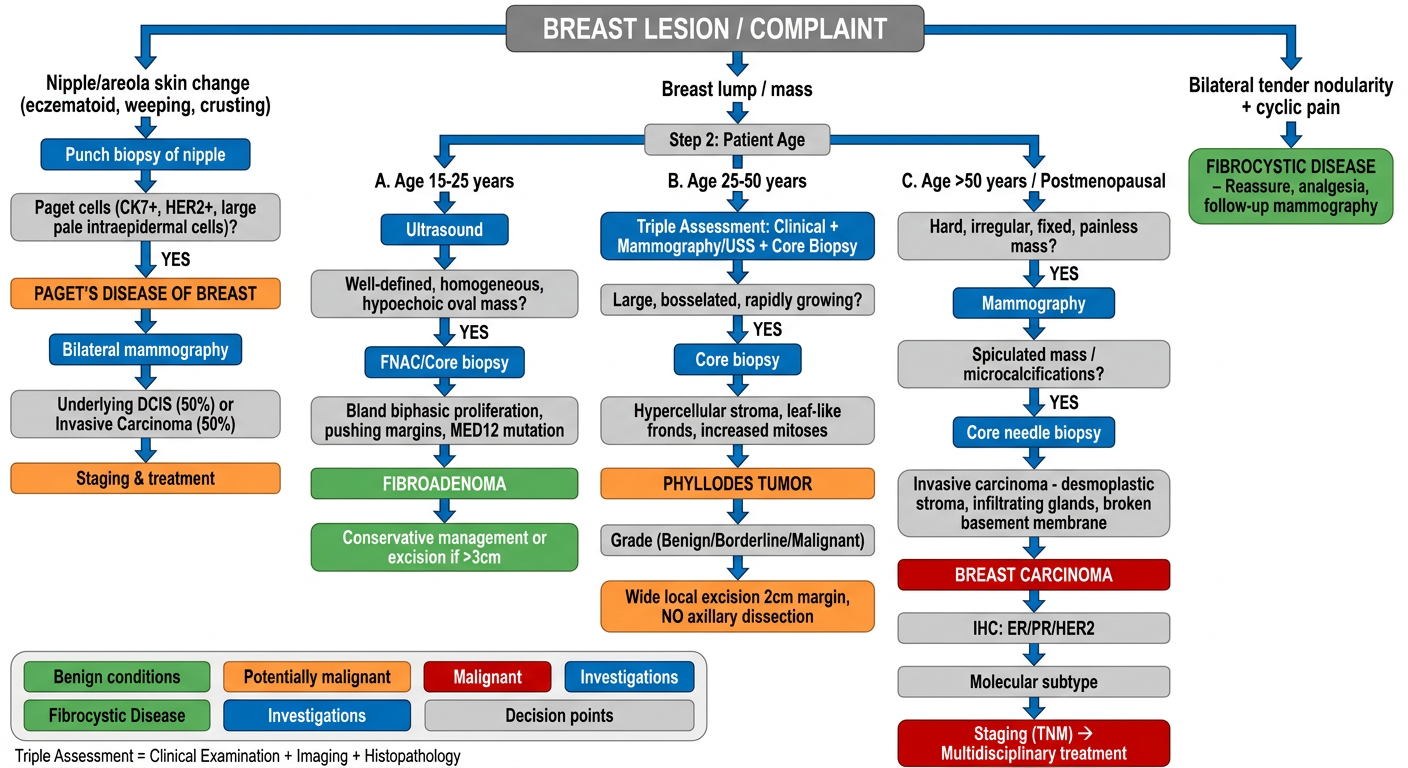
CLINICAL DECISION FLOWCHART: BREAST LESION DIAGNOSIS
                        BREAST LESION / COMPLAINT
                               |
          ┌────────────────────┼──────────────────────┐
          ▼                    ▼                       ▼
  Nipple/Areola          Breast Lump/Mass         Bilateral Tender
  Skin Change                                     Nodularity + Cyclic
  (eczematoid,                                    Pain / Mastalgia
  weeping, crusting)                                    │
          │                                             ▼
          ▼                                    FIBROCYSTIC DISEASE
  Punch Biopsy Nipple                         Reassure + analgesia
          │                                   Mammography follow-up
          ▼
  Paget cells? (CK7+, HER2+,
  large pale intraepidermal
  vacuolated cells)
          │
    YES ──┤
          ▼
  PAGET'S DISEASE OF BREAST
  → Bilateral Mammography
  → Palpable mass?
    - NO: Underlying DCIS → BCS + RT
    - YES: Underlying Invasive Ca → Staging + MDT
              BREAST LUMP - TRIPLE ASSESSMENT
        (Clinical + Imaging + Histopathology)
                          │
         ┌────────────────┼────────────────┐
         ▼                ▼                ▼
      Age 15-25         Age 25-50       Age >50 / PMB

         │                │                │
         ▼                ▼                ▼
    Ultrasound       Triple Assessment   Mammogram
    (preferred        (MMG + USS +       (spiculated
    over MMG in       core biopsy)       mass /
    young women)                         microcalcifications?)
         │                │                │
         ▼                ▼                ▼
   Well-defined,    Large, bosselated,   Hard, irregular,
   smooth,          rapidly growing,     fixed, painless
   homogeneous      mobile mass          mass
   oval mass?
         │                │                │
    FNAC/Core          Core Biopsy       Core Needle Biopsy
    Biopsy                │              (MANDATORY)
         │                │                │
         ▼                ▼                ▼
   Bland biphasic    Hypercellular     Invasive carcinoma
   proliferation,    stroma, leaf-     (broken BM,
   pushing margins,  like fronds,      desmoplastic stroma,
   MED12 mutation    mitoses counted   infiltrating glands)
         │                │                │
         ▼                ▼                ▼
   FIBROADENOMA      PHYLLODES TUMOR   BREAST CARCINOMA
         │                │                │
    <3 cm: observe   Grade:            IHC: ER / PR / HER2
    >3 cm: excise    Benign (<4/HPF)          │
    Giant: enucleate Border (4-9/HPF)   ┌────┴─────────┐
    Complex + FHx:   Malignant(>10/HPF) │              │
    close follow-up        │          Luminal      HER2 / TNBC
                           ▼            │              │
                  WLE (2 cm margin)   Endocrine    Chemo ±
                  NO axillary LN      therapy +    Targeted
                  dissection          surgery      therapy
                  Malignant: monitor              + surgery
                  for hematogenous mets
Key Rule - Triple Assessment
| Arm | Tool |
|---|---|
| 1. Clinical | Inspection + palpation (size, mobility, skin, LN) |
| 2. Imaging | USS (<35 yrs) / Mammography (>35 yrs) / MRI (high-risk) |
| 3. Histology | Core needle biopsy (preferred) or FNAC |
Concordance of all three = definitive diagnosis. Discordance = repeat biopsy / excision.
Lung carcinoma differential diagnosis
Lung Carcinoma: Differential Diagnosis - Clinical Features & Pathology
TABLE 1: OVERVIEW & EPIDEMIOLOGY
| Parameter | Squamous Cell Carcinoma | Adenocarcinoma | Large Cell Carcinoma | Small Cell Carcinoma (SCLC) | Carcinoid Tumor | Large Cell Neuroendocrine Carcinoma (LCNEC) |
|---|---|---|---|---|---|---|
| Frequency | ~25-30% | Most common (~40%) | ~10-15% | ~13% | ~1-2% | ~3% of NSCLC |
| WHO 2021 Group | NSCLC | NSCLC | NSCLC | SCLC / Neuroendocrine | Neuroendocrine (low/intermediate grade) | High-grade neuroendocrine |
| Smoking association | Strongest (almost exclusively smokers) | Moderate (also non-smokers, women, young) | Strong | Very strong (reconsider diagnosis if non-smoker) | None / Weak | Strong |
| Peak age | 50-70 yrs | 50-70 yrs (also <45 yrs) | 50-70 yrs | 50-70 yrs | Any age (typical carcinoid: young adults) | 50-70 yrs |
| Sex predilection | Male > Female | Female ≥ Male (rising in women); most common in never-smokers | Male > Female | Male > Female | No strong sex predilection | Male > Female |
TABLE 2: LOCATION & GROSS PATHOLOGY
| Parameter | Squamous Cell Carcinoma | Adenocarcinoma | Large Cell Carcinoma | Small Cell Carcinoma | Carcinoid Tumor | LCNEC |
|---|---|---|---|---|---|---|
| Location | Central (hilar, main/lobar bronchi) | Peripheral (subpleural, distal bronchioles/alveoli) | Peripheral (usually) | Central (perihilar, mediastinal) | Central (80%) or peripheral | Central or peripheral |
| Gross appearance | Gray-white, firm, cavitating mass at hilum; necrotic center; may involve bronchus with mucosal thickening | Gray-white peripheral nodule, "scar cancer" appearance; may have pleural puckering; no cavitation (usually) | Large, fleshy, necrotic peripheral mass; no distinctive gross features | Soft, white, fish-flesh perihilar mass; extensive mediastinal involvement; marked nodal enlargement | Well-circumscribed, tan-yellow, intrabronchial polypoid lesion; smooth surface; rarely necrotic | Large fleshy mass, necrosis, may cavitate |
| Cavitation | Common (central necrosis) | Rare | Rare | Rare (too rapid growth) | Absent | Occasional |
| Pleural involvement | Less common (central) | Common (peripheral - pleural puckering, effusion) | Common (peripheral) | Pericardial/pleural effusion (mediastinal spread) | Rare | Variable |
| Size | Large central mass | 1-5 cm (peripheral); lepidic type may be diffuse | Large (>3 cm usually) | Large mediastinal mass with hilar widening | Small (1-3 cm), well-defined | Variable, often large |
TABLE 3: MICROSCOPIC PATHOLOGY
| Parameter | Squamous Cell Carcinoma | Adenocarcinoma | Large Cell Carcinoma | Small Cell Carcinoma | Carcinoid Tumor | LCNEC |
|---|---|---|---|---|---|---|
| Cell of origin | Bronchial squamous epithelium (via squamous metaplasia) | Bronchiolar/alveolar epithelium, Clara cells, bronchioalveolar stem cells | Undifferentiated large epithelial cells | Neuroendocrine cells (Kulchitsky cells) of bronchial epithelium | Neuroendocrine cells (Kulchitsky cells) | Neuroendocrine cells |
| Cell size | Large, polygonal | Variable; columnar/cuboidal | Large (>3 lymphocyte diameters) | Small (<3 lymphocyte diameters); oval/spindle | Medium-sized, uniform, round-oval | Large (>3 lymphocyte diameters) |
| Cytoplasm | Abundant, eosinophilic, keratinized | Variable; may be vacuolated (mucin-containing) | Abundant | Scant (key feature) | Moderate, granular, eosinophilic | Moderate to abundant |
| Nuclei | Large, hyperchromatic; intercellular bridges | Vesicular; prominent nucleoli | Large, vesicular, prominent nucleoli | Finely granular "salt & pepper" chromatin; nuclear molding; absent/inconspicuous nucleoli | Round, uniform, "salt & pepper" chromatin; no nucleoli | Vesicular chromatin; frequent prominent nucleoli (key distinction from SCLC) |
| Key histologic pattern | Keratinization (keratin pearls, individual cell keratinization); intercellular bridges; stratified squamous growth | Acinar, papillary, micropapillary, solid, or lepidic (growth along alveolar walls without invasion - AIS pattern); mucin production | Sheets/nests of large undifferentiated cells; no glands, keratin, neuroendocrine features | Sheets of small cells; nuclear molding; crush artifact (Azzopardi effect - DNA encrustation of vessel walls); extensive necrosis | Organoid nesting, trabecular, insular patterns; uniform "salt & pepper" chromatin; ≤2 mitoses/2mm² (typical) or 2-10 (atypical) | Organoid nesting, trabecular, rosette-like, palisading patterns with LARGE cells; necrosis; >10 mitoses/2mm² |
| Necrosis | Central necrosis (causes cavitation) | Occasional | Present | Extensive, coagulative | Absent (typical); may be present in atypical | Prominent |
| Mitotic rate | Variable (moderate) | Variable | High | Very high (avg >60 mitoses/2mm²) | Typical: ≤2/2mm²; Atypical: 2-10/2mm² | >10 mitoses/2mm² (defining criterion) |
| Precursor lesion | Squamous dysplasia → CIS (central airways) | Atypical adenomatous hyperplasia (AAH) → AIS → minimally invasive adenocarcinoma → invasive adenocarcinoma | No defined precursor | DIPNECH (diffuse idiopathic pulmonary neuroendocrine cell hyperplasia) | DIPNECH | - |
TABLE 4: IHC & MOLECULAR MARKERS
| Marker | Squamous Cell Ca | Adenocarcinoma | Large Cell Ca | Small Cell Ca | Carcinoid | LCNEC |
|---|---|---|---|---|---|---|
| CK5/6 | + | - | - | - | - | - |
| p40 / p63 | + | - | - | - | - | - |
| TTF-1 | - | + (75-85%) | - | + (90%) | +/- | +/- |
| Napsin A | - | + | - | - | - | - |
| Synaptophysin | - | - | - | + | + | + |
| Chromogranin A | - | - | - | + | + | + |
| CD56 | - | - | - | + | + | + |
| Ki-67 | Intermediate | Intermediate | High | Very high (>50-80%) | Low (<20% typical; 20-40% atypical) | High (>40%) |
| p53 | Frequent | Less common | - | Very common | Rare | Common |
| Key driver mutations | TP53, CDKN2A, SOX2, FGFR1 | EGFR (10-15% Western; 30-40% Asian), KRAS (30%), ALK fusion (4-6%), ROS1, HER2, MET, BRAF | TP53, various | TP53, RB1, MYC amplification | MEN1 (familial), sporadic | RB1, TP53 (similar to SCLC) |
| Molecular subtype relevance | PDL1 expression (immunotherapy); FGFR1 amplification (targeted) | EGFR/ALK/ROS1/KRAS targeted therapy | PDL1 high in some | Rarely targetable | MEN-1 gene (familial) | SCLC-like treatment often used |
TABLE 5: CLINICAL FEATURES & PRESENTATION
| Parameter | Squamous Cell Ca | Adenocarcinoma | Large Cell Ca | Small Cell Ca | Carcinoid | LCNEC |
|---|---|---|---|---|---|---|
| Typical presentation | Cough, hemoptysis, post-obstructive pneumonia (central) | Often asymptomatic initially (peripheral); pleural effusion; dyspnea | Late-presenting large peripheral mass; non-specific symptoms | Rapidly progressive cough, dyspnea, wheezing, hemoptysis, mediastinal symptoms | Episodic hemoptysis, recurrent pneumonia, wheeze (central); incidental nodule (peripheral) | Aggressive like SCLC; cough, dyspnea, weight loss |
| Hemoptysis | Common (central, ulcerates bronchus) | Less common | Variable | Common | Common (vascular polypoid lesion) | Variable |
| Obstructive symptoms | Common (endobronchial growth -> lobar collapse, post-obstructive pneumonitis) | Less common | Rare | Very common (massive mediastinal adenopathy) | Common (central intrabronchial polyp) | Variable |
| Superior vena cava syndrome | Occasional | Rare | Rare | Classic (massive mediastinal involvement) | Rare | Rare |
| Pancoast tumor | Can occur (apical) | Can occur (apical) | Can occur | Rare | Rare | Rare |
| Horner syndrome | Apical (Pancoast) | Apical (Pancoast) | Rare | Mediastinal invasion | Rare | Rare |
| Pleural effusion | Less common | Common (peripheral, pleural seeding) | Common | Pericardial + pleural | Rare | Variable |
| Metastasis pattern | Regional LN, bone, liver, brain, adrenal | Bone, brain, liver, adrenal, pleural seeding | Brain, liver, bone | Widespread early: brain, bone, liver, adrenal (>50% at presentation) | Rare (typical); liver mets (atypical) | Brain, liver, bone |
TABLE 6: PARANEOPLASTIC SYNDROMES
| Syndrome | Squamous Cell Ca | Adenocarcinoma | Large Cell Ca | Small Cell Ca | Carcinoid | LCNEC |
|---|---|---|---|---|---|---|
| Hypercalcemia (PTHrP) | Classic | - | Rare | - | - | - |
| SIADH (ADH secretion) | Rare | Rare | Rare | Classic | - | - |
| Cushing syndrome (ectopic ACTH) | Rare | Rare | Rare | Classic | Rare | Rare |
| Eaton-Lambert syndrome (anti-VGCC) | - | - | - | Classic | - | - |
| Carcinoid syndrome (5-HT, histamine) | - | - | - | - | Classic (liver mets required for typical carcinoid) | - |
| Hypertrophic pulmonary osteoarthropathy | Common | Common | Variable | Rare | Rare | Rare |
| Gynecomastia (HCG) | - | - | Classic (large cell) | - | - | - |
| Trousseau syndrome (migratory thrombophlebitis) | - | Classic (adenocarcinoma, mucin) | - | - | - | - |
TABLE 7: RADIOLOGY & STAGING
| Parameter | Squamous Cell Ca | Adenocarcinoma | Large Cell Ca | Small Cell Ca | Carcinoid | LCNEC |
|---|---|---|---|---|---|---|
| CXR pattern | Central hilar mass, lobar collapse, cavitating lesion | Peripheral nodule/mass, pleural effusion, "ground glass" (AIS) | Large peripheral opacification | Hilar/mediastinal widening, bilateral | Coin lesion, well-defined oval/round endobronchial mass | Large peripheral mass |
| CT features | Spiculated or lobulated central mass; luminal narrowing; post-obstructive consolidation | Spiculated peripheral mass; "ground glass" with solid component (part-solid); pleural retraction | Large necrotic peripheral mass | Large central mass with extensive mediastinal/hilar LN; may have SVC compression | Well-defined, homogeneous, non-calcified nodule; may see "sail sign" (intralobar atelectasis) | Large mass, often with necrosis |
| PET-FDG | High avidity | High avidity | High avidity | High avidity | Low/variable avidity (low-grade carcinoids may be FDG-negative; use octreotide scan/68Ga-DOTATATE instead) | High avidity |
| Staging system | TNM (8th ed AJCC/IASLC) | TNM | TNM | TNM (also IASLC limited vs extensive stage for treatment) | TNM | TNM |
| Resectability | If NSCLC stages I-IIIA: resectable | If NSCLC stages I-IIIA: resectable | If NSCLC stages I-IIIA: resectable | Rarely resectable (<10%); chemotherapy is primary treatment | Resectable (most typical carcinoids); surgical cure common | Often not resectable |
TABLE 8: PROGNOSIS & TREATMENT
| Parameter | Squamous Cell Ca | Adenocarcinoma | Large Cell Ca | Small Cell Ca | Carcinoid | LCNEC |
|---|---|---|---|---|---|---|
| 5-year survival (all stages) | ~15-20% | ~20-25% (better with targeted therapy) | ~10-15% | <5% (extensive); ~20% (limited) | Typical: >90%; Atypical: ~60-70% | ~15-30% |
| Primary treatment | Surgery (I-III); RT + Chemo (III); Immunotherapy (PDL1+) | Surgery (I-IIIA); EGFR/ALK/ROS1 inhibitors; Immunotherapy; Chemotherapy | Surgery (early); Chemo ± RT (advanced) | Chemotherapy (etoposide + cisplatin/carboplatin) + RT; PCI (prophylactic cranial irradiation if limited stage) | Surgical resection (lobectomy/sleeve resection); somatostatin analogues (symptomatic/metastatic) | SCLC-like chemo regimens; surgery if resectable |
| Targeted therapy | FGFR1, MET (limited); Pembrolizumab | EGFR (gefitinib, erlotinib, osimertinib); ALK (crizotinib, alectinib); ROS1 (crizotinib); KRAS G12C (sotorasib); BRAF (dabrafenib) | Immunotherapy (PD-L1) | Atezolizumab + chemo (1st line) | Somatostatin analogues (octreotide, lanreotide); everolimus | Limited |
| Key distinguishing prognostic factor | Stage + PDL1 expression | Driver mutation status (EGFR/ALK) | Stage | Extremely chemosensitive but almost always relapses | Grade (typical vs atypical carcinoid) | Poor prognosis overall |
QUICK SUMMARY TABLE: KEY DIFFERENTIATING FEATURES
| Feature | SCC | Adeno | Large Cell | SCLC | Carcinoid | LCNEC |
|---|---|---|---|---|---|---|
| Location | Central | Peripheral | Peripheral | Central | Central (80%) | Any |
| Smoking | +++ | ++ (also non-smokers) | ++ | +++ | - | ++ |
| Cavitation | Yes | No | No | No | No | Occasional |
| Keratin pearls | Yes | No | No | No | No | No |
| Nuclear molding | No | No | No | Yes | No | No |
| "Salt & pepper" chromatin | No | No | No | Yes | Yes | Coarse |
| Mucin production | No | Yes | No | No | No | No |
| TTF-1 | - | + | - | + | +/- | +/- |
| Synaptophysin/CD56 | - | - | - | + | + | + |
| Key paraneoplastic | Hypercalcemia | Trousseau | Gynecomastia | SIADH, Cushing, Eaton-Lambert | Carcinoid syndrome | - |
| Key mutation | TP53, SOX2 | EGFR, KRAS, ALK | TP53 | RB1, TP53 | MEN1 | RB1, TP53 |
| Prognosis | Intermediate | Best (targeted Rx) | Poor | Worst | Best | Poor |